. 2017 Jan 23;35(4):672-679.
doi: 10.1016/j.vaccine.2016.11.073. Epub 2016 Dec 21.

B cell and antibody responses in mice induced by a putative cell surface peptidase of Pneumocystis murina protect against experimental infection

Affiliations

B cell and antibody responses in mice induced by a putative cell surface peptidase of Pneumocystis murina protect against experimental infection

Sanbao Ruan et al. Vaccine. .

Abstract

Rationale: Pneumocystis pneumonia is a major cause of morbidity and mortality in HIV-infected subjects, cancer patients undergoing chemotherapy and solid organ transplant recipients. No vaccine is currently available. By chemical labeling coupled with proteomic approach, we have identified a putative surface protein (SPD1, Broad Institute gene accession number PNEG_01848) derived from single suspended P. murina cysts. SPD1 was expressed in an insect cell line and tested for vaccine development.

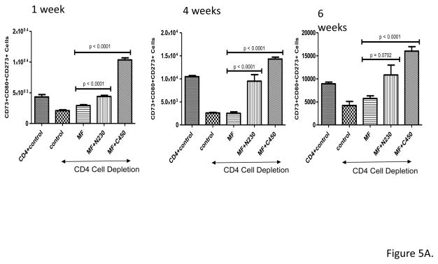
Methods: Mice were immunized with SPD1 plus adjuvant MF-59 by subcutaneous injection. Three weeks after the last immunization, CD4+ cells were depleted with anti-CD4 antibody GK1.5. The mice were then challenged with 2×10⁵Pneumocystis organisms. Mice were sacrificed at 4 and 6weeks after PC challenge. Spleen/lung cells and serum were harvested. B cells and memory B cells were assessed via flow cytometry. Specific Pneumocystis IgG antibody was measured by ELISA before and after challenge. Infection burden was measured as real-time PCR for P. murina rRNA.

Results: Normal mice infected with Pneumocystis mounted a serum IgG antibody response to SPD1. Serum from rhesus macaques exposed to Pneumocystis showed a similar serum IgG response to purified SPD1. SPD1 immunization increased B cell and memory B cell absolute cell counts in CD4-depleted Balb/c mice post Pneumocystis challenge in spleen and lung. Immunization with SPD1 significantly increased specific Pneumocystis IgG antibody production before and after challenge. Mice immunized with SPD1 showed significantly decreased P. murina copy number compared with mice that did not receive SPD1 at 6weeks after challenge.

Conclusion: Immunization with SPD1 provides protective efficacy against P. murina infection. SPD1 protection against Pneumocystis challenge is associated with enhanced memory B cell production and higher anti-Pneumocystis IgG antibody production. SPD1 is a potential vaccine candidate to prevent or treat pulmonary infection with Pneumocystis.

Keywords: CD4 T-cell deficiency; Pneumocystis pneumonia; Pneumocystis vaccine.


Conflict of interest statement

STATEMENT Conflicts of interest: none

Figures

Figure 1
Figure 1
Putative sequence of SPD1 (Broad Institute accession no. PNEG_01848). Peptide segments (in red letters) were identified by chemical labeling and LC-MS based proteomic approach and supposed to be the exposed regions of the protein. N-terminal of matured SPD1 was determined by the labeling of A34 in peptide “A34TTATLL TENIKKPEGD HYSYR55”. C-terminal of SPD1 was also confirmed by the detection of peptide “N1050SLKLSPVPLP VMKWSSYY1068”. Signal peptide of protein SPD1 is underlined. Fragment SPD1-N230 (AA 34-262) contains the experimentally identified N-terminus of the matured protein, and fragment SPD1-C450 (AA 627-1068) contains the C terminus. SPD1-N230 and C450 are selected as targets for vaccination
Figure 2
Figure 2
Control mice and simian/human immunodeficiency virus (SHIV)-infected rhesus macaques exposed to Pneumocystis have serum IgG antibodies to SPD1 N230 and C450. Serum from control mice challenged with Pneumocystis (PC serum) and from three rhesus macaques containing high titers of antibody to PC kexin (1791, 3613, and R697) were tested by ELISA for IgG antibodies to SPD1 N230 and C450. −serum represent sera from mice that were not immunized. Data represent mean ± SD, n=6 for mice, n=3 for nonhuman primates.
Figure 3
Figure 3
Antibody responses to SPD1 after immunization with SPD1. Mice were immunized with N230 or C450 plus MF59 adjuvant or adjuvant alone 3 times at 3 week intervals. −control mice were not immunized. +control mice represent sera from mice that cleared a PC challenge. Two weeks after the last immunization, serum was tested for IgG antibodies to the respective immunogen. Panel A. N230 immunization. Panel B. C450 immunization. Results are compared to animals vaccinated with adjuvant alone and to serum from unexposed mice (−control) and from mice who cleared an experimental challenge with Pneumocystis (+control). Note that control mice show an antibody response to SPD1 but that vaccination with SPD1 produces a greater serum IgG response. Data represent mean ± SD, n=10.
Figure 4
Figure 4
Tissue B cell responses after SPD1 immunization and challenge with Pneumocystis. Mice were immunized with N230 + MF59 adjuvant or C450 + MF59 adjuvant and then challenged with Pneumocystis. Some mice received adjuvant alone (MF) or no vaccine (CD4+ control and control). CD4 depletion was started 3 weeks after the last immunization (2 days prior to PC challenge) and continued weekly, except for the CD4+ control mice. Total B cells were assayed by flow cytometry at 1,4, and 6 weeks post PC challenge. Panel A. Lung B cell responses (1,4,6 weeks), Panel B. Spleen B cell responses (1, 4, 6 weeks). Data represent mean ± SD, n=10 at each time interval.
Figure 4
Figure 4
Tissue B cell responses after SPD1 immunization and challenge with Pneumocystis. Mice were immunized with N230 + MF59 adjuvant or C450 + MF59 adjuvant and then challenged with Pneumocystis. Some mice received adjuvant alone (MF) or no vaccine (CD4+ control and control). CD4 depletion was started 3 weeks after the last immunization (2 days prior to PC challenge) and continued weekly, except for the CD4+ control mice. Total B cells were assayed by flow cytometry at 1,4, and 6 weeks post PC challenge. Panel A. Lung B cell responses (1,4,6 weeks), Panel B. Spleen B cell responses (1, 4, 6 weeks). Data represent mean ± SD, n=10 at each time interval.
Figure 5
Figure 5
Tissue memory B cell responses after SPD1 immunization and challenge with Pneumocystis. Mice were immunized with N230 + MF59 adjuvant or C450 + MF59 adjuvant and then challenged with Pneumocystis. Some mice received adjuvant alone (MF) or no vaccine (CD4+ control and control). CD4 depletion was started 3 weeks after the last immunization (2 days prior to PC challenge) and continued weekly, except for the CD4+ control mice. Memory B cells (CD45R+, CD73+, CD80+, CD273+) were assayed by flow cytometry at 1, 4, and 6 weeks post PC challenge. Panel A. Lung memory B cell responses (1, 4, 6 weeks), Panel B. Spleen memory B cell responses (1, 4, 6 weeks). Data represent mean ± SD, n=10.
Figure 5
Figure 5
Tissue memory B cell responses after SPD1 immunization and challenge with Pneumocystis. Mice were immunized with N230 + MF59 adjuvant or C450 + MF59 adjuvant and then challenged with Pneumocystis. Some mice received adjuvant alone (MF) or no vaccine (CD4+ control and control). CD4 depletion was started 3 weeks after the last immunization (2 days prior to PC challenge) and continued weekly, except for the CD4+ control mice. Memory B cells (CD45R+, CD73+, CD80+, CD273+) were assayed by flow cytometry at 1, 4, and 6 weeks post PC challenge. Panel A. Lung memory B cell responses (1, 4, 6 weeks), Panel B. Spleen memory B cell responses (1, 4, 6 weeks). Data represent mean ± SD, n=10.
Figure 6
Figure 6
Clearance of infection in SPD1 immunized mice. Mice were immunized with N230 + MF59 adjuvant or C450 + MF59 adjuvant and then challenged with Pneumocystis. Some mice received adjuvant alone (MF) or no vaccine (CD4+ control and control). CD4 depletion was started 3 weeks after the last immunization (2 days prior to PC challenge) and continued weekly, except for the CD4+ control mice. Lung tissue burden of Pneumocystis was assayed by real time PCR at 1, 4, and 6 weeks after infectious challenge. Data represent mean ± SD, n=10/experimental group, experiment repeated x1.

Similar articles

Cited by

References

    1. Brown GD, Denning DW, Gow NA, Levitz SM, Netea MG, TCW Hidden killers: human fungal infections. Sci Transl Med. 2012;4:165rv113. - PubMed
    1. Qian J, Cutler JE, Cole RB, Cai Y. MALDI-TOF mass signatures for differentiation of yeast species, strain grouping and monitoring of morphogenesis markers. Anal Bioanal Chem. 2008;392(3):439–449. - PubMed
    1. Zheng M, Cai Y, Eddens T, Ricks DM, Kolls JK. Novel pneumocystis antigen discovery using fungal surface proteomics. Infect Immun. 2014;82(6):2417–2423. - PMC - PubMed
    1. Ruan S, McKinley L, Zheng M, et al. Interleukin-12 and host defense against murine Pneumocystis pneumonia. Infect Immun. 2008;76(5):2130–2137. - PMC - PubMed
    1. Shelllito J, Suzara J, Blumenfeld W, Beck J, Steger H, Ermak T. A new model of Pneumocystis carinii pneumonia in mice selectively depleted of helper T lymphocytes. J Clin Invest. 1990;85:1686–1693. - PMC - PubMed

Publication types

MeSH terms