

Pathogenic conversion of coagulase-negative staphylococci
- PMID: 28012900
- PMCID: PMC5274588
- DOI: 10.1016/j.micinf.2016.12.002
Pathogenic conversion of coagulase-negative staphylococci
Abstract
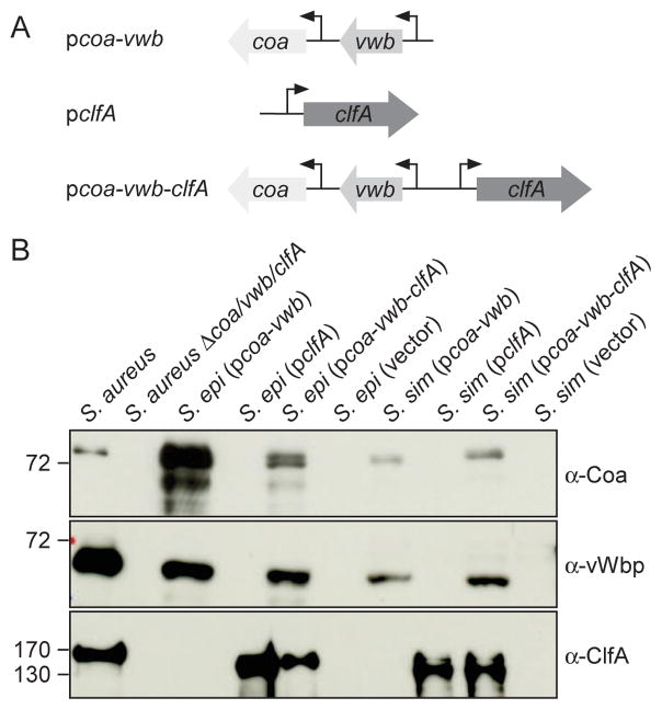
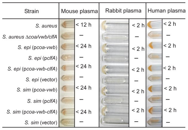
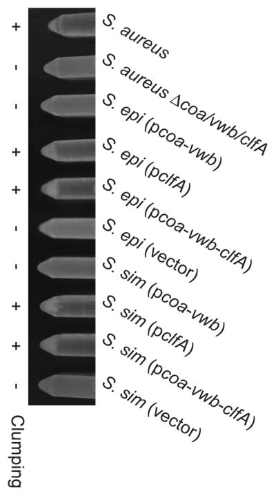
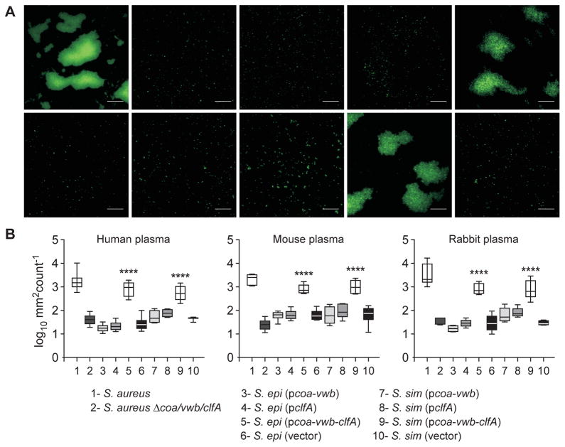
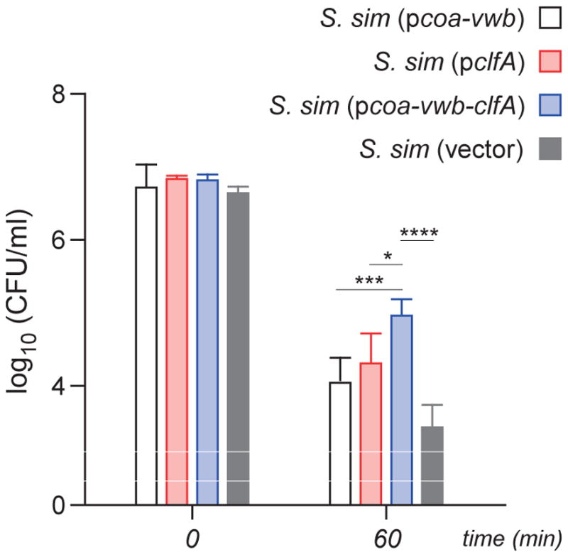
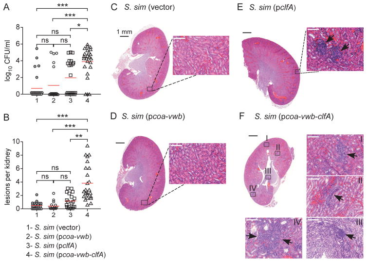
Humans and animals are colonized by members of the genus Staphylococcus, however only some of these species evolved to cause invasive disease. The genetic basis for conversion of commensal staphylococci into pathogens is not known. We hypothesized that Staphylococcus aureus genes for coagulation and agglutination in vertebrate blood (coa, vwb and clfA) may support pathogenic conversion. Expression of coa and vwb in Staphylococcus epidermidis or Staphylococcus simulans supported a coagulase-positive phenotype but not the ability to cause disease in a mouse model of bloodstream infection. However, the simultaneous expression of coa, vwb and clfA in coagulase-negative staphylococci enabled bacterial agglutination in plasma and enhanced survival of S. simulans in human whole blood. Agglutination of S. simulans in the bloodstream of infected mice upon expression of coa, vwb and clfA provided also a mean for dissemination and replication in distal organs. Thus, the acquisition of genes for bacterial agglutination with fibrin appear sufficient for the conversion of commensal staphylococci into invasive pathogens.
Keywords: Abscess; Agglutination; Clumping; Coagulation; Staphylococcus; Virulence.
Copyright © 2016 Institut Pasteur. Published by Elsevier Masson SAS. All rights reserved.
Conflict of interest statement
HKK, DM and OS declare a conflict of interest as inventors of patent applications related to the development of S. aureus vaccines.
Figures
Similar articles
-
Evolutionary and Functional Analysis of Coagulase Positivity among the Staphylococci.mSphere. 2021 Aug 25;6(4):e0038121. doi: 10.1128/mSphere.00381-21. Epub 2021 Aug 4. mSphere. 2021. PMID: 34346700 Free PMC article.
-
Preventing Staphylococcus aureus sepsis through the inhibition of its agglutination in blood.PLoS Pathog. 2011 Oct;7(10):e1002307. doi: 10.1371/journal.ppat.1002307. Epub 2011 Oct 20. PLoS Pathog. 2011. PMID: 22028651 Free PMC article.
-
Contribution of Staphylococcus aureus Coagulases and Clumping Factor A to Abscess Formation in a Rabbit Model of Skin and Soft Tissue Infection.PLoS One. 2016 Jun 23;11(6):e0158293. doi: 10.1371/journal.pone.0158293. eCollection 2016. PLoS One. 2016. PMID: 27336691 Free PMC article.
-
Coagulase-Negative Staphylococci Pathogenomics.Int J Mol Sci. 2019 Mar 11;20(5):1215. doi: 10.3390/ijms20051215. Int J Mol Sci. 2019. PMID: 30862021 Free PMC article. Review.
-
Coagulase negative staphylococci and their participation in pathogenesis of human infections.Bratisl Lek Listy. 2006;107(11-12):448-52. Bratisl Lek Listy. 2006. PMID: 17425165 Review.
Cited by
-
Antimicrobial resistance patterns and biofilm formation of coagulase-negative Staphylococcus species isolated from subclinical mastitis cow milk samples submitted to the Onderstepoort Milk Laboratory.BMC Vet Res. 2019 Nov 26;15(1):420. doi: 10.1186/s12917-019-2175-3. BMC Vet Res. 2019. PMID: 31771575 Free PMC article.
-
Nutritional Regulation of the Sae Two-Component System by CodY in Staphylococcus aureus.J Bacteriol. 2018 Mar 26;200(8):e00012-18. doi: 10.1128/JB.00012-18. Print 2018 Apr 15. J Bacteriol. 2018. PMID: 29378891 Free PMC article.
-
Time-Dependent Changes in the Intestinal Microbiome of Gilts Exposed to Low Zearalenone Doses.Toxins (Basel). 2019 May 24;11(5):296. doi: 10.3390/toxins11050296. Toxins (Basel). 2019. PMID: 31137638 Free PMC article.
-
Prevalence and antimicrobial resistance of coagulase negative staphylococci clinical isolates from Ethiopia: a meta-analysis.BMC Microbiol. 2018 May 25;18(1):43. doi: 10.1186/s12866-018-1188-6. BMC Microbiol. 2018. PMID: 29801462 Free PMC article.
-
CodY controls the SaeR/S two-component system by modulating branched-chain fatty acid synthesis in Staphylococcus aureus.J Bacteriol. 2024 Nov 21;206(11):e0019124. doi: 10.1128/jb.00191-24. Epub 2024 Oct 9. J Bacteriol. 2024. PMID: 39382300 Free PMC article.
References
-
- Lowy FD. Staphylococcus aureus infections. New Engl J Med. 1998;339:520–32. - PubMed
-
- Götz F, Bannerman T, Schleifer K-H. The Genera Staphylococcus and Macrococcus. In: Dworkin M, Falkow S, Rosenberg E, Schleifer K-H, Stackebrandt E, editors. The Prokaryotes. Springer; New York: 2006. pp. 5–75.
Publication types
MeSH terms
Substances
Grants and funding
LinkOut - more resources
Full Text Sources
Other Literature Sources
Molecular Biology Databases
