. 2017 Mar 24;429(6):858-872.
doi: 10.1016/j.jmb.2016.12.014. Epub 2016 Dec 21.

Interaction of E. coli Hsp90 with DnaK Involves the DnaJ Binding Region of DnaK

Affiliations

Interaction of E. coli Hsp90 with DnaK Involves the DnaJ Binding Region of DnaK

Andrea N Kravats et al. J Mol Biol. .

Abstract

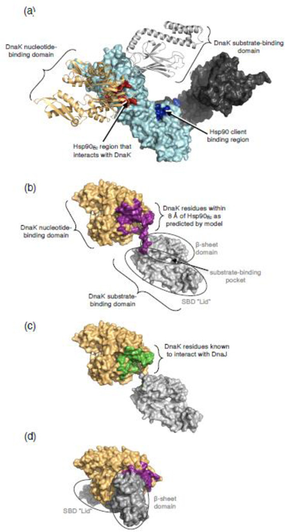
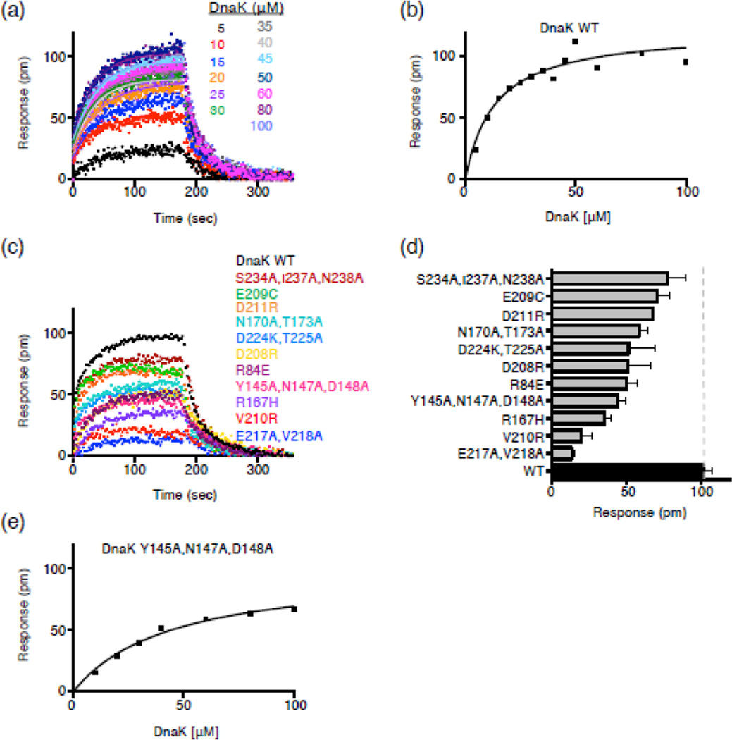
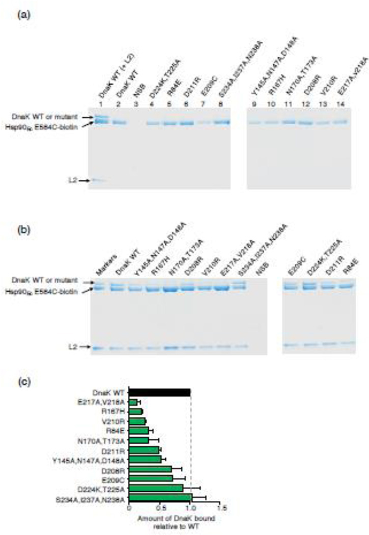
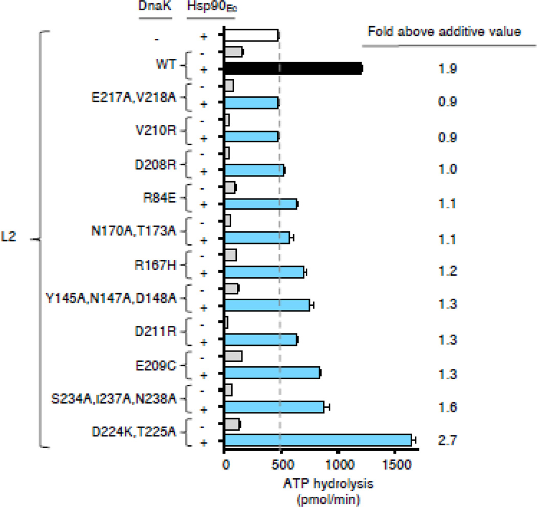
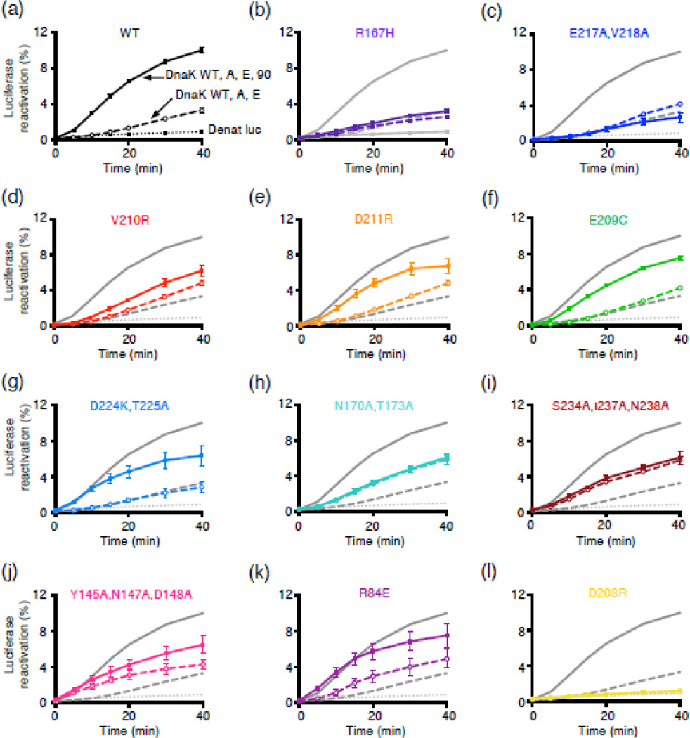
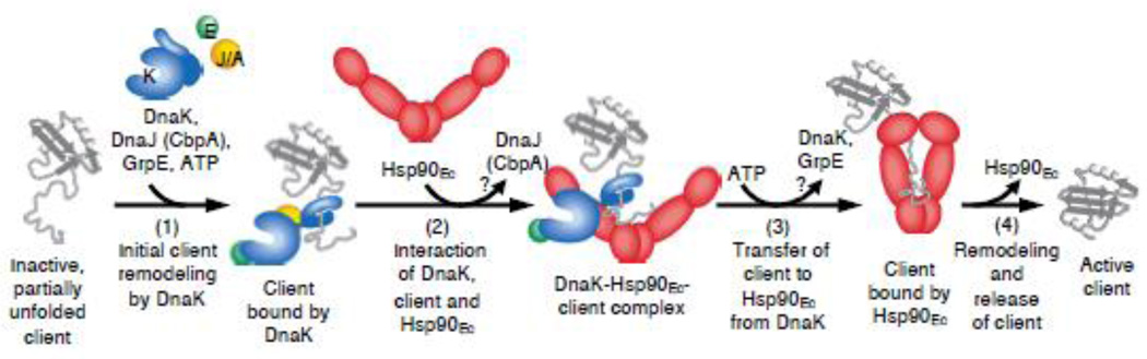
The 90-kDa heat shock protein (Hsp90) is a widely conserved and ubiquitous molecular chaperone that participates in ATP-dependent protein remodeling in both eukaryotes and prokaryotes. It functions in conjunction with Hsp70 and the Hsp70 cochaperones, an Hsp40 (J-protein) and a nucleotide exchange factor. In Escherichia coli, the functional collaboration between Hsp90Ec and Hsp70, DnaK, requires that the two chaperones directly interact. We used molecular docking to model the interaction of Hsp90Ec and DnaK. The top-ranked docked model predicted that a region in the nucleotide-binding domain (NBD) of DnaK interacted with a region in the middle domain of Hsp90Ec. We then made substitution mutants in DnaK residues suggested by the model to interact with Hsp90Ec. Of the 12 mutants tested, 11 were defective or partially defective in their ability to interact with Hsp90Ecin vivo in a bacterial two-hybrid assay and in vitro in a bio-layer interferometry assay. These DnaK mutants were also defective in their ability to function collaboratively in protein remodeling with Hsp90Ec but retained the ability to act with DnaK cochaperones. Taken together, these results suggest that a specific region in the NBD of DnaK is involved in the interaction with Hsp90Ec, and this interaction is functionally important. Moreover, the region of DnaK that we found to be necessary for Hsp90Ec binding includes residues that are also involved in J-protein binding, suggesting a functional interplay among DnaK, DnaK cochaperones, and Hsp90Ec.

Keywords: CbpA; Hsp40; HtpG; molecular chaperone; protein remodeling.


Figures

Fig. 1
Fig. 1
Regions of interaction on DnaK and Hsp90Ec. (a) Docked model of the apo structure of Hsp90Ec [21] and ADP-bound DnaK [31] as determined using ZDOCK and ZRANK and described in Materials and Methods. Hsp90Ec is shown as a surface rendering with one protomer in dark gray and one protomer in light cyan. The DnaK interacting region of Hsp90Ec [30] is shown in red while the client binding region is in blue [14]. DnaK in the ADP-bound conformation is shown as a ribbon model with the NBD in light orange and the SBD in light gray. (b) DnaK in the ADP-bound conformation [31] showing residues (purple) on DnaK within 8 Å of Hsp90Ec as predicted by the docked model in (a). In (b-d) DnaK is shown as a surface rendering with the NBD in light orange and the SBD in light gray. (c) DnaK in the ADP-bound conformation [31] showing residues (green) experimentally identified as interacting with DnaJ [–40]. (d) DnaK in the ATP-bound conformation [32] showing residues (purple) on DnaK within 8 Å of Hsp90Ec as predicted by the docked model in (a). In the ATP-bound conformation, only some of the DnaK residues within 8 Å of Hsp90Ec in the model are surface exposed. Images in (a-d) were made using PyMOL (Schrodinger, LLC; www.pymol.org).
Fig. 2
Fig. 2
Identification of DnaK amino acid residues involved in Hsp90Ec interaction in vivo. (a) Model of E. coli DnaK in the ADP-bound conformation [31] with the mutated residues used in this study shown as CPK models. The NBD is colored light orange and the SBD is gray. DnaK was rendered using PyMOL. (b, c) Interaction between DnaK wild-type or mutant and Hsp90Ec in a bacterial two-hybrid system in vivo, as described in Materials and Methods. Interaction was measured by monitoring β-galactosidase activity on MacConkey indicator plates (b) and in liquid assays (c). In (b), a representative plate from three independent experiments is shown with colonies labeled 1 through 16. For colonies 2 through 13, all DnaK substitution mutants, named by substituted residues, have been constructed in T25-DnaK and are present in reactions with the T18-Hsp90Ec construct. In (c), β-galactosidase activity is shown as mean ± SEM (n = 3) and are also presented in Supplemental Table S5.
Fig. 3
Fig. 3
DnaK NBD substitution mutants are defective in direct interaction with Hsp90Ec in vitro. (a) Bio-Layer Interferometry (BLI) was used to monitor the kinetics of association and dissociation between biotinylated Hsp90Ec and DnaK wild-type as described in Materials and Methods. Representative curves are shown for numerous concentrations of DnaK as indicated. The single-exponential fit of the association step was used to obtain the response value (pm) for the plateau plotted in (b). (b) Steady-state analysis of the DnaK wild-type-Hsp90Ec interaction. The response value (pm) for the plateau obtained in (a) is plotted vs. each DnaK concentration and fit as described in Materials and Methods. The Kd and Bmax for the Hsp90Ec interaction with DnaK wild-type are 13.4 ± 3.3 µM and 0.12 ± 0.01 nm, respectively. (c) Curves showing association and dissociation of 50 µM DnaK wild-type (black) or mutant (colored) and biotinylated Hsp90Ec using BLI. (d) Average plateau response value for the interaction between DnaK wild-type or mutant and biotinylated Hsp90Ec. Data are plotted as mean ± SEM (n=2 or more) and are also presented in Supplemental Table S5. (e) Steady-state analysis of the DnaKY145A,N147A,D148A-Hsp90Ec interaction as described in (b). The Kd and Bmax for the Hsp90Ec interaction with DnaKY145A,N147A,D148A are 44.4 ± 7.5 µM and 0.10 ± 0.01 nm, respectively.
Fig. 4
Fig. 4
DnaK mutants exhibit defective interaction with Hsp90Ec in the presence of L2. Hsp90Ec-biotin was incubated with DnaK wild-type or mutant, without or with L2. Hsp90Ec-biotin associated proteins were monitored using a pull-down assay and analyzed by Coomassie blue staining following SDS-PAGE as described in Materials and Methods. (a) In control experiments, Hsp90Ec-biotin was incubated with DnaK wild-type with L2 (lane 1) or without L2 (lane 2) or with DnaK mutant proteins without L2 (lanes 4-14). DnaK wild-type is seen in association with Hsp90Ec-biotin in the presence of L2 (lane 1), but not in the absence of L2 by this analysis (lane 2); however, the weak association between Hsp90Ec and DnaK is seen by Western blot analysis [30]. NSB indicates non-specific binding of DnaK wild-type in the absence of Hsp90Ec-biotin. (b) Interaction between Hsp90Ec-biotin and DnaK wild-type or mutant in the presence of L2 was determined as in a. Pure proteins are shown in the first lane as markers. NSB indicates non-specific binding of DnaK wild-type and L2 in the absence of Hsp90Ec-biotin. The gels shown are representative of at least three independent experiments. (c) Quantification of DnaK wild-type or mutant associated with Hsp90Ec-biotin in the presence of L2. The results were normalized to Hsp90Ec-biotin and the ratio of DnaK mutant to wild-type was plotted. Data from three or more replicates are presented as mean ± SEM and are also presented in Supplemental Table S5. The gray dashed line indicates DnaK wild-type binding and is meant to aid the eye.
Fig. 5
Fig. 5
DnaK mutants defective in Hsp90Ec interaction are defective in synergistic stimulation of ATP hydrolysis with Hsp90Ec and a client protein, L2. ATP hydrolysis by DnaK wild-type or mutant in the presence of L2 was determined in the absence and presence of Hsp90Ec as described in Materials and Methods. The fold above additive is calculated by dividing the rate of ATP hydrolysis by Hsp90Ec and DnaK in the presence of L2 by the sum of the rate for Hsp90Ec in the presence of L2 and the rate for DnaK in the presence of L2. Data from three or more replicates are presented as mean ± SEM and are also presented in Supplemental Table S5. The dashed line indicates the rate of ATP hydrolysis by Hsp90Ec with L2 and is meant to aid the eye.
Fig. 6
Fig. 6
DnaK NBD mutants defective in Hsp90Ec interaction are defective in functional collaboration with Hsp90Ec in luciferase reactivation. Heat-inactivated luciferase was reactivated as described in Materials and Methods using DnaK wild-type or mutant, CbpA, GrpE and Hsp90Ec as indicated. (a-l) Luciferase reactivation by DnaK wild-type (black/gray) or mutant (color), as indicated in panels a-l in combination with CbpA and GrpE (open symbols and dashed lines) or CbpA, GrpE and Hsp90Ec (filled symbols and solid lines) as a function of time. Dotted lines indicate luciferase alone control. In a-l, data from three or more replicates are presented as mean ± SEM. For some points, the symbols obscure the error bars.
Fig. 7
Fig. 7
Working model for the mechanism of action of the DnaK system in collaboration with Hsp90Ec. First, the client protein is bound by DnaK for initial ATP-dependent remodeling, a process that requires DnaJ/CbpA and GrpE (step 1). Next, DnaK, through a direct interaction of the DnaK NBD with the Hsp90Ec middle domain, recruits Hsp90Ec to the client, which further stabilizes the interaction between DnaK and Hsp90Ec (step 2). DnaJ/CbpA may be released at this time. Binding and hydrolysis of ATP by Hsp90Ec triggers conformational changes in the chaperone that lead to client transfer and stabilization of client binding to Hsp90Ec (step 3). DnaK and GrpE may be released at this step. Hsp90Ec promotes further client remodeling and releases the active native client (step 4). Client proteins that do not attain the active conformation may reenter the chaperone cycle.

Similar articles

Cited by

References

    1. Johnson JL. Evolution and function of diverse Hsp90 homologs and cochaperone proteins. Biochim. Biophys. Acta. - Molecular Cell Research. 2012;1823:607–613. - PubMed
    1. Mayer MP. Gymnastics of molecular chaperones. Mol. Cell. 2010;39:321–331. - PubMed
    1. Röhl A, Rohrberg J, Buchner J. The chaperone Hsp90: Changing partners for demanding clients. Trends Biochem. Sci. 2013;38:253–262. - PubMed
    1. Taipale M, Jarosz DF, Lindquist S. HSP90 at the hub of protein homeostasis: emerging mechanistic insights. Nat. Rev. Mol. Cell Biol. 2010;11:515–528. - PubMed
    1. Li J, Soroka J, Buchner J. The Hsp90 chaperone machinery: Conformational dynamics and regulation by co-chaperones. Biochim. Biophys. Acta. 2012;1823:624–635. - PubMed

MeSH terms

LinkOut - more resources