

doi: 10.4132/jptm.2016.09.30.
Epub 2016 Dec 24.
Human Herpes Virus 8/Epstein-Barr Virus-Copositive, Plasmablastic Microlymphoma Arising in Multicentric Castleman's Disease of an Immunocompetent Patient
Affiliations
- PMID: 28013535
- PMCID: PMC5267541
- DOI: 10.4132/jptm.2016.09.30
Item in Clipboard
Human Herpes Virus 8/Epstein-Barr Virus-Copositive, Plasmablastic Microlymphoma Arising in Multicentric Castleman's Disease of an Immunocompetent Patient
J Pathol Transl Med.
2017 Jan.
No abstract available
Conflict of interest statement
No potential conflict of interest relevant to this article was reported.
Figures
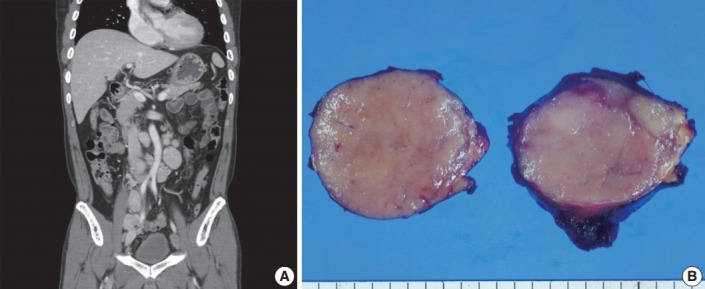
Imaging and macroscopic findings of the lymph node. (A) Abdominal computed tomography showing multiple enlarged retroperitoneal lymph nodes along the axis. (B) A 5.0-cm round and tan to pinkish contexture demonstrated with vague lobulation on cut section.
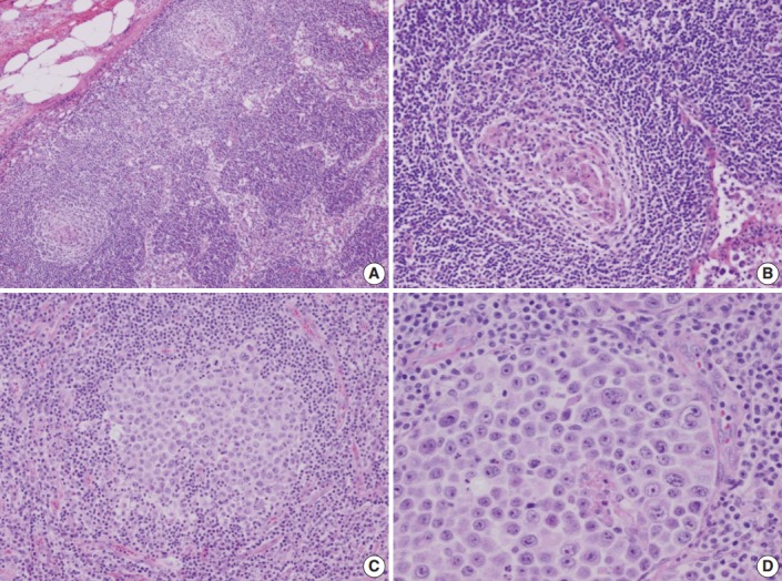
Microscopic findings of the lymph node. (A) Low-magnification showing multiple, well-formed germinal centers. (B) The hyalinized vascular proliferations surrounding follicles characteristic of Castleman’s disease. (C) The confluent aggregates of plasmablasts in the germinal centers indicating the germinotrophism. (D) High-magnification showing the invasion of plasmablasts to the interfollicular spaces with sinusoidal pattern.
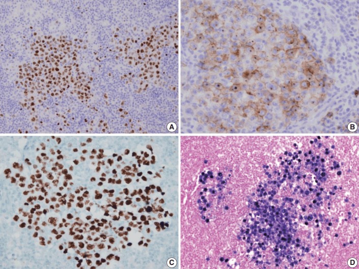
Immunohistochemical staining results of the lymph node. (A) MUM-1/IRF4-stain highlighting sheets of germinotrophic plasmablasts. (B) Epithelial membrane antigen–stain demonstrating confluent aggregation of plasmablasts with sinusoidal pattern. (C) Human herpes virus 8 encoded latency associated nuclear antigen 1 highlighting the atypical plasmablasts. (D) In situ hybridization for Epstein-Barr virus (EBV) encoded ribonucleic acids showing coinfection of EBV in the atypical plasmablasts.
References
-
- Du MQ, Liu H, Diss TC, et al. Kaposi sarcoma-associated herpesvirus infects monotypic (IgM lambda) but polyclonal naive B cells in Castleman disease and associated lymphoproliferative disorders. Blood. 2001;97:2130–6. - PubMed
-
- Seliem RM, Griffith RC, Harris NL, et al. HHV-8+, EBV+ multicentric plasmablastic microlymphoma in an HIV+ Man: the spectrum of HHV-8+ lymphoproliferative disorders expands. Am J Surg Pathol. 2007;31:1439–45. - PubMed
-
- Dong HY, Wang W, Uldrick TS, Gangi M. Human herpesvirus 8- and Epstein-Barr virus-associated solitary B cell lymphoma with a T cell immunophenotype. Leuk Lymphoma. 2013;54:1560–3. - PubMed
-
- Chadburn A, Cesarman E, Nador RG, Liu YF, Knowles DM. Kaposi’s sarcoma-associated herpesvirus sequences in benign lymphoid proliferations not associated with human immunodeficiency virus. Cancer. 1997;80:788–97. - PubMed
LinkOut - more resources
Full Text Sources
Other Literature Sources
