Case Reports
. 2017 Jan 5;100(1):128-137.
doi: 10.1016/j.ajhg.2016.11.018. Epub 2016 Dec 22.

A Syndromic Neurodevelopmental Disorder Caused by De Novo Variants in EBF3

Collaborators, Affiliations
Case Reports

A Syndromic Neurodevelopmental Disorder Caused by De Novo Variants in EBF3

Hsiao-Tuan Chao et al. Am J Hum Genet. .

Abstract

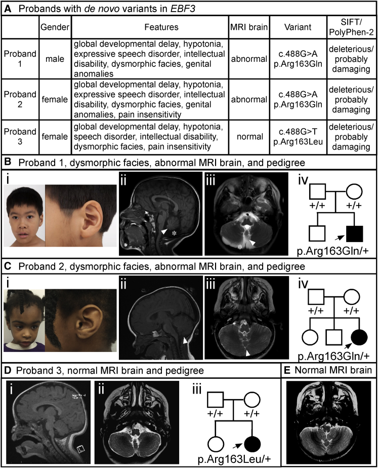
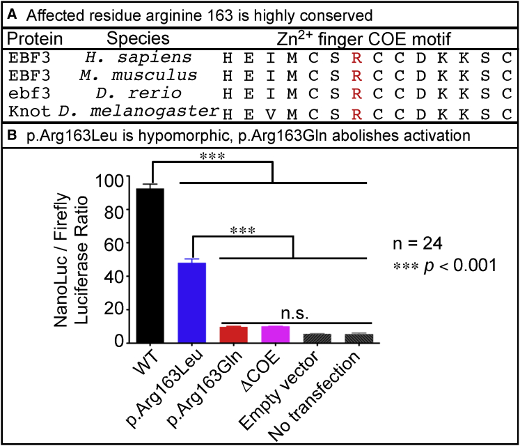
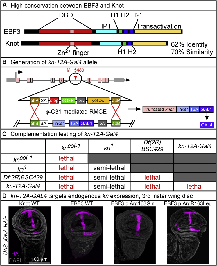
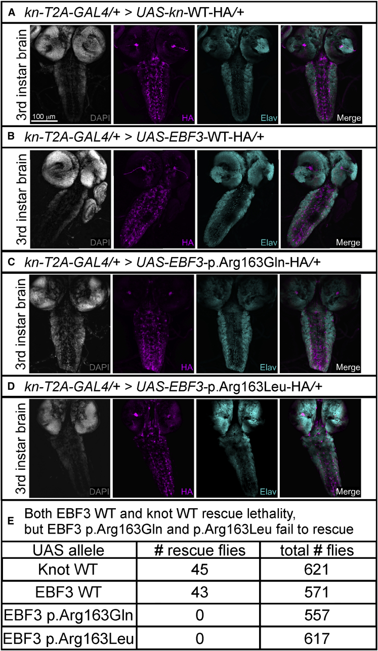
Early B cell factor 3 (EBF3) is a member of the highly evolutionarily conserved Collier/Olf/EBF (COE) family of transcription factors. Prior studies on invertebrate and vertebrate animals have shown that EBF3 homologs are essential for survival and that loss-of-function mutations are associated with a range of nervous system developmental defects, including perturbation of neuronal development and migration. Interestingly, aristaless-related homeobox (ARX), a homeobox-containing transcription factor critical for the regulation of nervous system development, transcriptionally represses EBF3 expression. However, human neurodevelopmental disorders related to EBF3 have not been reported. Here, we describe three individuals who are affected by global developmental delay, intellectual disability, and expressive speech disorder and carry de novo variants in EBF3. Associated features seen in these individuals include congenital hypotonia, structural CNS malformations, ataxia, and genitourinary abnormalities. The de novo variants affect a single conserved residue in a zinc finger motif crucial for DNA binding and are deleterious in a fly model. Our findings indicate that mutations in EBF3 cause a genetic neurodevelopmental syndrome and suggest that loss of EBF3 function might mediate a subset of neurologic phenotypes shared by ARX-related disorders, including intellectual disability, abnormal genitalia, and structural CNS malformations.

Keywords: COE3; Drosophila; ataxia; expressive speech disorder; hypotonia; inhibitory GABAergic neurons; intellectual disability; knot; transcription factor; vermian hypoplasia.


Figures

Figure 1
Figure 1
Probands and Phenotypic Features (A) Summary of phenotypic features, brain MRI findings, gene variants, and SIFT and PolyPhen-2 predictions for the three probands with the de novo EBF3 p.Arg163Gln and p.Arg163Leu variants. (B) Proband 1. Representative images show (i) mild facial dysmorphisms including oval-shaped myopathic facies, short anteverted nostrils, and overfolding of the superior helices, (ii) mid-sagittal T1-weighted and (iii) axial T2-weighted images depicting vermian hypoplasia (white arrows) and reduced cerebellar hemispheres volume (white asterisk), and (iv) a pedigree showing the de novo p.Arg163Gln variant. (C) Proband 2. Representative images show (i) mild facial dysmorphisms including triangular myopathic facies and overfolding of the superior helices, (ii) mid-sagittal T1-weighted and (iii) axial T2-weighted images depicting vermian hypoplasia (white arrows) with normal cerebellar hemispheres, and (iv) a pedigree showing the de novo p.Arg163Gln variant. (D) Proband 3. Brain MRI shows (i) mid-sagittal T1-weighted and (ii) axial T2-weighted images depicting normal cerebellar vermis and hemispheres. A pedigree (iii) shows the de novo p.Arg163Leu variant. (E) A representative axial T2-weighted image from the normal brain MRI of a 23-month-old control individual is shown for comparison. Note the typical cerebellar hemispheres and vermian structures.
Figure 2
Figure 2
EBF3 p.Arg163Gln and p.Arg163Leu Impair Transcriptional Activation (A) Affected residue Arg163 in the Zn2+ finger COE motif is highly conserved across vertebrates and invertebrates. (B) Activation of reporter-gene expression in HEK293 cells was assessed as the ratio of NanoLuc to firefly luciferase according to the Promega NanoGlo Dual Reporter protocol and was measured on the TD 20/20 Luminometer. The cDNAs encoding WT EBF3 and the EBF3 p.Arg163Gln and p.Arg163Leu variants had a stop codon to ensure that the proteins were untagged to minimize off-target effects from a protein tag. The cDNAs were subcloned into the mammalian expression vector, pcDNA-DEST40. Two synthetic oligonucleotides containing the imperfect palindromic COE binding sequence were used to generate six concatamerized COE binding sites in the NanoLuc vector, pNL3.1. The pGL4.53 firefly luciferase vector was used as an internal transfection control. Additional experimental controls included EBF3 with deletion of the Zn2+ finger COE motif (denoted as ΔCOE). As transfection background controls, we either transfected only the pNL3.1 with six COE binding sites and pGL4.53 but no cDNA expression vectors (denoted as “empty vector”) or did not transfect any vectors (denoted as “no transfection”). A 92-fold induction was observed with WT EBF3 (black). However, EBF3 p.Arg163Leu caused only a 45-fold induction (blue), indicating a partial loss of transcriptional activation. EBF3 p.Arg163Gln showed a very poor induction of transcription (red) similar in level to the transfection background (gray) and that of EBF3 ΔCOE (magenta) controls, indicating severe loss of activity. Data represent the mean ± SEM. n = 24 (six replicates per four separate transfections per experimental condition); ∗∗∗p < 0.001 via one-way ANOVA with Tukey’s post hoc analysis; n.s., no significant difference.
Figure 3
Figure 3
Generation and Characterization of the Fly kn-T2A-GAL4 Allele (A) High conservation of protein structure and amino acid sequence between human EBF3 and fly Knot with 62% identity and 70% similarity. Conserved domains include the DNA-binding domain (DBD, red), Zn2+ finger COE motif (pink), Ig-like/plexins/transcription factors (IPT) domain (cyan), helix-loop-helix dimerization motif with α helices H1 (green) and H2 or H2′ (blue), and C-terminal transactivation domain (yellow). (B) Conversion of the MI15480 line with a MiMIC transposable-element insertion in the fourth coding intron of knot via ϕC31-mediated RMCE for generation of the kn-T2A-GAL4 allele, which expresses GAL4 transactivator in the pattern of kn. This allele also creates a loss-of-function allele of knot by prematurely truncating the transcript by a ribosomal skipping signal (T2A) and a premature polyadenylation signal (pA). (C) Complementation testing shows that kn-T2A-GAL4 with both the amorphic kncol-1 and genomic deficiency Df(2R)BSC429, encompassing the entire kn locus, fails to complement the lethality. Complementation with hypomorphic kn1 is semi-lethal with <10% viability. (D) For expression analysis, yw/y; kn-T2A-GAL4/CyO, Kr-GAL4, UAS-GFP males were crossed with yw; UAS-cDNA/TM3 Sb, Kr-GAL4, UAS-GFP virgin females, and double heterozygotes were selected by loss of GFP expression. Images were acquired on a Leica Sp8 laser-scanning confocal microscope. The same settings for laser power and detector gain were used for all genotypes. Images were acquired as a z stack with a z-step of 1 μm and line average of 4 at 400 Hz with a 25× water objective at 1024 × 1024 pixel resolution. Maximum intensity projections were created from the stack in ImageJ. Immunolabeling revealed that kn-T2A-GAL4 recapitulates the endogenous knot expression pattern in third instar wing disc, as shown with the HA-tagged UAS fly lines for WT Knot, WT EBF3, and the EBF3 p.Arg163Gln and p.Arg163Leu variants. Images show HA (magenta) and DAPI (gray). The scale bar represents 100 μm.
Figure 4
Figure 4
WT EBF3 and knot Rescue Lethality, but EBF3 p.Arg163Gln and p.Arg163Leu Fail to Rescue Lethality in Flies (A–D) For expression analysis of third instar brain and ventral nerve cord, yw/y; kn-T2A-GAL4/CyO, Kr-GAL4, UAS-GFP males were crossed with yw; UAS-cDNA/TM3 Sb, Kr-GAL4, UAS-GFP virgin females, and double heterozygotes were selected by loss of GFP expression in the wandering third instar larval stage. Images were acquired on a Leica Sp8 laser-scanning confocal microscope. The same settings for laser power and detector gain were used for all genotypes. Third instar larval brain images were acquired as a z stack with a z-step of 1.51 μm and line average of 4 at 400 Hz with a 20× objective at 1024 × 1024 pixel resolution. Maximum intensity projections were created from the stack in ImageJ. Immunolabeling revealed that kn-T2A-GAL4 drives expression of HA-tagged UAS fly lines for WT Knot (A), WT EBF3 (B), EBF3 p.Arg163Gln (C), and EBF3 p.Arg163Leu (D) in the third instar brain and ventral nerve cord (pan-neuronal marker Elav in cyan; nuclei labeled with DAPI in gray). The scale bar represents 100 μm. (E) Fly in vivo rescue analysis using the UAS-GAL4 system. For generating the rescue flies, w1118/y; Df(2R)BSC429/Sp; UAS-cDNA-WT-HA/+ males were crossed with yw; kn-T2A-GAL4/SM6a virgin females to produce rescue animals with Knot or EBF3 produced solely from the UAS allele under the control of the kn-T2A-GAL4 driver. The genotypes of the rescued flies are yw/y; Df(2R)BSC429/kn-T2A-GAL4; UAS-cDNA-HA/+ males and w1118/yw; ; Df(2R)BSC429/kn-T2A-GAL4; UAS-cDNA-HA/+ females. For each UAS-cDNA line, >550 adult flies were scored; data represent the number of observed rescue flies and the total number of flies scored. UAS fly lines expressing WT Knot or WT EBF3 rescued embryonic lethality in viable adults. UAS fly lines expressing EBF3 variant p.Arg163Gln or p.Arg163Leu completely failed to rescue the lethality such that no rescue animals were observed as adults or pupae.

References

    1. Alwan A., Modell B. Recommendations for introducing genetics services in developing countries. Nat. Rev. Genet. 2003;4:61–68. - PubMed
    1. American Psychiatric Association, editor. Diagnostic and statistical manual of mental disorders. Fifth Edition. American Psychiatric Association Publishing; 2013.
    1. Boivin M.J., Kakooza A.M., Warf B.C., Davidson L.L., Grigorenko E.L. Reducing neurodevelopmental disorders and disability through research and interventions. Nature. 2015;527:S155–S160. - PubMed
    1. Yang Y., Muzny D.M., Reid J.G., Bainbridge M.N., Willis A., Ward P.A., Braxton A., Beuten J., Xia F., Niu Z. Clinical whole-exome sequencing for the diagnosis of mendelian disorders. N. Engl. J. Med. 2013;369:1502–1511. - PMC - PubMed
    1. Zhu X., Petrovski S., Xie P., Ruzzo E.K., Lu Y.F., McSweeney K.M., Ben-Zeev B., Nissenkorn A., Anikster Y., Oz-Levi D. Whole-exome sequencing in undiagnosed genetic diseases: interpreting 119 trios. Genet. Med. 2015;17:774–781. - PMC - PubMed

Publication types

MeSH terms