. 2017 Jan 3;25(1):146-156.
doi: 10.1016/j.str.2016.11.014. Epub 2016 Dec 22.

The Ciliopathy-Associated Cep104 Protein Interacts with Tubulin and Nek1 Kinase

Affiliations

The Ciliopathy-Associated Cep104 Protein Interacts with Tubulin and Nek1 Kinase

Caezar Al-Jassar et al. Structure. .

Abstract

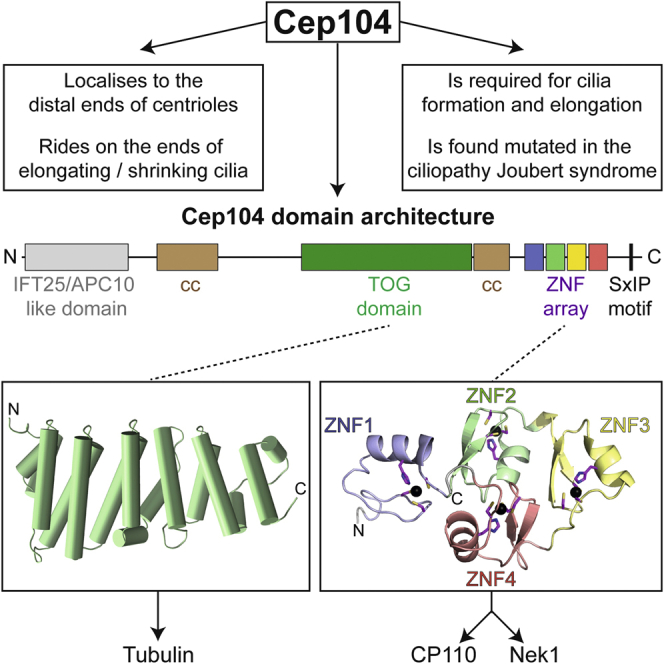
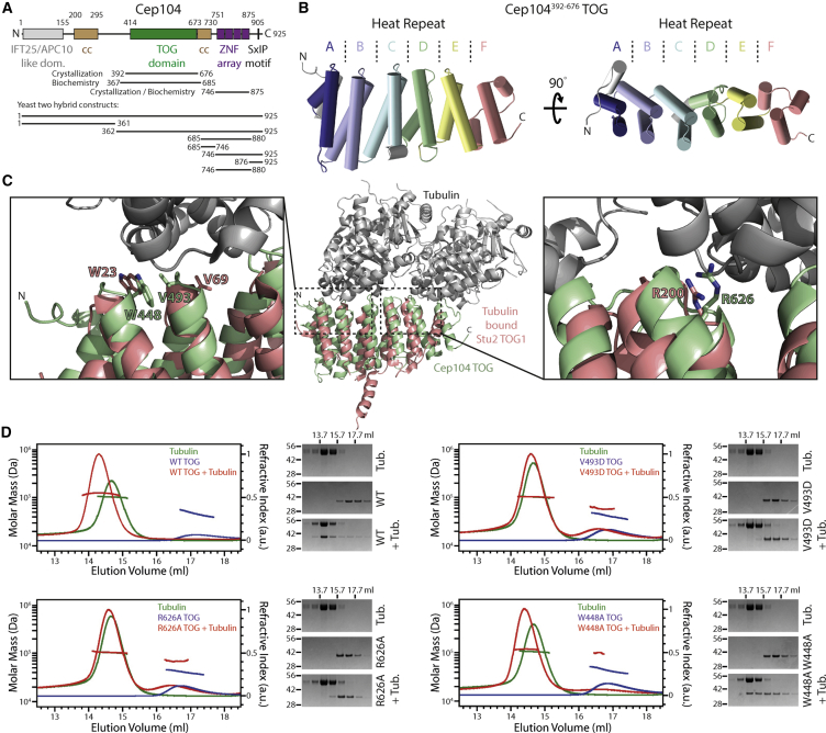
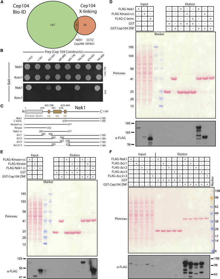
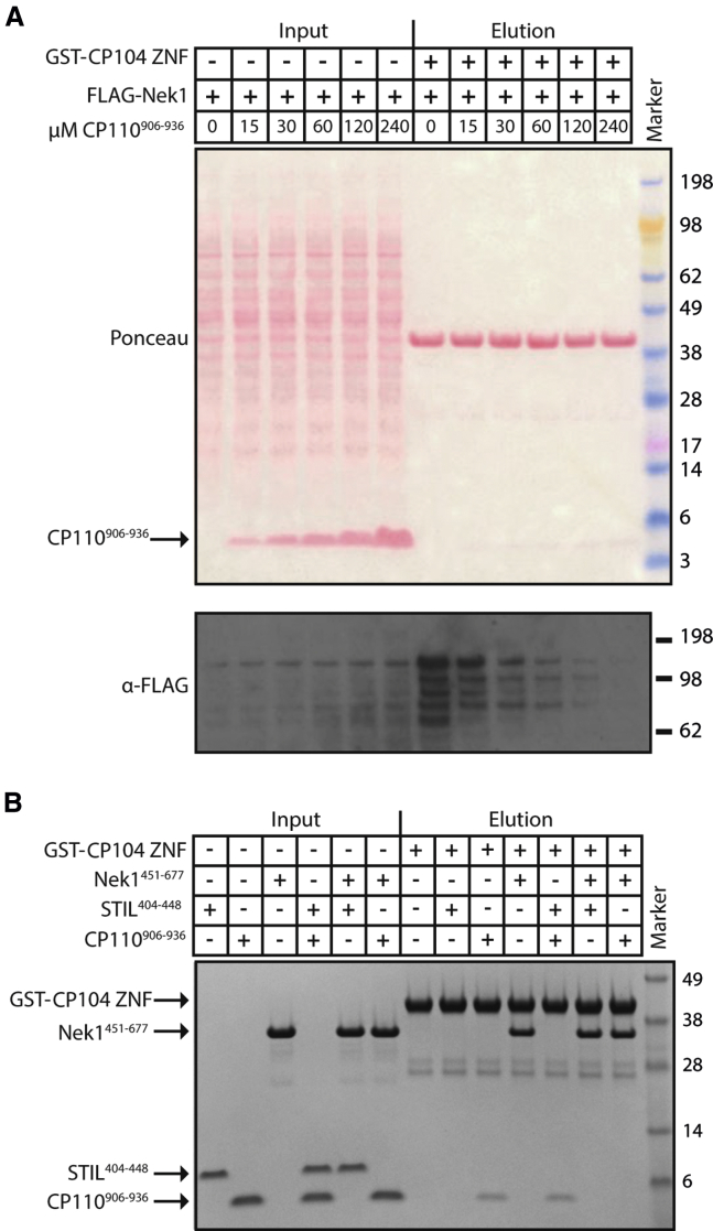
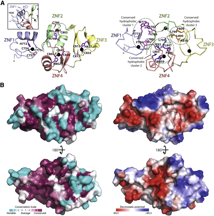
Cilia are thin cell projections with essential roles in cell motility, fluid movement, sensing, and signaling. They are templated from centrioles that dock against the plasma membrane and subsequently extend their peripheral microtubule array. The molecular mechanisms underpinning cilia assembly are incompletely understood. Cep104 is a key factor involved in cilia formation and length regulation that rides on the ends of elongating and shrinking cilia. It is mutated in Joubert syndrome, a genetically heterogeneous ciliopathy. Here we provide structural and biochemical data that Cep104 contains a tubulin-binding TOG (tumor overexpressed gene) domain and a novel C2HC zinc finger array. Furthermore, we identify the kinase Nek1, another ciliopathy-associated protein, as a potential binding partner of this array. Finally, we show that Nek1 competes for binding to Cep104 with the distal centriole-capping protein CP110. Our data suggest a model for Cep104 activity during ciliogenesis and provide a novel link between Cep104 and Nek1.

Keywords: CP110; Cep104; Nek1; TOG; basal body; centriole; cilia; tubulin; zinc finger.


Figures

None
Graphical abstract
Figure 1
Figure 1
Cep104 Contains a Canonical Tubulin-Binding TOG Domain (A) Domain overview of human Cep104. Lines indicate constructs that were used in this work. (B) Ribbon representation of the Cep104 TOG structure. Helices are displayed as cylinders. The six HEAT repeats that constitute the TOG domain are colored individually. Note the partial distortion of HEAT repeat D and F and the slight curvature of the TOG domain. (C) Middle: ribbon representation of TOG1 of yeast Stu2 (red) bound to a tubulin dimer (gray), PDB: 4ffb. Stu2 TOG1 is overlaid with our Cep104 TOG structure (green), showing the overall similarity of the TOG domain fold. Left and right: detailed views of the Stu2 TOG1-tubulin-binding interfaces with those residues labeled that were previously found to be critical for tubulin binding by Stu2. Note that these residues are conserved in Cep104 TOG and found in similar positions. (D) Cep104 TOG binds tubulin in solution. SEC-MALS chromatograms of human Cep104 TOG constructs, run alone or in the presence of tubulin. The horizontal lines indicate the molar masses derived from the refractive index and light-scattering signals as described in Experimental Procedures. On the right side of the chromatograms, Coomassie-stained SDS-PAGE gels show the corresponding peak fractions. The approximate elution volumes are indicated above the gels. The tubulin-alone run is identical in all chromatograms and gels and is only shown repetitively to allow an easier comparison. See also Figures S1 and S2.
Figure 2
Figure 2
Nek1 Is a Potential Binding Partner of Cep104 (A) Summary of the (proximity) interactome of Cep104. Green circle, number of Cep104 proximity interactors obtained from mass-spectrometric identification of biotinylated proteins in Hek293 cells expressing BirA-Cep104-GFP (BioID). Orange circle, number of hits from mass-spectrometric analysis of a pull-down experiment from Hek293 cell lysates expressing Avitag-BirA(WT)-Cep104-GFP, crosslinked with DTSSP. In both cases only hits with an enrichment of at least 10-fold compared with the corresponding negative control were considered. The overlap between the two circles shows the hits shared between both experiments. (B) Nek1 interacts with Cep104 in a yeast two-hybrid assay. Yeast plates showing growth of yeast expressing the indicated Bait and Prey proteins. SC -Leu/-Trp plates select for the presence of Bait and Prey plasmid only, while SC -Ura plates select for positive yeast two-hybrid interactions. (C) Domain overview of human Nek1, isoform 4. Lines indicate constructs that were used in the pull-down experiments probing the Nek1-Cep104 interaction shown in (D) to (F). (D–F) The Nek1 interaction with Cep104 maps to its coiled-coil domain. Western blots showing pull-down experiments with GST or GST-Cep104 ZNF and lysates from Hek293 cells transiently overexpressing the 3×FLAG-tagged human Nek1 constructs indicated above the blot. See also Figures S3 and S4.
Figure 3
Figure 3
CP110906−936 and Nek1 Binding to the Cep104 ZNF Array Is Mutually Exclusive (A) Western blot showing a pull-down experiment with GST-Cep104 ZNF beads and lysates from Hek293 cells transiently overexpressing 3×FLAG-tagged human Nek1 in the presence of increasing concentrations of recombinant CP110906−936. (B) Coomassie-stained SDS-PAGE gel showing the results of an in vitro pull-down assay with recombinantly produced GST-Cep104-ZNF and Nek1451−677 in the presence of CP110906−936 or Danio rerio STIL404−448. See also Figure S5.
Figure 4
Figure 4
High-Resolution Structure of the Zn Finger Array of Cep104 (A) The Cep104 ZNFs adopt an overall globular domain. Left: ribbon presentation of the human Cep104 ZNF array structure (Cep104746−875 S763E). The individual Zn fingers are colored distinctly. The Zn-coordinating side chains in the ZNFs are shown as sticks and labeled, the coordinated Zn ions are displayed as black spheres. Inset: close-up of ZNF1 with side chains displayed as sticks. Note that the S763E mutation that was used to improve solubility of the Cep104 ZNF domain is located in a loop and does not make contact with the rest of the domain. This residue is poorly conserved (Figure S6A). Right: similar view of the Cep104 ZNF domain. Labeled and shown as sticks are the side chains of the three conserved hydrophobic clusters in the interfaces between the individual ZNFs (Figure S6A). These clusters maintain the overall globular packing of the ZNF array. (B) Top: similar view as in (A) but as a molecular surface colored according to CONSURF evolutionary conservation score (left) from unconserved (cyan) to highly conserved (burgundy), or colored according to in vacuo electrostatic potential (right) from positive (blue) to negative potential (red). Bottom: rotated 180° as indicated. See also Figure S6.

References

    1. Afonine P.V., Grosse-Kunstleve R.W., Echols N., Headd J.J., Moriarty N.W., Mustyakimov M., Terwilliger T.C., Urzhumtsev A., Zwart P.H., Adams P.D. Towards automated crystallographic structure refinement with phenix.refine. Acta Crystallogr. D Biol. Crystallogr. 2012;68:352–367. - PMC - PubMed
    1. Akhmanova A., Steinmetz M.O. Control of microtubule organization and dynamics: two ends in the limelight. Nat. Rev. Mol. Cell Biol. 2015;16:711–726. - PubMed
    1. Altschul S.F., Madden T.L., Schaffer A.A., Zhang J., Zhang Z., Miller W., Lipman D.J. Gapped BLAST and PSI-BLAST: a new generation of protein database search programs. Nucleic Acids Res. 1997;25:3389–3402. - PMC - PubMed
    1. Ashkenazy H., Erez E., Martz E., Pupko T., Ben-Tal N. ConSurf 2010: calculating evolutionary conservation in sequence and structure of proteins and nucleic acids. Nucleic Acids Res. 2010;38:W529–W533. - PMC - PubMed
    1. Au S.W., Leng X., Harper J.W., Barford D. Implications for the ubiquitination reaction of the anaphase-promoting complex from the crystal structure of the Doc1/Apc10 subunit. J. Mol. Biol. 2002;316:955–968. - PubMed

Publication types

LinkOut - more resources