. 2017 Jan 10;8(1):30-38.
doi: 10.1016/j.stemcr.2016.11.012. Epub 2016 Dec 22.

Primary Human Testicular Cells Self-Organize into Organoids with Testicular Properties

Affiliations

Primary Human Testicular Cells Self-Organize into Organoids with Testicular Properties

Yoni Baert et al. Stem Cell Reports. .

Abstract

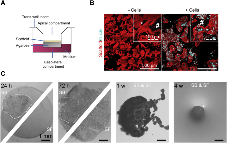
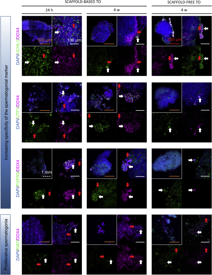
So far, successful de novo formation of testicular tissue followed by complete spermatogenesis in vitro has been achieved only in rodents. Our findings reveal that primary human testicular cells are able to self-organize into human testicular organoids (TOs), i.e., multi-cellular tissue surrogates, either with or without support of a biological scaffold. Despite lacking testis-specific topography, these mini-tissues harbored spermatogonia and their important niche cells, which retained specific functionalities during long-term culture. These observations indicate the posibility of in vitro re-engineering of a human testicular microenvironment from primary cells. Human TOs might help in the development of a biomimetic testicular model that would exert a tremendous impact on research and development, clinical treatment of infertility, and screening in connection with drug discovery and toxicology.

Keywords: immunofluorescence; inhibin B; in vitro spermatogenesis; organoid; scaffold; secretome; spermatogonia; testis; testosterone; whole mount.


Figures

Figure 1
Figure 1
Formation of Scaffold-Based and Scaffold-Free TOs (A) Schematic illustration of the culture system involving inoculation of testicular cells onto a scaffold to prepare scaffold-based (SB) TOs. Testicular cells were cultured in the apical compartment of the insert without scaffold support to form scaffold-free (SF) TOs. (B) A testicular scaffold (red) before (left panel) and 24 hr after (right panel) cell seeding. DAPI was used to stain cell nuclei blue. The inserts show higher magnifications of a seminiferous tubule () and the interstitial space (#). (C) Morphological transformation of TOs viewed through a stereomicroscope. The dotted lines delineate the re-cellularized testis scaffold. See also Table S1.
Figure 2
Figure 2
Characterization of Somatic Testicular Niche Cells in the TOs by Immunofluorescent Staining of Whole Mounts Double staining for STAR/3βHSD (top row), ACTA2/COL1 (middle row), and SOX9/ZO1 (bottom row) applied to scaffold-based (left and middle columns) and scaffold-free (right column) adult TOs (n = 3 TOs derived from different donors per staining). Representative photographs of TOs following short-term (left column) and long-term (middle and right columns) culture are shown. Steroidogenic Leydig cells (white arrows) stained positive for both STAR (green) and 3βHSD (purple), PTMCs (white/red arrows) stained for ACTA2 (green), scaffold () and COL1-producing cells (red arrows) stained for COL1 (purple), COL1-producing PTMCs (white arrow) stained for both ACTA and COL1, and tight-junction protein-producing Sertoli cells (white arrow) stained for both SOX9 (green) and ZO1 (purple). Cell nuclei were stained blue with DAPI. The inserts depict several z stacks at low magnification merged with maximum intensity projection to give an overview. See also Figure S1 and Table S1.
Figure 3
Figure 3
Characterization of Spermatogonia in the TOs by Immunofluorescent Staining of Whole Mounts Immunostaining for UCHL1/DDX4 (first row), UTF1/DDX4 (second row), FGFR3/DDX4 (third row), and KI67/DDX4 (fourth row) in adult TOs (n = 3 TOs derived from different donors per staining) derived with (left and middle column) and without (right column) scaffold support following 24 hr (left column) and 4 weeks (middle and right column) of culture. Representative images show cells that stained positive for UCHL1, UTF1, or FGFR3 (green), and DDX4 (purple), which are unmistakably spermatogonia (white arrow). Cells positive for DDX4 alone represent a different germ-cell type (red arrow). The DDX4+ (purple) cells staining for KI67 (green) are dividing germ cells (white arrow). Cell nuclei are stained blue by DAPI. The inserts depict low-magnification overviews of the TOs generated using maximum intensity projection of z stacks. See also Figure S2 and Table S1.
Figure 4
Figure 4
Secretory Profile of Hormones and Cytokines of Testicular Cells in the TOs The levels of (A) T and (B) InhB in the medium were measured after 24 and 72 hr, and 1, 2, 3, and 4 weeks of culturing scaffold-based and scaffold-free TOs under different hormonal conditions (−/−; rFSH/−; −/hCG; rFSH/hCG). Cytokine profiles were obtained by incubating array membranes with medium from scaffold-based and scaffold-free TOs after (C) 1 week and (D) 4 weeks of culture. The data are presented as means ± SDs (n = 3 TOs derived from different donors per condition). For an explanation of the protein abbreviations, see Table S2. hCG, human chorionic gonadotrophin; MPD, mean pixel density; rFSH, recombinant follicle-stimulating hormone; SB, scaffold based; SF, scaffold free. See also Figure S3 for the hormone and cytokine secretion profiles of pubertal TOs and a representative cytokine antibody array.

References

    1. Baert Y., Stukenborg J.-B., Landreh M., De Kock J., Jörnvall H., Söder O., Goossens E. Derivation and characterization of a cytocompatible scaffold from human testis. Hum. Reprod. 2015;30:256–267. - PubMed
    1. Berensztein E., Saraco N., Belgorosky A., Rivarola M.A. Enhancement of long-term testosterone secretion and steroidogenic enzyme expression in human Leydig cells by co-culture with human Sertoli cell-enriched preparations. Eur. J. Endocrinol. 2000;142:481–485. - PubMed
    1. Brown B.N., Badylak S.F. Extracellular matrix as an inductive scaffold for functional tissue reconstruction. Transl. Res. 2014;163:268–285. - PMC - PubMed
    1. Chada M., Průša R., Bronsky J., Pechová M., Kotaška K., Lisá L. Inhibin B, follicle stimulating hormone, luteinizing hormone, and estradiol and their relationship to the regulation of follicle development in girls during childhood and puberty. Physiol. Res. 2003;52:341–346. - PubMed
    1. Chapin R.E., Boekelheide K., Cortvrindt R., van Duursen M.B.M., Gant T., Jegou B., Marczylo E., van Pelt A.M.M., Post J.N., Roelofs M.J.E. Assuring safety without animal testing: the case for the human testis in vitro. Reprod. Toxicol. 2013;39:63–68. - PubMed

Publication types

LinkOut - more resources