Review
. 2017 Apr;22(4):665-680.
doi: 10.1016/j.drudis.2016.12.009. Epub 2016 Dec 23.

Recent advances in hyaluronic acid-decorated nanocarriers for targeted cancer therapy

Affiliations
Review

Recent advances in hyaluronic acid-decorated nanocarriers for targeted cancer therapy

Jennifer M Wickens et al. Drug Discov Today. 2017 Apr.

Abstract

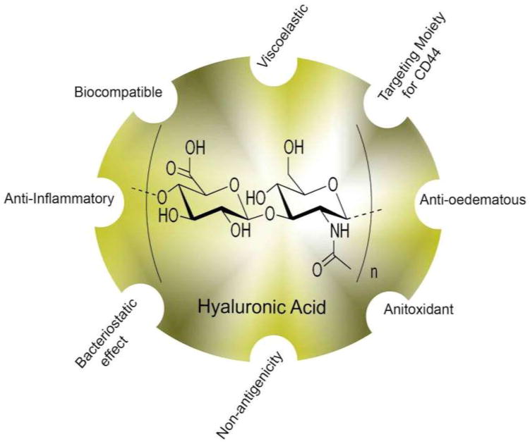
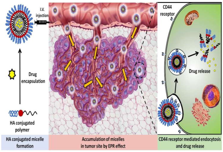
The cluster-determinant 44 (CD44) receptor has a high affinity for hyaluronic acid (HA) binding and is a desirable receptor for active targeting based on its overexpression in cancer cells compared with normal body cells. The nanocarrier affinity can be increased by conjugating drug-loaded carriers with HA, allowing enhanced cancer cell uptake via the HA-CD44 receptor-mediated endocytosis pathway. In this review, we discuss recent advances in HA-based nanocarriers and micelles for cancer therapy. In vitro and in vivo experiments have repeatedly indicated HA-based nanocarriers to be a target-specific drug and gene delivery platform with great promise for future applications in clinical cancer therapy.


Figures

Figure 1
Figure 1
Hyaluronic acid (HA) structure and function. HA is a natural mucopolysaccharide that was discovered to have excellent properties for utilization in drug delivery as a targeted moiety.
Figure 2
Figure 2
Hyaluronic acid (HA)-decorated nanomicelle-mediated active and passive targeting. The accumulation of targeted nanomicelles at the tumor site occurs via cluster-determinant 44 (CD44) receptor-mediated endocytosis. This results from the speci c binding of HA to CD44 receptors overexpressed on cancer cells. This represents active or passive targeting through the enhanced permeation and retention (EPR) effect. Abbreviation: IV, intravenous.
Figure 3
Figure 3
In vivo antitumor efficacy assay. (a) Tumor volumes of tumor-bearing mice as a function of time (day). The arrows indicate the time points of the drug injection. (b) Tumor-bearing mice at the end of the tests (the 20th day post-treatment) imaged by the IFLUOR™ in vivo imaging system (inset: excised tumor tissues after imaging). Paclitaxel (PTX)-hyaluronic acid (HA)-ss- deoxycholic acid (DOCA) micelles (iv) exhibited the highest tumor growth inhibition efficacy compared with saline (i), Taxol® (ii) and PTX-HA-DOCA micelles (iii). Reproduced, with permission from [65].
Figure 4
Figure 4
In vivo tumor inhibition study. (a) Balb/c nude mice were injected with PBS, free paclitaxel (PTX) and hyaluronic acid (HA)-cholanic acid (CA)-PTX at Day 0 (n = 3). Comparison of tumor size on Day 8 of (b) Control, (c) PTX, (d) HA-CA-PTX (2 mg/kg), and (e) HA-CA-PTX (5 mg/kg). Tumor inhibition data show mean tumor volume, of triplicate samples ± SD. *P <0.05 relative to HA-CA-PTX (2 mg/kg) and **P <0.05 relative to control group. Reproduced, with permission, from [72].
Figure 5
Figure 5
(a) In vivo imaging of tumor-bearing mice after administration of DiR/FAM-small interfering RNA (siRNA) co-loaded HSOP micelles (i) and HOP micelles (ii) at 2 h, 12 h, and 24 h under DiR channel (720 nm for excitation and 790 nm for emission). (b) Ex vivo fluorescence images of tissues including: lung (1), heart (2), tumor (3), liver (4), spleen (5), and kidneys (6) collected at 2 h, 12 h and 24 h post-injection of DiR/FAM-siRNA co-loaded HSOP micelles (DiRHSOPFAM-siRNA) and HOP micelles (DiRHOPFAM-siRNA) under DiR channel [(i) 720 nm for excitation and 790 nm for emission) and FAM channel [(ii) 470 nm for excitation and 530 nm for emission), respectively. Reproduced, with permission, from [95].
Figure 6
Figure 6
(a) Whole-body optical imaging. The distribution of indocyanine green-encapsulated hyaluronic acid (HA)-poly(ethylenimine)/HA-poly(ethylene glycol) self-assembled nanoparticles (ICG/HA-PEI/PEG NP) in A549/A549DDP non-small cell lung cancer- (i) and H69/H69AR small cell lung cancer- (ii) bearing mice. Mice bearing A549 andA549DDP and H69/H69AR tumors were injected with ICG/HA-PEI/PEG NPs and imaged at different time points using the IVIS live imaging system. To see the half-life of ICG alone in circulation, the free dye ICG was injected into A549 tumor-bearing mice and imaged at different time points (iii). (b) Biodistribution of NPs throughout different organs in the mice tissues. Reproduced, with permission, from [98].

References

    1. Siegel RL, et al. Cancer statistics. CA Cancer J Clin. 2016;66:7–30. - PubMed
    1. Fox ME, et al. Soluble polymer carriers for the treatment of cancer: the importance of molecular architecture. ACC Chem Res. 2009;42:1141–1151. - PMC - PubMed
    1. Mayol L, et al. Amphiphilic hyaluronic acid derivatives toward the design of micelles for the sustained delivery of hydrophobic drugs. Carbohydr Polym. 2014;102:110–116. - PubMed
    1. Thomas RG, et al. Effectiveness of losartan-loaded hyaluronic acid (HA) micelles for the reduction of advanced hepatic fibrosis in C3H/HeN mice model. PLoS ONE. 2015;10:e0145512. - PMC - PubMed
    1. Matsumura Y, Maeda H. A New concept for macromolecular therapeutics in cancer chemotherapy: mechanism of tumoritropic accumulation of proteins and the antitumor agent Smancs. Cancer Res. 1986;46:6387–6392. - PubMed

Publication types