. 2016 Dec 15:8:299.
doi: 10.3389/fnagi.2016.00299. eCollection 2016.

Mitochondrial Functional Changes Characterization in Young and Senescent Human Adipose Derived MSCs

Affiliations

Mitochondrial Functional Changes Characterization in Young and Senescent Human Adipose Derived MSCs

Bernd R Stab 2nd et al. Front Aging Neurosci. .

Abstract

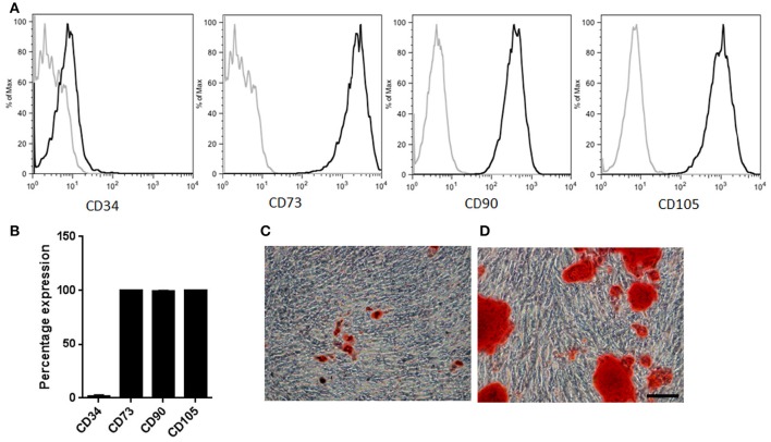
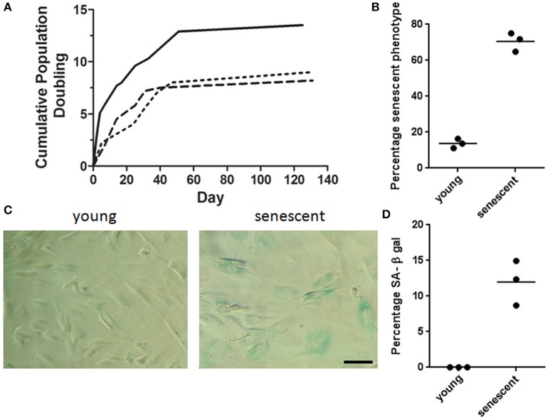
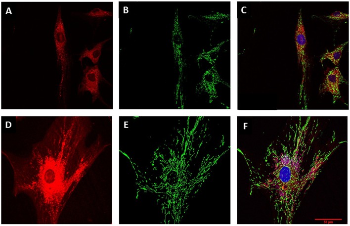
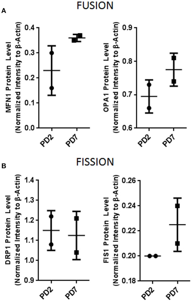
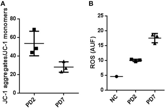
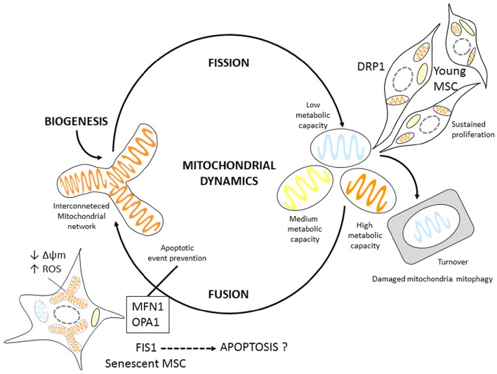
Mitochondria are highly dynamic organelles that in response to the cell's bio-energetic state continuously undergo structural remodeling fission and fusion processes. This mitochondrial dynamic activity has been implicated in cell cycle, autophagy, and age-related diseases. Adult tissue-derived mesenchymal stromal/stem cells present a therapeutic potential. However, to obtain an adequate mesenchymal stromal/stem cell number for clinical use, extensive in vitro expansion is required. Unfortunately, these cells undergo replicative senescence rapidly by mechanisms that are not well understood. Senescence has been associated with metabolic changes in the oxidative state of the cell, a process that has been also linked to mitochondrial fission and fusion events, suggesting an association between mitochondrial dynamics and senescence. In the present work, we studied the mitochondrial structural remodeling process of mesenchymal stromal/stem cells isolated from adipose tissue in vitro to determine if mitochondrial phenotypic changes were associated with mesenchymal stromal/stem cell senescence. For this purpose, mitochondrial dynamics and oxidative state of stromal/stem cell were compared between young and old cells. With increased cell passage, we observed a significant change in cell morphology that was associated with an increase in β-galactosidase activity. In addition, old cells (population doubling seven) also showed increased mitochondrial mass, augmented superoxide production, and decreased mitochondrial membrane potential. These changes in morphology were related to slightly levels increases in mitochondrial fusion proteins, Mitofusion 1 (MFN1), and Dynamin-related GTPase (OPA1). Collectively, our results showed that adipose tissue-derived MSCs at population doubling seven developed a senescent phenotype that was characterized by metabolic cell changes that can lead to mitochondrial fusion.

Keywords: adipose derived mesenchymal stromal cells; fission and fusion; mitochondria; reactive oxygen species; senescence.


Figures

Figure 1
Figure 1
MSCs characterization. (A) Representative sample illustrating immunophenotypic profile. Antibodies against CD34, CD73, CD90, and CD105 (thick gray lines) with isotype control (thin gray lines). (B) Percentage expression corresponding to the average profile of all cell types (n = 3). (C) Representative sample of MSCs induced into adipogenic lineage differentiation. Neutral fat lipid droplets were detected by Oil red O stain (D) Representative sample of MSCs induced into osteogenic differentiation. Extracellular calcium deposits were determined by Alizarin Red stain. Scale bar 100 μm.
Figure 2
Figure 2
MSCs senescent associated phenotype. (A) Cumulative Population doubling for adipose derived MSCs. Adipose tissue derived MSCs were cultured for a total of 131 days. Cumulative PD were calculated at the end of every passage in relation to cell number of the first passage (n = 3). (B) Percentage senescent cells determined by cell count/field. Over 60% of MSCs at PD7 acquired a flattened and widened phenotype. (C) Representative brightfield images of young and senescent MSCs. Young cells are spindle shaped and slender in contrast to senescent cells with a widened and flat morphology. Cells presenting blue staining indicate the presence of β-galactosidase activity in senescent cells. Scale bar 100 μm. (D) Quantification of β-galactosidase positive cells for young (PD2) and senescent cells (PD7).
Figure 3
Figure 3
Mitochondrial characterization. MSC cell morphology and mitochondrial mass evaluation by confocal microscopy. (A) MSC at PD2 cytoplasm revealed by CellTracker red. (B) Mitochondrial stain with Mito Tracker green for MSCs at PD2. (C) Merge for (A) and (C) with nucleus stained with Hoechst (blue). (D) Larger cytoplasm in MSC at PD7 revealed by CellTracker red. (E) Mitochondrial stain with Mito Tracker green for MSCs at PD7. (F) Merge for (A) and (C) with nucleus stained with Hoechst (blue). Scale bar 50 μm for all images.
Figure 4
Figure 4
Mitochondrial dynamics protein evaluation. (A) Mitochondrial fusion proteins MFN1 and OPA1. (B) Mitochondrial fission proteins DRP1 and FIS1. Bar graphs depict protein expression as an arbitrary unit when protein of interest was normalized to β-actin. MFN1, Mitofusion1; OPA1, Optic atrophy 1; DRP1, Dynamin-1-like protein; FIS1, Mitochondrial fission 1.
Figure 5
Figure 5
Mitochondrial membrane potential and Reactive oxygen species production in young and senescent MSCs. (A) FACS analysis to determine membrane potential changes by means of JC-1, a lipophilic cationic dye selectively entering into mitochondria. JC-1 accumulation in mitochondria, due to concentration-dependent formation of red fluorescent JC-1 aggregates was higher for MSCs cultured up to PD2 compared with PD7 MSCs. Results are presented as JC-1 aggregates/JC-1 monomer (B). Reactive oxygen species production in young and senescent MSCs was compared by Mitosox Red fluorescent intensity production quantified by flow cytometry and expressed as arbitrary units fluorescence ROS (AUF). As a positive control rotenone at 50 μM was used.
Figure 6
Figure 6
Mitochondrial dynamic Model proposal for MSCs. Young MSCs can readily undergo proliferation. Mitotic cells have slightly higher DRP1 expression, possibly regulating mitochondrial fission. Senescent MSCs display mitochondrial fusion. In addition they have larger mitochondrial volume; express proteins associated with fusion events most likely as an adaptation mechanism to energetic changes, such as increased ROS production, and decreased Δψm possibly to prevent apoptosis.

References

    1. Andreyev A. Y., Kushnareva Y. E., Starkov A. A. (2005). Mitochondrial metabolism of reactive oxygen species. Biochemistry 70, 200–214. - PubMed
    1. Bentivegna A., Roversi G., Riva G., Paoletta L., Redaelli S., Miloso M., et al. (2016). The effect of culture on human bone marrow mesenchymal stem cells: focus on DNA methylation profiles. Stem Cells Int. 2016:5656701. 10.1155/2016/5656701 - DOI - PMC - PubMed
    1. Brooks C., Cho S. G., Wang C. Y., Yang T., Dong Z. (2011). Fragmented mitochondria are sensitized to Bax insertion and activation during apoptosis. Am. J. Physiol. Cell Physiol. 300, C447–C455. 10.1152/ajpcell.00402.2010 - DOI - PMC - PubMed
    1. Caplan A. I. (1991). Mesenchymal stem cells. J. Orthop. Res. 9, 641–650. 10.1002/jor.1100090504 - DOI - PubMed
    1. Caplan A. I., Correa D. (2011). The MSC: an injury drugstore. Cell Stem Cell 9, 11–15. 10.1016/j.stem.2011.06.008 - DOI - PMC - PubMed

LinkOut - more resources