

Myofilament Calcium Sensitivity: Role in Regulation of In vivo Cardiac Contraction and Relaxation
- PMID: 28018228
- PMCID: PMC5159616
- DOI: 10.3389/fphys.2016.00562
Myofilament Calcium Sensitivity: Role in Regulation of In vivo Cardiac Contraction and Relaxation
Abstract
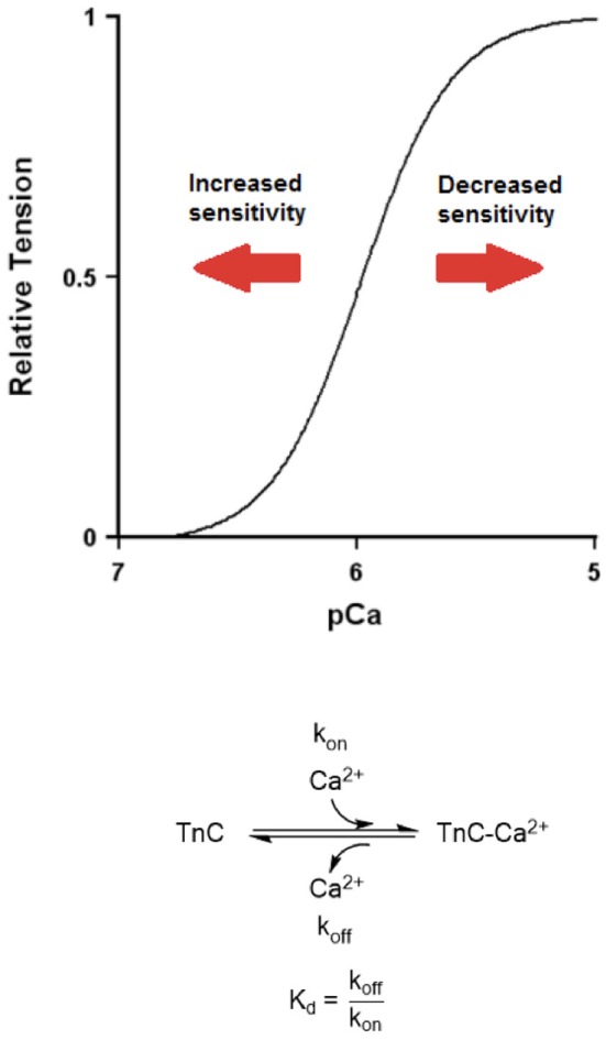
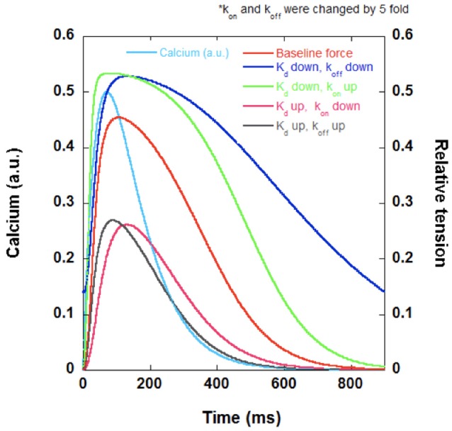
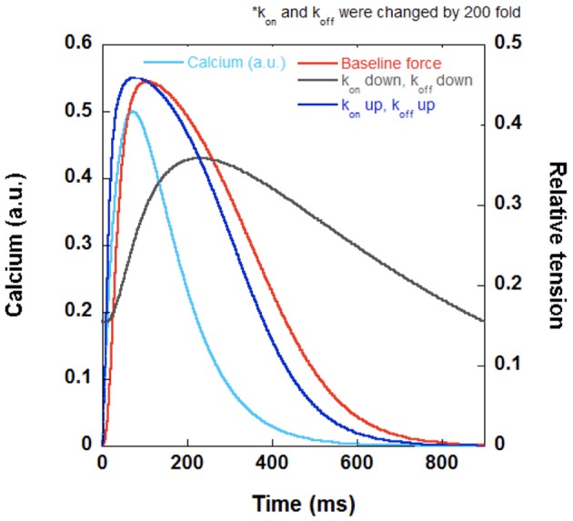
Myofilament calcium sensitivity is an often-used indicator of cardiac muscle function, often assessed in disease states such as hypertrophic cardiomyopathy (HCM) and dilated cardiomyopathy (DCM). While assessment of calcium sensitivity provides important insights into the mechanical force-generating capability of a muscle at steady-state, the dynamic behavior of the muscle cannot be sufficiently assessed with a force-pCa curve alone. The equilibrium dissociation constant (Kd) of the force-pCa curve depends on the ratio of the apparent calcium association rate constant (kon) and apparent calcium dissociation rate constant (koff) of calcium on TnC and as a stand-alone parameter cannot provide an accurate description of the dynamic contraction and relaxation behavior without the additional quantification of kon or koff, or actually measuring dynamic twitch kinetic parameters in an intact muscle. In this review, we examine the effect of length, frequency, and beta-adrenergic stimulation on myofilament calcium sensitivity and dynamic contraction in the myocardium, the effect of membrane permeabilization/mechanical- or chemical skinning on calcium sensitivity, and the dynamic consequences of various myofilament protein mutations with potential implications in contractile and relaxation behavior.
Keywords: desensitize; kinetics; muscle; sensitize; twitch.
Figures
Similar articles
-
Methods for assessing cardiac myofilament calcium sensitivity.Front Physiol. 2023 Dec 5;14:1323768. doi: 10.3389/fphys.2023.1323768. eCollection 2023. Front Physiol. 2023. PMID: 38116581 Free PMC article. Review.
-
Measurement of myofilament calcium sensitivity at physiological temperature in intact cardiac trabeculae.Am J Physiol Heart Circ Physiol. 2006 May;290(5):H2092-7. doi: 10.1152/ajpheart.01241.2005. Am J Physiol Heart Circ Physiol. 2006. PMID: 16603708
-
Contractile abnormalities and altered drug response in engineered heart tissue from Mybpc3-targeted knock-in mice.J Mol Cell Cardiol. 2013 Oct;63:189-98. doi: 10.1016/j.yjmcc.2013.07.011. Epub 2013 Jul 26. J Mol Cell Cardiol. 2013. PMID: 23896226
-
Selective phosphorylation of PKA targets after β-adrenergic receptor stimulation impairs myofilament function in Mybpc3-targeted HCM mouse model.Cardiovasc Res. 2016 May 15;110(2):200-14. doi: 10.1093/cvr/cvw026. Epub 2016 Jan 29. Cardiovasc Res. 2016. PMID: 26825555
-
Mutations in Troponin that cause HCM, DCM AND RCM: what can we learn about thin filament function?J Mol Cell Cardiol. 2010 May;48(5):882-92. doi: 10.1016/j.yjmcc.2009.10.031. Epub 2009 Nov 12. J Mol Cell Cardiol. 2010. PMID: 19914256 Review.
Cited by
-
Methods for assessing cardiac myofilament calcium sensitivity.Front Physiol. 2023 Dec 5;14:1323768. doi: 10.3389/fphys.2023.1323768. eCollection 2023. Front Physiol. 2023. PMID: 38116581 Free PMC article. Review.
-
Profiling of the muscle-specific dystroglycan interactome reveals the role of Hippo signaling in muscular dystrophy and age-dependent muscle atrophy.BMC Med. 2020 Jan 21;18(1):8. doi: 10.1186/s12916-019-1478-3. BMC Med. 2020. PMID: 31959160 Free PMC article.
-
Discrepancy in Myocardial Responses to Pressure Overload versus Volume Load Lesions.J Cardiovasc Echogr. 2025 Jan-Mar;35(1):50-54. doi: 10.4103/jcecho.jcecho_72_24. Epub 2025 Apr 30. J Cardiovasc Echogr. 2025. PMID: 40463754 Free PMC article.
-
Myofilament Calcium Sensitivity: Mechanistic Insight into TnI Ser-23/24 and Ser-150 Phosphorylation Integration.Front Physiol. 2016 Dec 15;7:567. doi: 10.3389/fphys.2016.00567. eCollection 2016. Front Physiol. 2016. PMID: 28018230 Free PMC article.
-
Ex vivo Methods for Measuring Cardiac Muscle Mechanical Properties.Front Physiol. 2021 Jan 8;11:616996. doi: 10.3389/fphys.2020.616996. eCollection 2020. Front Physiol. 2021. PMID: 33488406 Free PMC article. Review.
References
-
- Ait-Mou Y., Hsu K., Farman G. P., Kumar M., Greaser M. L., Irving T. C., et al. . (2016). Titin strain contributes to the frank-starling law of the heart by structural rearrangements of both thin- and thick-filament proteins. Proc. Natl. Acad. Sci. U.S.A. 113, 2306–2311. 10.1073/pnas.1516732113 - DOI - PMC - PubMed
-
- Bers D. M. (2001). Excitation-contraction coupling and cardiac contractile force, in Developments in Cardiovascular Medicine, 2nd Edn. (Dordrecht: Springer; ), 50, 326. - PubMed
Publication types
Grants and funding
LinkOut - more resources
Full Text Sources
Other Literature Sources
Miscellaneous