Review
. 2016 Dec 15:7:563.
doi: 10.3389/fphys.2016.00563. eCollection 2016.

The Calcium-Sensing Receptor and the Parathyroid: Past, Present, Future

Affiliations
Review

The Calcium-Sensing Receptor and the Parathyroid: Past, Present, Future

Arthur D Conigrave. Front Physiol. .

Abstract

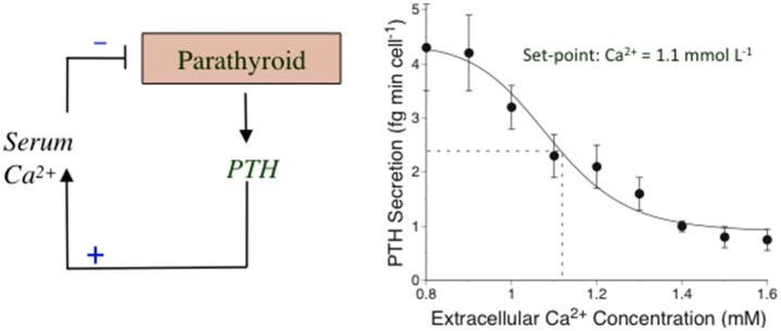
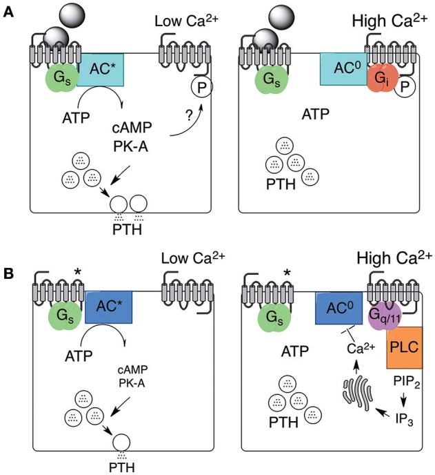
Parathyroid hormone (PTH) defends the extracellular fluid from hypocalcemia and has powerful and well-documented actions on the skeleton and renal tubular system. To achieve a satisfactory stable plasma calcium level, the secretion of PTH, and the resulting serum PTH level, is titrated carefully to the prevailing plasma ionized Ca2+ concentration via a Ca2+ sensing mechanism that mediates feedback inhibition of PTH secretion. Herein, I consider the properties of the parathyroid Ca2+ sensing mechanism, the identity of the Ca2+ sensor, the intracellular biochemical mechanisms that it controls, the manner of its integration with other components of the PTH secretion control mechanism, and its modulation by other nutrients. Together the well-established, recently elucidated, and yet-to-be discovered elements of the story constitute the past, present, and future of the parathyroid and its calcium-sensing receptor (CaSR).

Keywords: Calcimimetics; adenylate cyclase; calcilytics; calcium-sensing receptor; heterotrimeric G proteins; mineral metabolism; parathyroid; phospholipase C.


Figures

Figure 1
Figure 1
The calciostat in parathyroid cells. Left: A representation of the feedback mechanism by which PTH elevates the serum* Ca2+ concentration and Ca2+ feeds back on the parathyroid to suppress PTH secretion in a process mediated by the CaSR. Right: Human parathyroid cells were perifused with HEPES-buffered physiological saline solutions containing various Ca2+ concentrations and samples of perifusate were collected at various times and subsequently analyzed for PTH1–84 as described in Conigrave et al. (2004). The results have been re-drawn. *Total and ionized calcium concentrations are comparable in serum and plasma since the major calcium-binding protein, albumin is present in similar concentrations in both these fluids.
Figure 2
Figure 2
Stimulated and spontaneous mechanisms in support of PTH secretion and its inhibition by high Ca2+o. PTH secretion and its inhibition by high Ca2+o arises from two distinct mechanisms. One mechanism is supported by exogenous agonists, including neurotransmitters or hormones, that activate Gs-coupled GPCRs as shown in (A) (left and right). PTH secretion continues provided Ca2+o remains low but is promptly inhibited by Gi-dependent inhibition of adenylate cyclase in the presence of high Ca2+o. The mechanism by which the Ca2+o sensor, now known to be the CaSR, preferentially binds to Gi in this context is not known but might depend on local protein kinase-A (PK-A) activation. A second mechanism occurs spontaneously and may be supported by constitutive Gs-coupled GPCR activity (as shown in B; left and right) or by autocrine/paracrine production of receptor activators. PTH secretion via this second mechanism continues provided Ca2+o remains low but is inhibited by high Ca2+o-induced Gq/11-dependent activation of intracellular Ca2+ mobilization or Ca2+ influx (not shown). One possible mechanism by which increased intracellular free Ca2+ concentration (Ca2+i) suppresses PTH secretion is shown via Ca2+i-dependent inhibition of adenylate cyclase. *Receptor activated in the absence of neuronal or hormonal stimuli.

Similar articles

Cited by

References

    1. Alexander S., Hunter T., Walter S., Dong J., Maclean D., Baruch A., et al. . (2015). Critical Cysteine residues in both the calcium-sensing receptor and the allosteric activator AMG 416 underlie the mechanism of action. Mol. Pharmacol. 88, 853–865. 10.1124/mol.115.098392 - DOI - PubMed
    1. Ba J., Brown D., Friedman P. A. (2003). Calcium-sensing receptor regulation of PTH-inhibitable proximal tubule phosphate transport. Am. J. Physiol. 285, F1233–F1243. 10.1152/ajprenal.00249.2003 - DOI - PubMed
    1. Bai M., Trivedi S., Brown E. M. (1998). Dimerization of the extracellular calcium-sensing receptor (CaR) on the cell surface of CaR-transfected HEK293 cells. J. Biol. Chem. 273, 23605–23610. 10.1074/jbc.273.36.23605 - DOI - PubMed
    1. Bai M., Trivedi S., Kifor O., Quinn S. J., Brown E. M. (1999). Intermolecular interactions between dimeric calcium-sensing receptor monomers are important for its normal function. Proc. Natl. Acad. Sci. U.S.A. 96, 2834–2839. 10.1073/pnas.96.6.2834 - DOI - PMC - PubMed
    1. Bell G., Huang S., Martin K. J., Block G. A. (2015). A randomized, double-blind, phase 2 study evaluating the safety and efficacy of AMG 416 for the treatment of secondary hyperparathyroidism in hemodialysis patients. Curr. Med. Res. Opin. 31, 943–952. 10.1185/03007995.2015.1031731 - DOI - PubMed