

Key Brainstem Structures Activated during Hypoxic Exposure in One-day-old Mice Highlight Characteristics for Modeling Breathing Network in Premature Infants
- PMID: 28018238
- PMCID: PMC5145891
- DOI: 10.3389/fphys.2016.00609
Key Brainstem Structures Activated during Hypoxic Exposure in One-day-old Mice Highlight Characteristics for Modeling Breathing Network in Premature Infants
Abstract
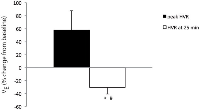
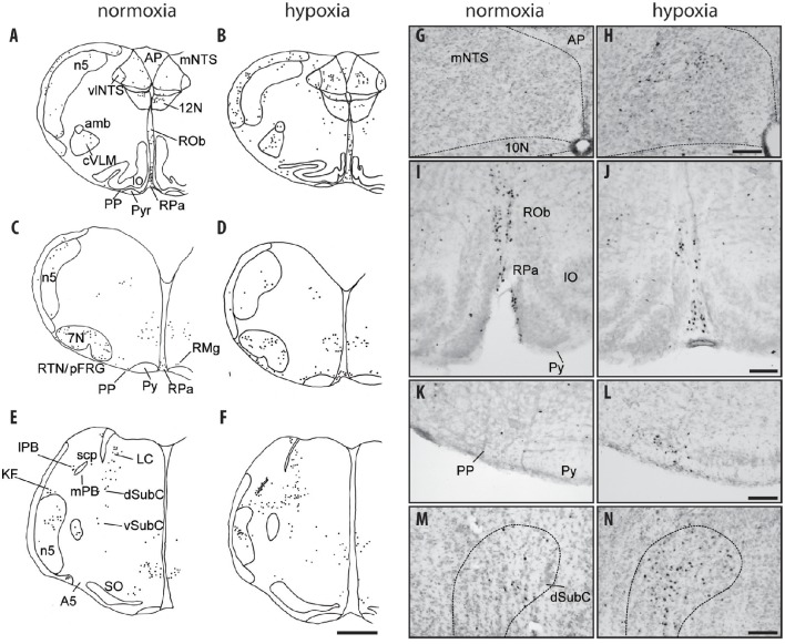
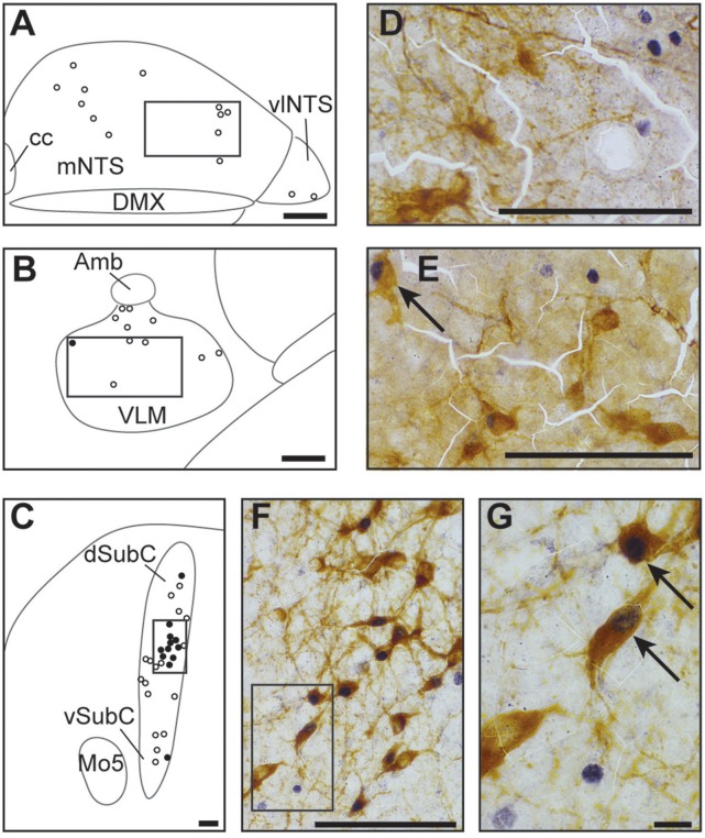
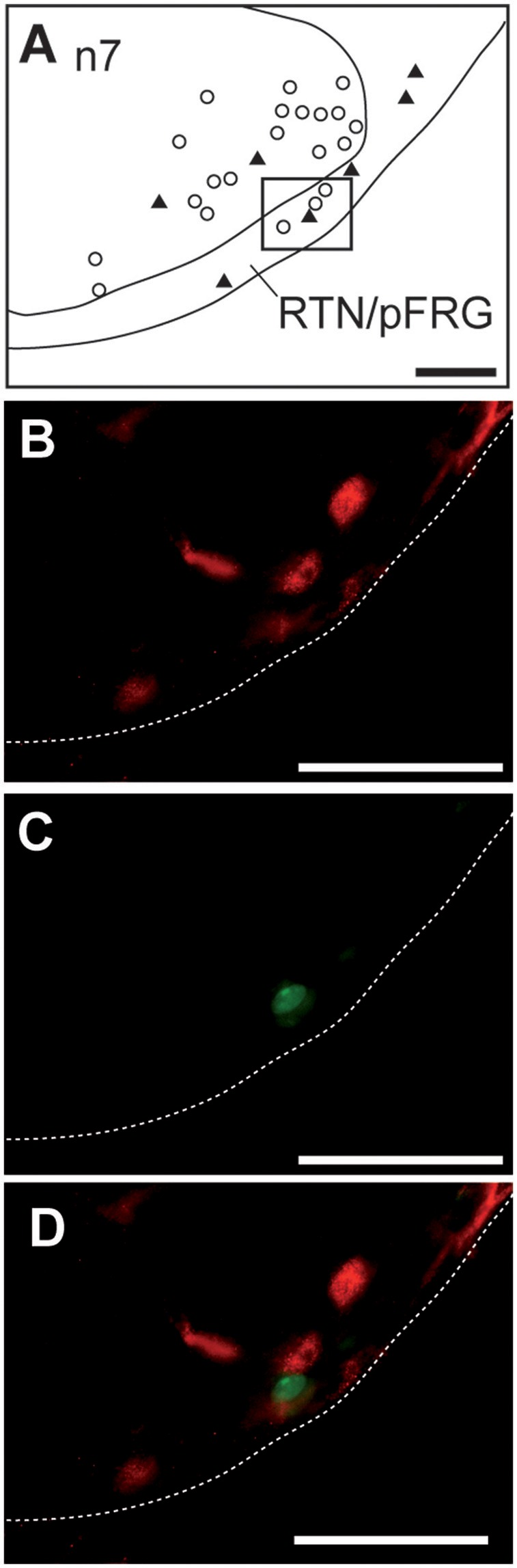
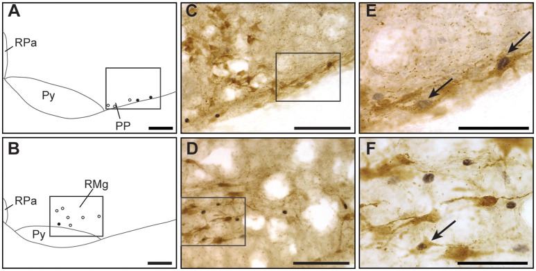
We mapped and characterized changes in the activity of brainstem cell groups under hypoxia in one-day-old newborn mice, an animal model in which the central nervous system at birth is particularly immature. The classical biphasic respiratory response characterized by transient hyperventilation, followed by severe ventilation decline, was associated with increased c-FOS immunoreactivity in brainstem cell groups: the nucleus of the solitary tract, ventral reticular nucleus of the medulla, retrotrapezoid/parafacial region, parapyramidal group, raphe magnus nucleus, lateral, and medial parabrachial nucleus, and dorsal subcoeruleus nucleus. In contrast, the hypoglossal nucleus displayed decreased c-FOS immunoreactivity. There were fewer or no activated catecholaminergic cells activated in the medulla oblongata, whereas ~45% of the c-FOS-positive cells in the dorsal subcoeruleus were co-labeled. Approximately 30% of the c-FOS-positive cells in the parapyramidal group were serotoninergic, whereas only a small portion were labeled for serotonin in the raphe magnus nucleus. None of the c-FOS-positive cells in the retrotrapezoid/parafacial region were co-labeled for PHOX2B. Thus, the hypoxia-activated brainstem neuronal network of one-day-old mice is characterized by (i) the activation of catecholaminergic cells of the dorsal subcoeruleus nucleus, a structure implicated in the strong depressive pontine influence previously reported in the fetus but not in newborns, (ii) the weak activation of catecholaminergic cells of the ventral reticular nucleus of the medulla, an area involved in hypoxic hyperventilation, and (iii) the absence of PHOX2B-positive cells activated in the retrotrapezoid/parafacial region. Based on these results, one-day-old mice could highlight characteristics for modeling the breathing network of premature infants.
Keywords: PHOX2B; c-FOS; catecholamine; hypoxic ventilatory depression; serotonin; subcoeruleus nucleus.
Figures
Similar articles
-
The kreisler mutation leads to the loss of intrinsically hypoxia-activated spots in the region of the retrotrapezoid nucleus/parafacial respiratory group.Neuroscience. 2011 Oct 27;194:95-111. doi: 10.1016/j.neuroscience.2011.07.062. Epub 2011 Jul 31. Neuroscience. 2011. PMID: 21839147
-
Hypoxia and electrical stimulation of the carotid sinus nerve induce Fos-like immunoreactivity within catecholaminergic and serotoninergic neurons of the rat brainstem.J Comp Neurol. 1994 Oct 8;348(2):161-82. doi: 10.1002/cne.903480202. J Comp Neurol. 1994. PMID: 7814687
-
Identification of brainstem neurons responding to hypoxia in fetal and newborn sheep.Brain Res. 1997 Feb 14;748(1-2):107-21. doi: 10.1016/s0006-8993(96)01273-5. Brain Res. 1997. PMID: 9067451
-
Afferent projections to the rat nuclei raphe magnus, raphe pallidus and reticularis gigantocellularis pars alpha demonstrated by iontophoretic application of choleratoxin (subunit b).J Chem Neuroanat. 1997 Jun;13(1):1-21. doi: 10.1016/s0891-0618(97)00019-7. J Chem Neuroanat. 1997. PMID: 9271192
-
In Transgenic Erythropoietin Deficient Mice, an Increase in Respiratory Response to Hypercapnia Parallels Abnormal Distribution of CO2/H+-Activated Cells in the Medulla Oblongata.Front Physiol. 2022 Apr 19;13:850418. doi: 10.3389/fphys.2022.850418. eCollection 2022. Front Physiol. 2022. PMID: 35514353 Free PMC article.
Cited by
-
Prenatal Hypoxia Induces Cl- Cotransporters KCC2 and NKCC1 Developmental Abnormality and Disturbs the Influence of GABAA and Glycine Receptors on Fictive Breathing in a Newborn Rat.Front Physiol. 2022 Feb 16;13:786714. doi: 10.3389/fphys.2022.786714. eCollection 2022. Front Physiol. 2022. PMID: 35250609 Free PMC article.
-
The Role of Maternal Smoking in Sudden Fetal and Infant Death Pathogenesis.Front Neurol. 2020 Oct 23;11:586068. doi: 10.3389/fneur.2020.586068. eCollection 2020. Front Neurol. 2020. PMID: 33193050 Free PMC article. Review.
-
Sleep-Disordered Breathing and Central Respiratory Control in Children: A Comprehensive Review.Children (Basel). 2025 Feb 25;12(3):279. doi: 10.3390/children12030279. Children (Basel). 2025. PMID: 40150562 Free PMC article. Review.
-
Galaninergic and hypercapnia-activated neuronal projections to the ventral respiratory column.Brain Struct Funct. 2024 Jun;229(5):1121-1142. doi: 10.1007/s00429-024-02782-8. Epub 2024 Apr 5. Brain Struct Funct. 2024. PMID: 38578351 Free PMC article.
-
Serotonin and the ventilatory effects of etonogestrel, a gonane progestin, in a murine model of congenital central hypoventilation syndrome.Front Endocrinol (Lausanne). 2023 Feb 21;14:1077798. doi: 10.3389/fendo.2023.1077798. eCollection 2023. Front Endocrinol (Lausanne). 2023. PMID: 36896185 Free PMC article.
References
LinkOut - more resources
Full Text Sources
Other Literature Sources