. 2016 Dec 15:7:1981.
doi: 10.3389/fmicb.2016.01981. eCollection 2016.

Outer Membrane Vesicles and Soluble Factors Released by Probiotic Escherichia coli Nissle 1917 and Commensal ECOR63 Enhance Barrier Function by Regulating Expression of Tight Junction Proteins in Intestinal Epithelial Cells

Affiliations

Outer Membrane Vesicles and Soluble Factors Released by Probiotic Escherichia coli Nissle 1917 and Commensal ECOR63 Enhance Barrier Function by Regulating Expression of Tight Junction Proteins in Intestinal Epithelial Cells

Carina-Shianya Alvarez et al. Front Microbiol. .

Abstract

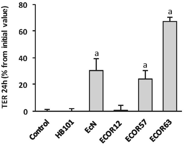
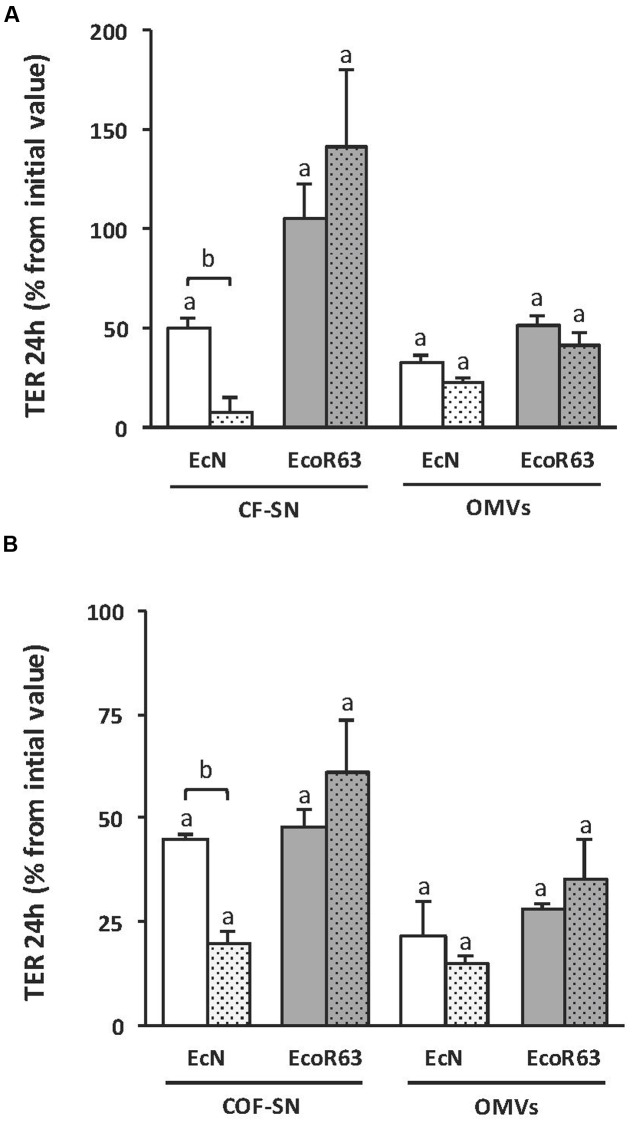
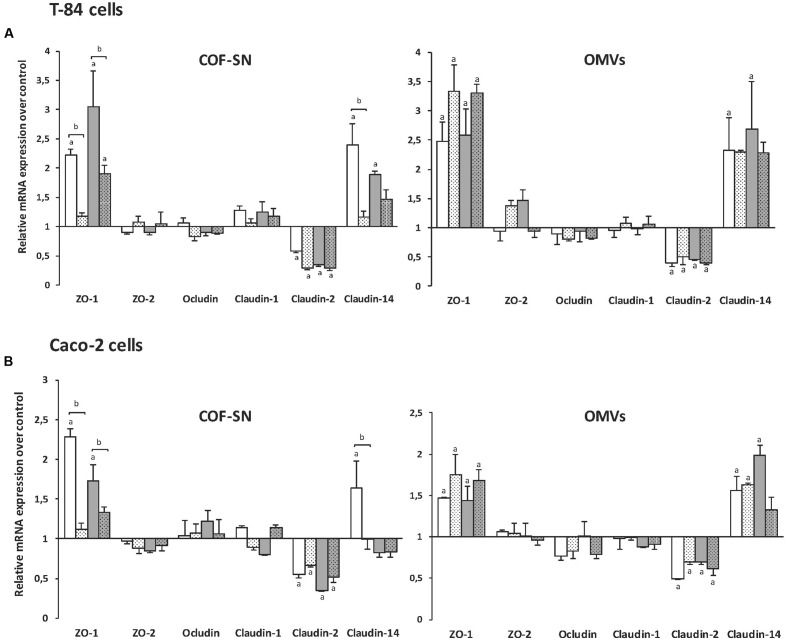
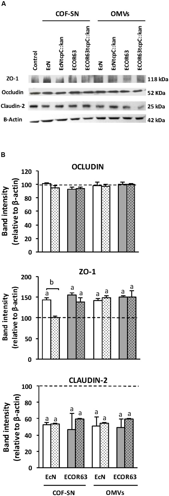
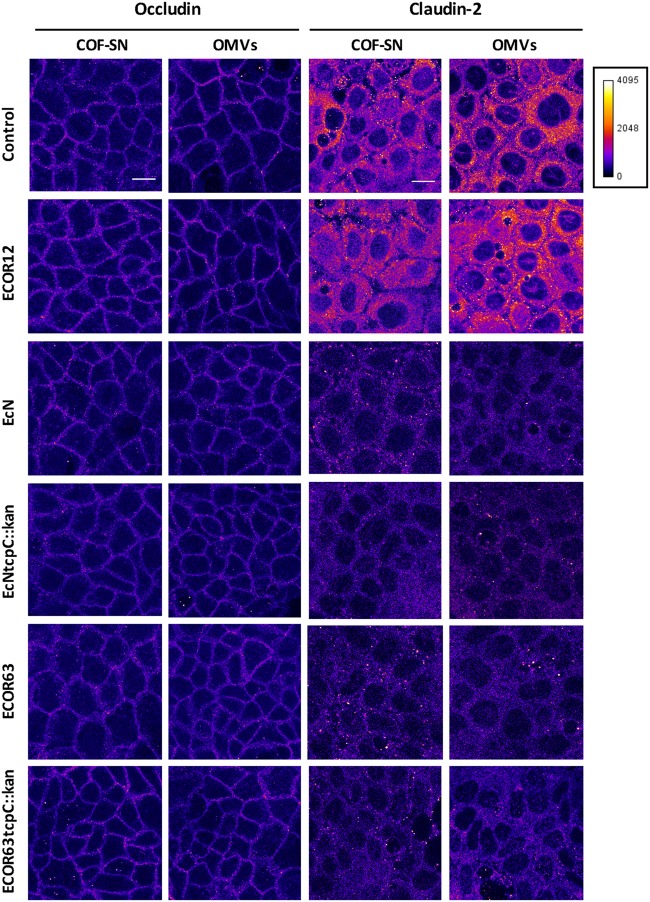
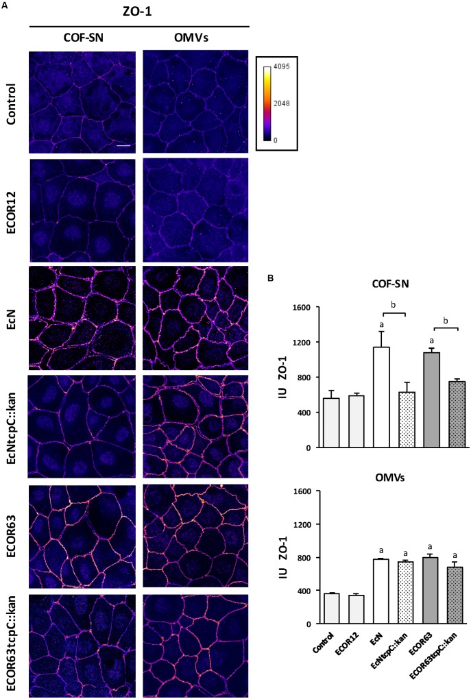
The gastrointestinal epithelial layer forms a physical and biochemical barrier that maintains the segregation between host and intestinal microbiota. The integrity of this barrier is critical in maintaining homeostasis in the body and its dysfunction is linked to a variety of illnesses, especially inflammatory bowel disease. Gut microbes, and particularly probiotic bacteria, modulate the barrier integrity by reducing gut permeability and reinforcing tight junctions. Probiotic Escherichia coli Nissle 1917 (EcN) is a good colonizer of the human gut with proven therapeutic efficacy in the remission of ulcerative colitis in humans. EcN positively modulates the intestinal epithelial barrier through upregulation and redistribution of the tight junction proteins ZO-1, ZO-2 and claudin-14. Upregulation of claudin-14 has been attributed to the secreted protein TcpC. Whether regulation of ZO-1 and ZO-2 is mediated by EcN secreted factors remains unknown. The aim of this study was to explore whether outer membrane vesicles (OMVs) released by EcN strengthen the epithelial barrier. This study includes other E. coli strains of human intestinal origin that contain the tcpC gene, such as ECOR63. Cell-free supernatants collected from the wild-type strains and from the derived tcpC mutants were fractionated into isolated OMVs and soluble secreted factors. The impact of these extracellular fractions on the epithelial barrier was evaluated by measuring transepithelial resistance and expression of several tight junction proteins in T-84 and Caco-2 polarized monolayers. Our results show that the strengthening activity of EcN and ECOR63 does not exclusively depend on TcpC. Both OMVs and soluble factors secreted by these strains promote upregulation of ZO-1 and claudin-14, and down-regulation of claudin-2. The OMVs-mediated effects are TcpC-independent. Soluble secreted TcpC contributes to the upregulation of ZO-1 and claudin-14, but this protein has no effect on the transcriptional regulation of claudin-2. Thus, in addition to OMVs and TcpC, other active factors released by these microbiota strains contribute to the reinforcement of the epithelial barrier.

Keywords: Escherichia coli; TcpC; gut microbes; intestinal barrier; membrane vesicles; phylogenetic group B2; probiotics; tight junctions.


Figures

FIGURE 1
FIGURE 1
Escherichia coli reference collection (ECOR) strains bearing tcpC gene increase TER in T-84 cell monolayers. The effect on TER of the indicated Escherichia coli strains from the ECOR collection was analyzed in T-84 monolayers in comparison with the probiotic EcN and the laboratory strain HB101. TER values were measured before and after 24 h incubation with cell-free supernatants (CF-SN) (2 mg protein/ml) collected from LB cultures (n = 3 independent biological replicates). Data are presented as percentage of increase in TER from the initial value. asignificance against untreated control cells (p ≤ 0.001).
FIGURE 2
FIGURE 2
The impact of ECOR63 and EcN on TER depends on both released soluble factors and OMVs. (A) TER analysis of T-84 monolayers after 24 h incubation with CF-SN (2 mg/ml) (containing both released soluble mediators and OMVs) or isolated OMVs (0.1 mg/ml) from overnight cultures in LB medium of the wild-type strains EcN (white bars) and ECOR63 (gray bars) and from their corresponding tcpC mutants (doted bars). (B) TER analysis of T-84 monolayers after 24 h incubation with COF-SN (0.5 mg/ml) (containing only released soluble mediators) or isolated OMVs (0.1 mg/ml) from DMEM exponential cultures of the same wild-type strains (plain bars) and tcpC mutants (doted bars). In both panels, data are presented as changes in TER (%) from the initial value (n = 3 independent biological replicates). asignificance against untreated control cells (p ≤ 0.05); bsignificance tcpC mutant vs. wild-type (p ≤ 0.002).
FIGURE 3
FIGURE 3
Gene expression levels of tight junction proteins in the intestinal epithelial cell lines T-84 (A) or Caco-2 (B). Cell monolayers were challenged for 4 h with COF-SN (0.5 mg/ml) or OMVs (0.1 mg/ml) from EcN (white bars), EcN tcpC::kan (doted white bars), ECOR63 (gray bars) or ECOR63 tcpC::kan (doted gray bars). Relative mRNA levels of the indicated proteins were measured by RT-qPCR, using β-actin as the reference gene. Data are presented as fold-change compared to untreated control cells (n = 3 independent biological replicates). aSignificance against untreated control cells (p ≤ 0.04); bsignificance tcpC mutant vs. wild-type (p ≤ 0.02).
FIGURE 4
FIGURE 4
Western blot analysis of TJ proteins in T-84 cell monolayers treated with COF-SN or OMVs from the indicated bacterial strains. Cell monolayers were challenged for 24 h with COF-SN (0.5 mg/ml) orOMVs (0.1 mg/ml) from EcN (white bars), EcN tcpC::kan (doted white bars), ECOR63 (gray bars) or ECOR63 tcpC::kan (doted gray bars). Occludin, ZO-1 and claudin-2 were immunodetected with specific antibodies. (A) Representative Western blots of three independent experiments areshown. (B) Densitometric quantification of the TJ proteins (n = 3 independent biological replicates). Values were normalized to λ-actin. Normalized values from untreated control cells were set as 100% and indicated by a dashedline. aSignificance against untreated control cells (p ≤ 0.04); bsignificance tcpC mutant vs. wild-type (p = 0.001).
FIGURE 5
FIGURE 5
Immunofluorescence staining for occludin and claudin-2 in Caco-2 cells treated for 24 h with COF-SN or OMVs from the indicated bacterial strains. Immunostaining of occludin (left) and claudin-2 (right) was carried out in Caco-2 cells at 5 days after seeding (n = 3 independent biological replicates). Images are color coded with Fire look-up table and its calibration bar is shown on the right. Scale bar, 20 μm.
FIGURE 6
FIGURE 6
Immunofluorescence staining for ZO-1 in Caco-2 cells treated for 24 h with COF-SN or OMVs from the indicated bacterial strains. (A) Immunostaining of ZO-1 was carried out in Caco-2 cells at 5 days after seeding. Images are color coded with Fire look-up table and its calibration bar is shown on the right. Scale bar, 20 μm. (B) Quantification of the mean fluorescence intensity of ZO-1 labeling in tight junctions. See Supplementary Figure S2 for details in the tracing and processing of the images. Data are presented as mean ± SEM of relative intensity units (IU) (n = 5 independent biological replicates for COF-SN treated cells and n = 3 independent biological replicates for OMVs treated cells). Statistical differences were assessed by the t-test. aSignificance against untreated control cells (p < 0.015); bsignificance tcpC mutant vs. wild-type (p ≤ 0.05).

References

    1. Aguilera L., Toloza L., Giménez R., Odena A., Oliveira E., Aguilar J., et al. (2014). Proteomic analysis of outer membrane vesicles from the probiotic strain Escherichia coli Nissle 1917. Proteomics 14 222–229. 10.1002/pmic.201300328 - DOI - PubMed
    1. Arribas B., Rodríguez-Cabezas M. E., Camuesco D., Comalada M., Bailón E., Utrilla P., et al. (2009). A probiotic strain of Escherichia coli, Nissle 1917, given orally exerts local and systemic anti-inflammatory effects in lipopolysaccharide-induced sepsis in mice. Br. J. Pharmacol. 157 1024–1033. 10.1111/j.1476-5381.2009.00270.x - DOI - PMC - PubMed
    1. Cañas M. A., Giménez R., Fábrega M. J., Toloza L., Baldomà L., Badia J. (2016). Outer membrane vesicles from the probiotic Escherichia coli Nissle 1917 and the commensal ECOR12 enter intestinal epithelial cells via clathrin-dependent endocytosis and elicit differential effects on DNA damage. PLoS ONE 11:e0160374 10.1371/journal.pone.0160374 - DOI - PMC - PubMed
    1. Chibbar R., Dieleman L. A. (2015). Probiotics in the management of ulcerative colitis. J. Clin. Gastroenterol. 49 S50–S55. 10.1097/MCG.0000000000000368 - DOI - PubMed
    1. Cirl C., Wieser A., Yadav M., Duerr S., Schubert S., Fisher H., et al. (2008). Subversion of Toll-like receptor signaling by a unique family of bacterial Toll/interleukin-1 receptor domain-containing proteins. Nat. Med. 14 399–406. 10.1038/nm1734 - DOI - PubMed