Review
. 2016 Dec 12:7:550.
doi: 10.3389/fimmu.2016.00550. eCollection 2016.

The PD1:PD-L1/2 Pathway from Discovery to Clinical Implementation

Affiliations
Review

The PD1:PD-L1/2 Pathway from Discovery to Clinical Implementation

Kankana Bardhan et al. Front Immunol. .

Abstract

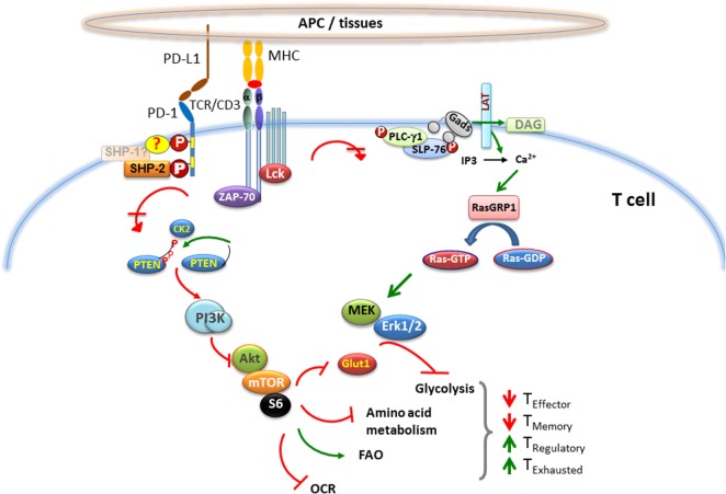
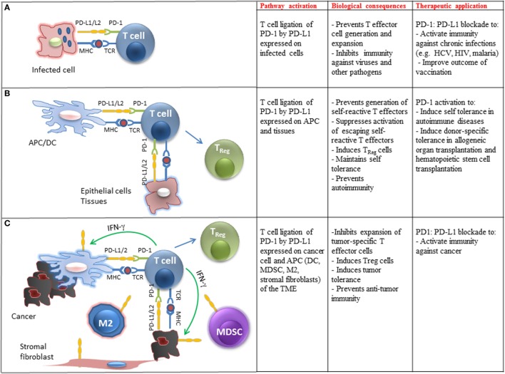
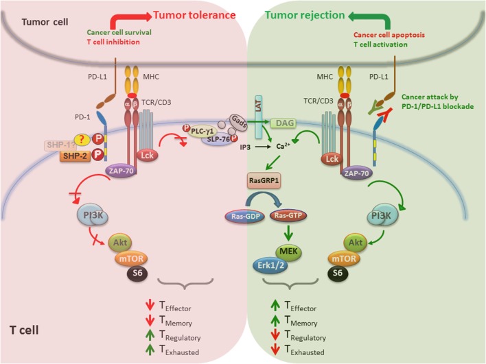
The immune system maintains a critically organized network to defend against foreign particles, while evading self-reactivity simultaneously. T lymphocytes function as effectors and play an important regulatory role to orchestrate the immune signals. Although central tolerance mechanism results in the removal of the most of the autoreactive T cells during thymic selection, a fraction of self-reactive lymphocytes escapes to the periphery and pose a threat to cause autoimmunity. The immune system evolved various mechanisms to constrain such autoreactive T cells and maintain peripheral tolerance, including T cell anergy, deletion, and suppression by regulatory T cells (TRegs). These effects are regulated by a complex network of stimulatory and inhibitory receptors expressed on T cells and their ligands, which deliver cell-to-cell signals that dictate the outcome of T cell encountering with cognate antigens. Among the inhibitory immune mediators, the pathway consisting of the programed cell death 1 (PD-1) receptor (CD279) and its ligands PD-L1 (B7-H1, CD274) and PD-L2 (B7-DC, CD273) plays an important role in the induction and maintenance of peripheral tolerance and for the maintenance of the stability and the integrity of T cells. However, the PD-1:PD-L1/L2 pathway also mediates potent inhibitory signals to hinder the proliferation and function of T effector cells and have inimical effects on antiviral and antitumor immunity. Therapeutic targeting of this pathway has resulted in successful enhancement of T cell immunity against viral pathogens and tumors. Here, we will provide a brief overview on the properties of the components of the PD-1 pathway, the signaling events regulated by PD-1 engagement, and their consequences on the function of T effector cells.

Keywords: PD-1; PD-L1; T cell exhaustion; T cell responses; T cell tolerance; cancer immunology; cancer immunotherapy.


Figures

Figure 1
Figure 1
Effect of PD-1 on major signaling pathways and subsequent metabolic reprograming in T cells. When T cell encounters a foreign antigen presented by MHC on the surface of APC, TCR gets phosphorylated upon oligomerization of TCR/CD3 chains, followed by recruitment of activated Lck and Zap-70 to the phosphorylated ITAM (tyrosine motifs) of TCR tail, leading to initiation of downstream TCR-signaling cascade. During TCR cross-linking, when PD-1 interacts with its ligands, the two tyrosine residues on PD-1 cytoplasmic tail also become phosphorylated, and SHP-2 is recruited to ITSM (also possibly SHP-1). As a consequence, Lck and Zap-70 become dephosphorylated. PD-1 ligation also causes inhibition of PI3K/Akt/mTOR and Ras/MAPK/Erk pathways, leading to downregulation of glycolysis and amino acid metabolism and increase in fatty acid oxidation in T cells. This alteration in T cell metabolic reprograming may change the course of T cell differentiation, leading to impaired differentiation of effector and memory T cells, while enhancing the differentiation of T regulatory cells and exhausted T cells.
Figure 2
Figure 2
Biological and clinical implications of PD-1 ligation on T cell immune function. (A) Engagement of PD-1 by PD-L1 expressed on pathogen-presenting cells inhibits differentiation, activation, and expansion of pathogen-specific T cells in chronic infections. Therapeutic blockade of this pathway can improve pathogen-specific immunity. (B) Engagement of PD-1 by PD-L1 expressed on tissues and APC-presenting self-antigens prevents the generation of self-reactive T effector cells, promotes the differentiation of TReg cells, suppresses expansion of escaping self-reactive T cells, and prevents autoimmunity. Therapeutic activation of this pathway may promote transplantation tolerance and induce self-tolerance in autoimmune diseases. (C) Engagement of PD-1 by PD-L1 expressed on cancer cells and immune cells infiltrating the tumor microenvironment (TME) inhibits expansion of tumor-specific T cells, promotes the generation of TReg cells, promotes tumor tolerance, and suppresses antitumor immunity. Therapeutic blockade of this pathway can activate antitumor immune responses.
Figure 3
Figure 3
PD-1/PD-L1 blockade enhances tumor rejection by activating T cells. (Left) When PD-1/PD-L1 pathway is active, promotes survival of cancer cells via antiapoptotic signals mediated via PD-L1 and inhibits signaling pathways that lead to activation and expansion of T cells that recognize tumor antigens. Together, these events lead to impaired generation of T effector and memory cells and preferentiation differentiation of TEX and TReg cells, which promote tumor tolerance. (Right) Blocking the PD-1/PD-L1 immune checkpoint pathway by anti-PD-1 or anti-PD-L1 antibodies suppresses cancer cell survival and enhances the antitumor responses of T cells, leading to tumor regression and rejection. In contrast to impaired TCR signaling induced by PD-1 engagement, PD-1/PD-L1 blockade causes activation of T cells by increasing PI3K/Akt or Ras/MAPK pathways, promoting differentiation of effector and memory T cells and suppression of TEX and TReg differentiation.

Similar articles

Cited by

References

    1. Bretscher P, Cohn M. A theory of self-nonself discrimination. Science (1970) 169:1042–9.10.1126/science.169.3950.1042 - DOI - PubMed
    1. Bretscher PA. A two-step, two-signal model for the primary activation of precursor helper T cells. Proc Natl Acad Sci U S A (1999) 96(1):185–90.10.1073/pnas.96.1.185 - DOI - PMC - PubMed
    1. Schwartz RH, Mueller DL, Jenkins MK, Quill H. T-cell clonal anergy. Cold Spring Harb Symp Quant Biol (1989) 54(Pt 2):605–10.10.1101/SQB.1989.054.01.072 - DOI - PubMed
    1. Appleman LJ, Boussiotis VA. T cell anergy and costimulation. Immunol Rev (2003) 192:161–80.10.1034/j.1600-065X.2003.00009.x - DOI - PubMed
    1. Tivol EA, Borriello F, Schweitzer AN, Lynch WP, Bluestone JA, Sharpe AH. Loss of CTLA-4 leads to massive lymphoproliferation and fatal multiorgan tissue destruction, revealing a critical negative regulatory role of CTLA-4. Immunity (1995) 3(5):541–7.10.1016/1074-7613(95)90125-6 - DOI - PubMed