. 2016 Dec 9:7:1856.
doi: 10.3389/fpls.2016.01856. eCollection 2016.

Narrow-Leafed Lupin (Lupinus angustifolius) β1- and β6-Conglutin Proteins Exhibit Antifungal Activity, Protecting Plants against Necrotrophic Pathogen Induced Damage from Sclerotinia sclerotiorum and Phytophthora nicotianae

Affiliations

Narrow-Leafed Lupin (Lupinus angustifolius) β1- and β6-Conglutin Proteins Exhibit Antifungal Activity, Protecting Plants against Necrotrophic Pathogen Induced Damage from Sclerotinia sclerotiorum and Phytophthora nicotianae

Jose C Jimenez-Lopez et al. Front Plant Sci. .

Abstract

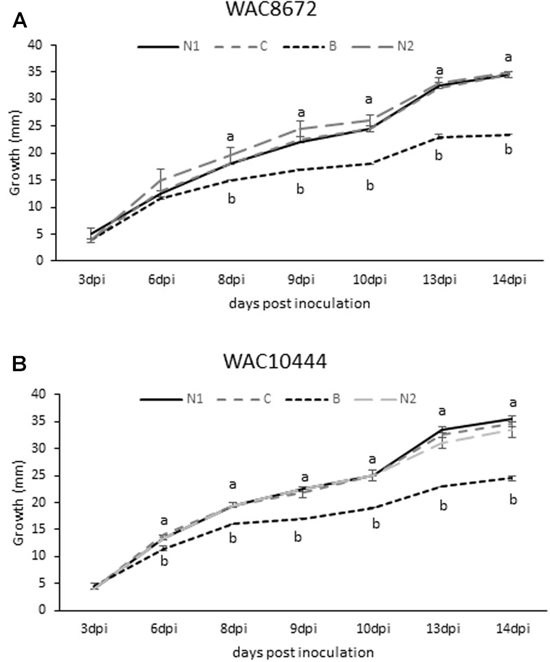
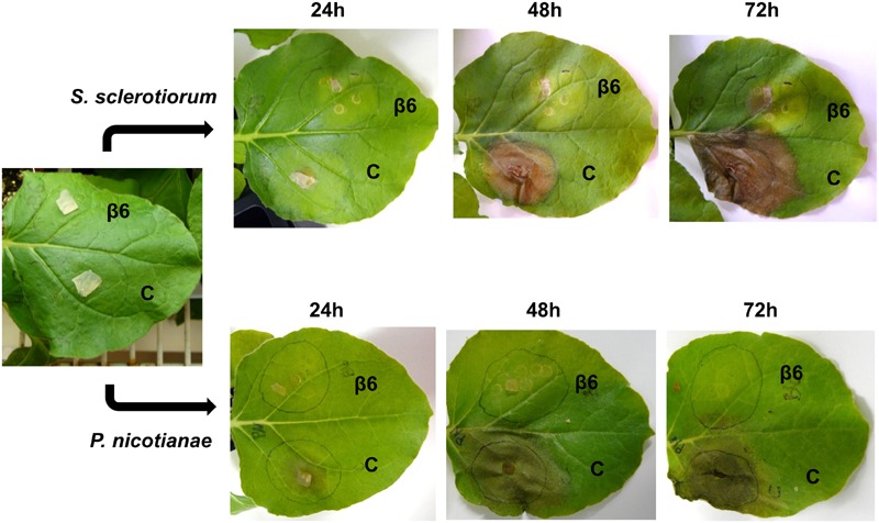
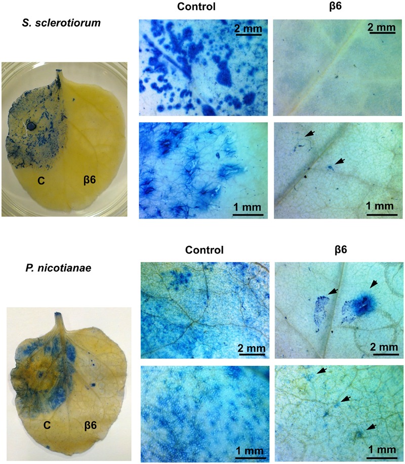
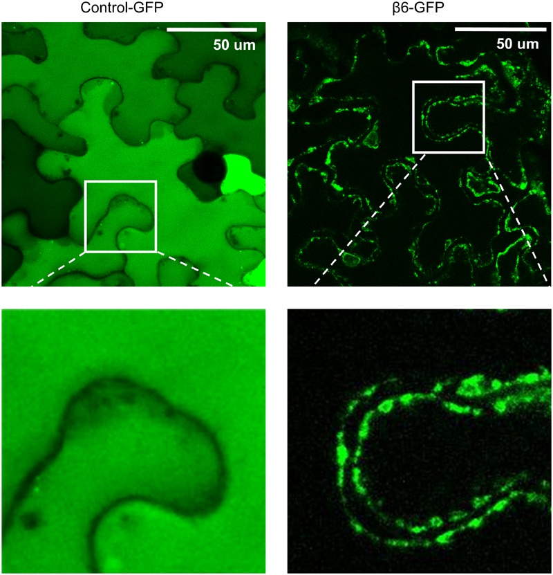
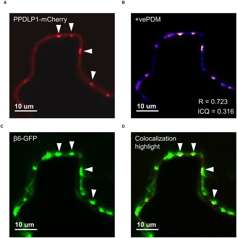
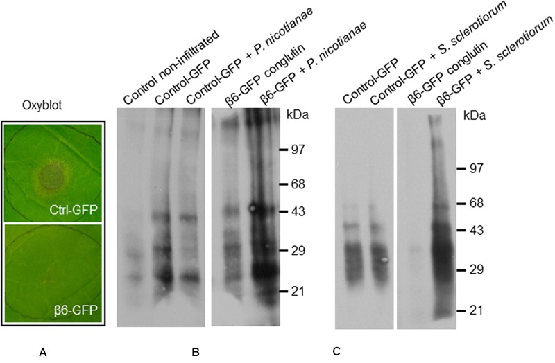
Vicilins (7S globulins) are seed storage proteins and constitute the main protein family in legume seeds, particularly in narrow-leafed lupin (Lupinus angustifolius L.; NLL), where seven vicilin genes, called β1- to β7-conglutin have been identified. Vicilins are involved in germination processes supplying amino acids for seedling growth and plant development, as well as in some cases roles in plant defense and protection against pathogens. The roles of NLL β-conglutins in plant defense are unknown. Here the potential role of five NLL β-conglutin family members in protection against necrotrophic fungal pathogens was investigated and it was demonstrated that recombinant purified 6xHis-tagged β1- and β6-conglutin proteins exhibited the strongest in vitro growth inhibitory activity against a range of necrotrophic fungal pathogens compared to β2, β3, and β4 conglutins. To examine activity in vivo, two representative necrotrophic pathogens, the fungus Sclerotinia sclerotiorum and oomycete Phytophthora nicotianae were used. Transient expression of β1- and β6-conglutin proteins in Nicotiana benthamiana leaves demonstrated in vivo growth suppression of both of these pathogens, resulting in low percentages of hyphal growth and elongation in comparison to control treated leaves. Cellular studies using β1- and β6-GFP fusion proteins showed these conglutins localized to the cell surface including plasmodesmata. Analysis of cellular death following S. sclerotiorum or P. nicotianae revealed both β1- and β6-conglutins suppressed pathogen induced cell death in planta and prevented pathogen induced suppression of the plant oxidative burst as determined by protein oxidation in infected compared to mock-inoculated leaves.

Keywords: 7S globulins; fungal pathogen; legume; oxidative stress; plant defense; seed storage protein; vicilins.


Figures

FIGURE 1
FIGURE 1
Antifungal activity of recombinant β6-conglutin in in vitro bioassays against the lupin pathogen Colletotrichum lupini. Significant growth inhibition toward β6-conglutin was observed from as early as (A) 8 dpi for isolate WAC8672 and (B) 6 dpi for isolate WAC10444. Means and standard error of three biological replicates. Letters indicate significant differences for a given day after inoculation (dpi) with the mycelial plug by student’s t-test. C, control containing 1 mg BSA; N1, no protein; B, 1 mg β6-conglutin; N2, no protein.
FIGURE 2
FIGURE 2
Recombinant β6-conglutin exhibits in planta anti-fungal and oomycete activity. Shown are representative images of Agrobacterium infiltrated N. benthamiana leaves expressing recombinant β6-conglutin proteins and subsequently inoculated with either S. sclerotiorum or P. nicotianae. The experiment was repeated three times with similar results. C, control agroinfiltration of the leaf area with Agrobacterium expressing GFP only; β6, agroinfiltration of the leaf area with Agrobacterium expressing GFP tagged β6-conglutin.
FIGURE 3
FIGURE 3
Recombinant β6-conglutin reduces pathogen growth and pathogen induced cell death in planta. Shown are representative images of Agrobacterium infiltrated N. benthamiana leaves expressing recombinant β6-conglutin proteins and subsequently inoculated either S. sclerotiorum or P. nicotianae. Trypan blue staining was performed to visualize hyphal growth and cell death. Arrows point to hyphae and hyphal damage.
FIGURE 4
FIGURE 4
β6-conglutin is localized to the cell surface. Confocal images of tobacco epidermis cell expressing GFP alone or β6-GFP shows GFP alone homogenously expressed throughout the cytoplasm while β6-conglutin localizes to the plasma membrane through the whole cell, with no expression in cytosol or intracellular organelles. Insert: β6-GFP shows punctate labeling at the cell surface.
FIGURE 5
FIGURE 5
β6-conglutin localizes to the plasmodesmata. Single-slice confocal images of co-expression GFP-β6 with the plasmodesmata marker PDLP1-mCherry after transient expression in N. benthamiana; (A) PPDLP1-mCherry, (B) β6-GFP, (C) Image showing pixel pairs that have a positive PDM value equal to the value (intensity of A- mean A intensity) (intensity of B-mean B intensity) as described in Li et al. (2004), (D) merge of (A,B) with highlighted co-localized pixels. ICQ, Intensity correlation quotient; R, Mandel’s overlap coefficient. 60× immersion objective.
FIGURE 6
FIGURE 6
β6-conglutin oxyblots assayes. Protein carbonyl formation in tobacco leaves 48 h after agroinfiltration with indicated constructs and 24 h after inoculation with pathogens. Protein carbonyls were assessed using an OxyBlot TM kit. (A) Typical levels of pathogen growth 24 h after inoculation of leaf samples used for assay. (B) Representative blot showing basal carbonylation levels in non-transformed leaves, control leaves expressing GFP mock-inoculated and after infection with P. nicotianae, and leaves expressing β6-GFP mock-inoculated and after infection. (C) Protein carbonylation levels after infection with S. sclerotiorum, same constructs as in (B).

Similar articles

Cited by

References

    1. Agrios G. N. (2005). “Plant diseases caused by fungi,” in Plant Pathology 5th Edn ed. Agrios G. N. (San Diego, CA: Academic Press; ) 385–614.
    1. Alscher R. G., Donahue J. L., Cramer C. L. (1997). Reactive oxygen species and antioxidants: Relationships in green cells. Physiol. Plant. 100 224–233. 10.1111/j.1399-3054.1997.tb04778.x - DOI
    1. Argos P., Narayana S. V., Nielsen N. C. (1985). Structural similarity between legumin and vicilin storage proteins from legumes. EMBO J. 4 1111–1117. - PMC - PubMed
    1. Bally J., Nakasugi K., Jia F., Hung H., Ho S. Y. W., Wong M., et al. (2015). The extremophile Nicotiana benthamiana has traded viral defence for early vigour. Nat. Plants 1:15165 10.1038/nplants.2015.165 - DOI - PubMed
    1. Barba-Espín G., Diaz-Vivancos P., Job D., Belghazi M., Job C., Hernández J. A. (2011). Understanding the role of H2O2 during pea seed germination: a combined proteomic and hormone profiling approach. Plant Cell Environ. 34 1907–1919. 10.1111/j.1365-3040.2011.02386.x - DOI - PubMed

LinkOut - more resources