Granular cell tumor of the esophagus in an adolescent
- PMID: 28018455
- PMCID: PMC5177722
- DOI: 10.3345/kjp.2016.59.11.S88
Granular cell tumor of the esophagus in an adolescent
Abstract
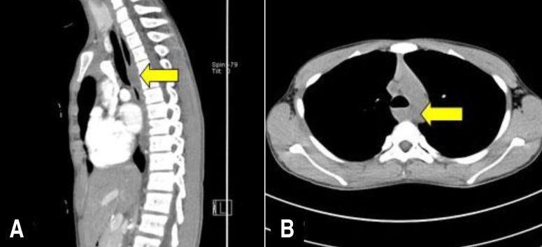
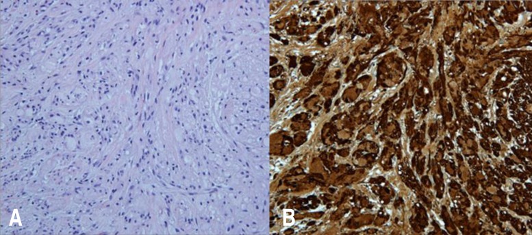
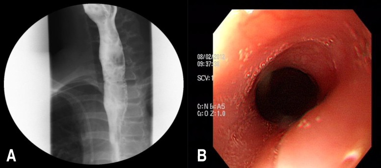
Esophageal granular cell tumor (GCT) is a rare neoplasm originating from the Schwann cells of the submucosal neuronal plexus. Histology is the gold standard for its diagnosis. Endoscopic resection or surgical excision should be considered, depending on the potential for malignancy. Here, we report a case of an esophageal GCT in an adolescent. A 12-year-old boy presented with a 1-year history of dysphagia and vomiting. Upper gastrointestinal endoscopic examination and esophagography showed narrowing of the midesophagus, and computed tomography angiography of the thoracic aorta revealed an esophageal or periesophageal mass posterior to the paratracheal segment of the esophagus. The tumor was surgically excised, and based on the pathological findings, esophageal GCT was diagnosed.
Keywords: Esophageal neoplasms; Granular cell tumor; Pediatrics; Vomiting.
Conflict of interest statement
No potential conflict of interest relevant to this article was reported.
Figures

Similar articles
-
Successful endoscopic submucosal dissection of an esophageal granular cell tumor in a pediatric patient: A case report and a therapeutic insight.JPGN Rep. 2024 Jul 4;5(3):384-388. doi: 10.1002/jpr3.12106. eCollection 2024 Aug. JPGN Rep. 2024. PMID: 39149202 Free PMC article.
-
Granular-cell tumor of the esophagus: report of a case with a cytologic diagnosis based on esophageal brushing.Diagn Cytopathol. 1998 Dec;19(6):455-7. doi: 10.1002/(sici)1097-0339(199812)19:6<455::aid-dc10>3.0.co;2-s. Diagn Cytopathol. 1998. PMID: 9839137
-
Esophageal Granular Cell Tumor: A Case Report and Review of Literature.Cureus. 2016 Sep 14;8(9):e782. doi: 10.7759/cureus.782. Cureus. 2016. PMID: 27752408 Free PMC article.
-
Novel diagnosis and treatment of esophageal granular cell tumor: report of 14 cases and review of the literature.Ann Thorac Surg. 2014 Jan;97(1):296-302. doi: 10.1016/j.athoracsur.2013.08.042. Epub 2013 Oct 17. Ann Thorac Surg. 2014. PMID: 24140217 Review.
-
Granular cell tumors of the esophagus: report of five cases and review of diagnostic and therapeutic techniques.Dis Esophagus. 2007;20(5):436-43. doi: 10.1111/j.1442-2050.2007.00692.x. Dis Esophagus. 2007. PMID: 17760659 Review.
Cited by
-
Successful endoscopic submucosal dissection of an esophageal granular cell tumor in a pediatric patient: A case report and a therapeutic insight.JPGN Rep. 2024 Jul 4;5(3):384-388. doi: 10.1002/jpr3.12106. eCollection 2024 Aug. JPGN Rep. 2024. PMID: 39149202 Free PMC article.
-
Eosinophilic esophagitis, Barrett's esophagus and esophageal neoplasms in the pediatric patient: a narrative review.Transl Gastroenterol Hepatol. 2021 Jul 25;6:32. doi: 10.21037/tgh-20-223. eCollection 2021. Transl Gastroenterol Hepatol. 2021. PMID: 34423153 Free PMC article. Review.
-
Esophageal Abrikossoff tumor.Indian J Gastroenterol. 2018 Nov;37(6):569-570. doi: 10.1007/s12664-018-0924-z. Epub 2019 Jan 8. Indian J Gastroenterol. 2018. PMID: 30617470 No abstract available.
-
Esophageal Granular Cell Tumors Can Be Differentiated from Leiomyomas Using Endoscopic Ultrasonography.Intern Med. 2018 Jun 1;57(11):1509-1515. doi: 10.2169/internalmedicine.9816-17. Epub 2018 Jan 11. Intern Med. 2018. PMID: 29321437 Free PMC article.
-
Rare case of granular cell tumor of perianal region: a case report and literature review.J Int Med Res. 2021 Jan;49(1):300060520982689. doi: 10.1177/0300060520982689. J Int Med Res. 2021. PMID: 33459105 Free PMC article. Review.
References
-
- Percinel S, Savas B, Yilmaz G, Erinanc H, Kupana Ayva S, Bektas M, et al. Granular cell tumor of the esophagus: three case reports and review of the literature. Turk J Gastroenterol. 2008;19:184–188. - PubMed
-
- Buratti S, Savides TJ, Newbury RO, Dohil R. Granular cell tumor of the esophagus: report of a pediatric case and literature review. J Pediatr Gastroenterol Nutr. 2004;38:97–101. - PubMed
-
- Mohammad S, Naiditch JA, Jaffar R, Rothstein D, Bass LM. Granular cell tumor of the esophagus in an adolescent girl. J Pediatr Gastroenterol Nutr. 2012;54:715. - PubMed
-
- Issaivanan M, Redner A, Weinstein T, Soffer S, Glassman L, Edelman M, et al. Esophageal carcinoma in children and adolescents. J Pediatr Hematol Oncol. 2012;34:63–67. - PubMed
-
- Ha J, Lee OJ, Cho HS, Jung TS, Yoon JH, Lee EJ, et al. Endoscopically removed granular cell tumor of the esophagus: a case report and review of Korean literature. Korean J Gastrointest Endosc. 2003;26:84–89.
LinkOut - more resources
Full Text Sources
Other Literature Sources