. 2016 Nov 15:8:ecurrents.outbreaks.9aa2e1fb61b0f632f58a098773008c4b.
doi: 10.1371/currents.outbreaks.9aa2e1fb61b0f632f58a098773008c4b.

Identifying Candidate Targets of Immune Responses in Zika Virus Based on Homology to Epitopes in Other Flavivirus Species

Affiliations

Identifying Candidate Targets of Immune Responses in Zika Virus Based on Homology to Epitopes in Other Flavivirus Species

Xiaojun Xu et al. PLoS Curr. .

Abstract

Introduction: The current outbreak of Zika virus has resulted in a massive effort to accelerate the development of ZIKV-specific diagnostics and vaccines. These efforts would benefit greatly from the definition of the specific epitope targets of immune responses in ZIKV, but given the relatively recent emergence of ZIKV as a pandemic threat, few such data are available.

Methods: We used a large body of epitope data for other Flaviviruses that was available from the IEDB for a comparative analysis against the ZIKV proteome in order to project targets of immune responses in ZIKV.

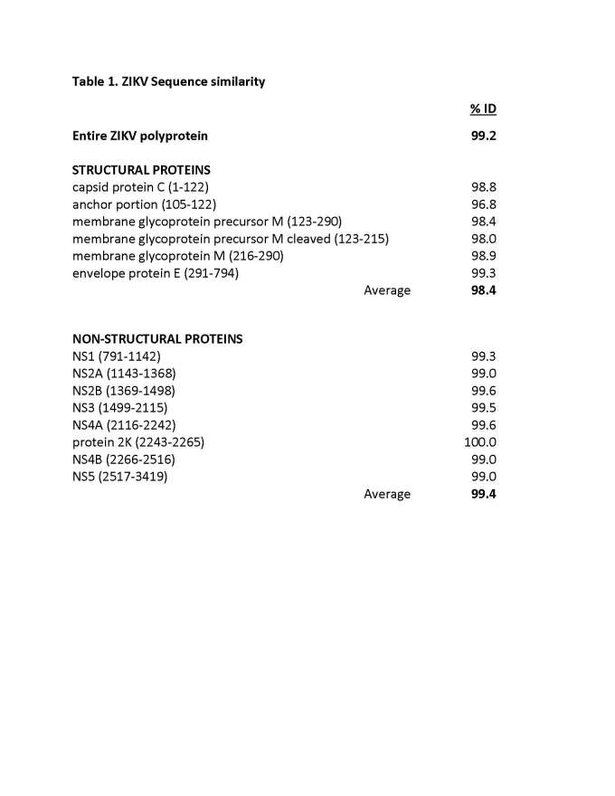
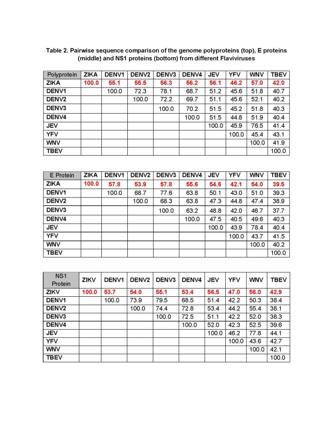
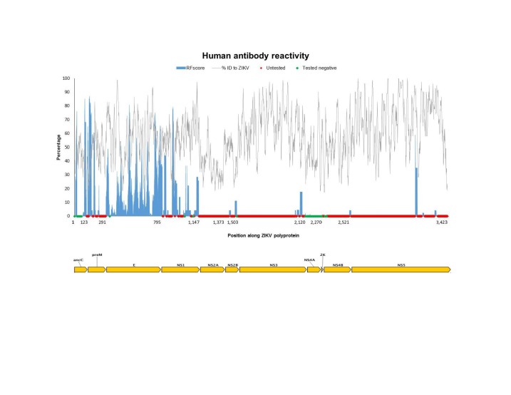
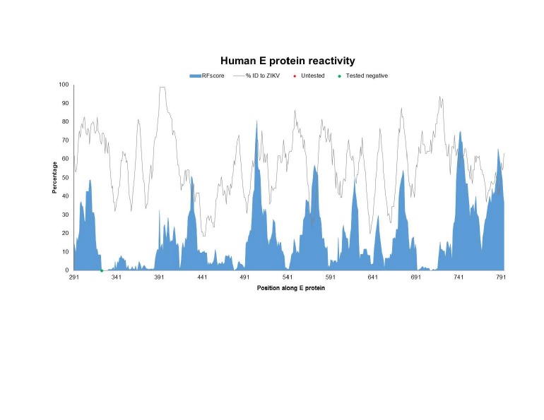
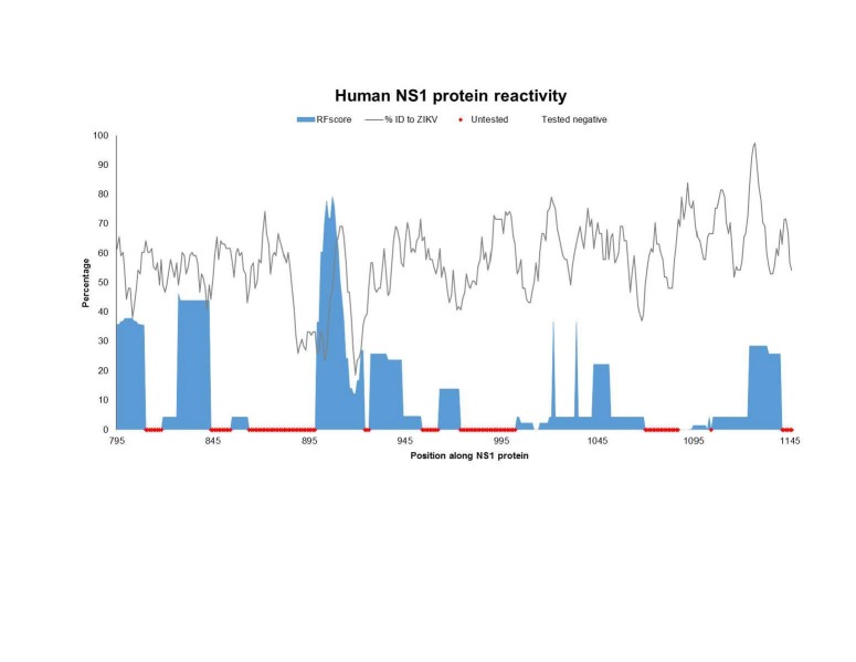
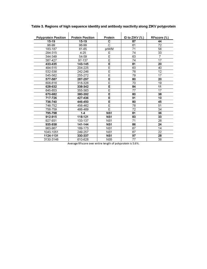
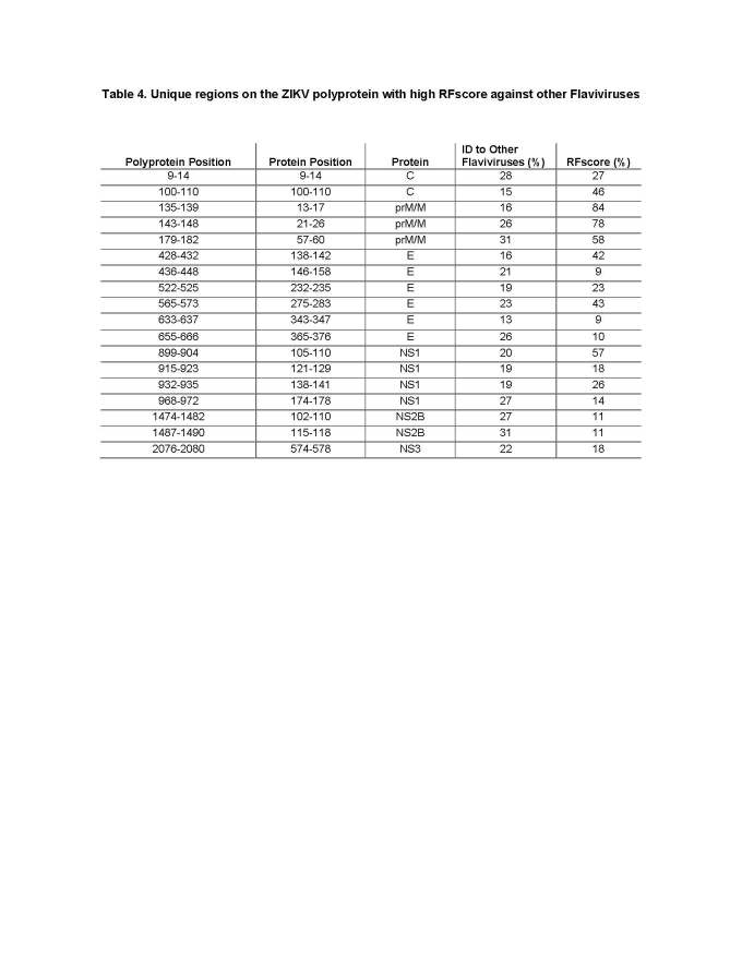
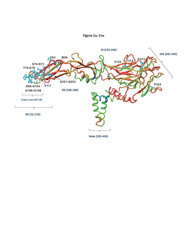
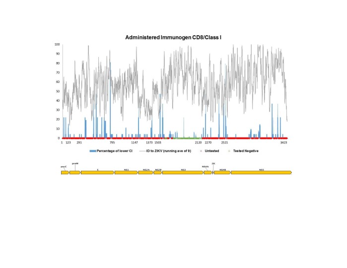
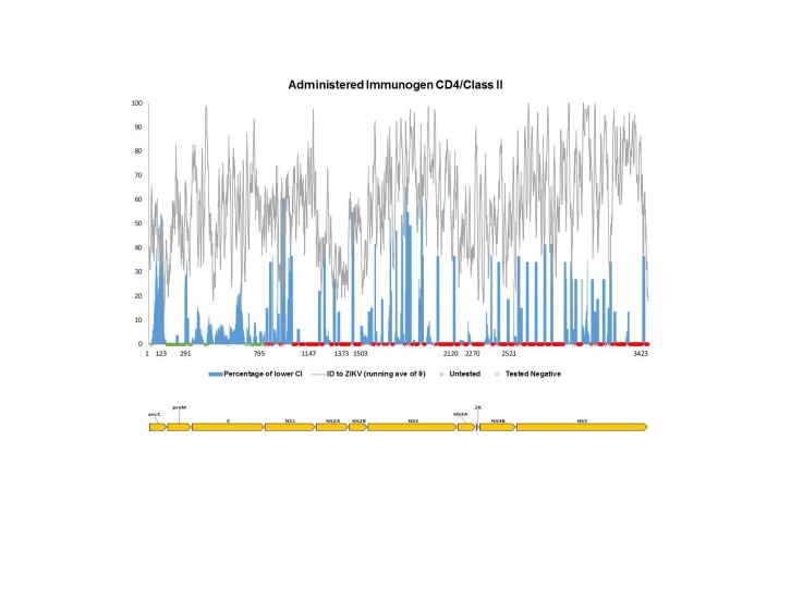
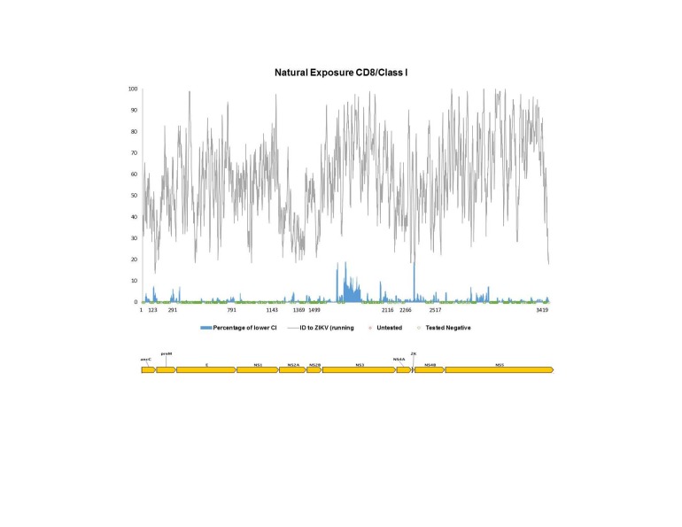
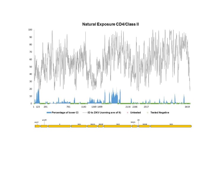
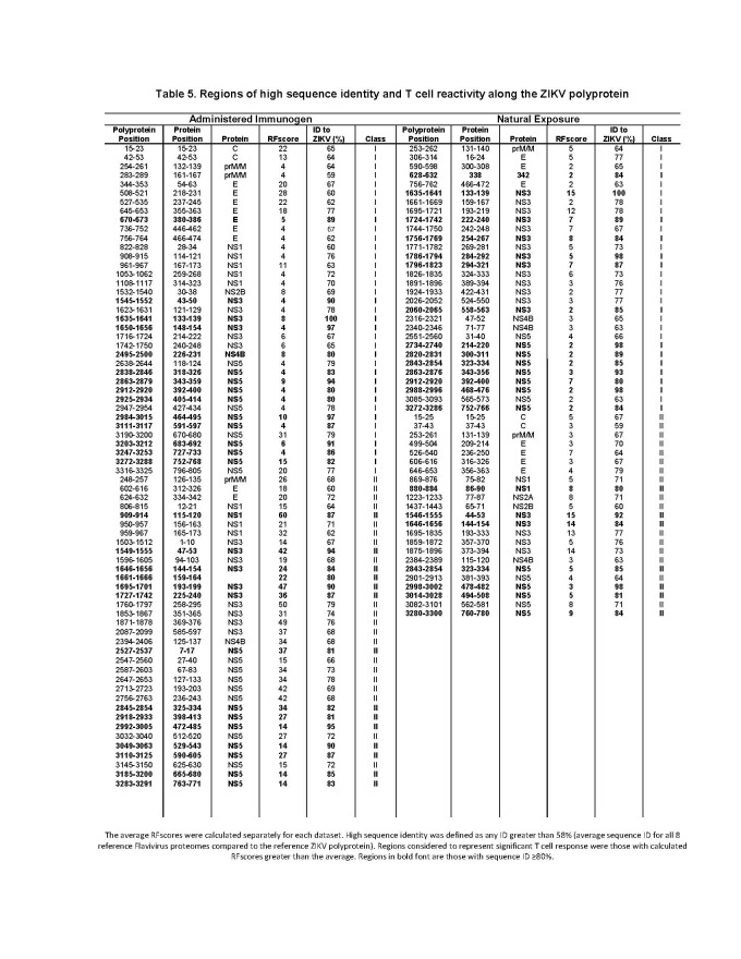
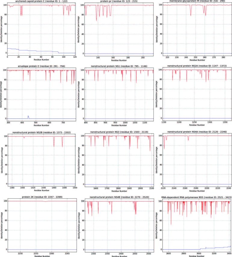
Results: We found a significant level of overlap between known antigenic sites from other Flavivirus proteins with residues on the ZIKV polyprotein. The E and NS1 proteins shared functional antibody epitope sites, whereas regions of T cell reactivity were conserved within NS3 and NS5 for ZIKV. Discussion: Our epitope based analysis provides guidance for which regions of the ZIKV polyprotein are most likely unique targets of ZIKV-specific antibodies, and which targets in ZIKV are most likely to be cross-reactive with other Flavivirus species. These data may therefore provide insights for the development of antibody- and T cell-based ZIKV-specific diagnostics, therapeutics and prophylaxis.


Figures

None
Table 1. ZIKV Sequence similarity
None
Table 2. Pairwise sequence comparison of the genome polyproteins (top), E proteins (middle) and NS1 proteins (bottom) from different Flaviviruses
None
Fig. 1A: The Flavivirus sequence identity and human antibody response frequency visualized along the entire ZIKV polyprotein. The sequence identity data (grey) shown represent a running average (window of 9 aa) of sequences for reference Flaviviruses to ZIKV, while the response frequency scores (blue) represent individual residues. Red and green dots along the x-axis represent untested and negative regions, respectively. Residues intervals along the x-axis of the polyprotein plot represent start sites of individual protein products in the polyprotein depicted also at the bottom of panel A.
None
Fig. 1B: The Flavivirus sequence identity and human antibody response frequency visualized along the E protein. The sequence identity data (grey) shown represent a running average (window of 9 aa) of sequences for reference Flaviviruses to ZIKV, while the response frequency scores (blue) represent individual residues. Red and green dots along the x-axis represent untested and negative regions, respectively. Residues intervals along the x-axis of the polyprotein plot represent start sites of individual protein products in the polyprotein depicted also at the bottom of panel A.
None
Fig. 1C: The Flavivirus sequence identity and human antibody response frequency visualized along the NS1 protein. The sequence identity data (grey) shown represent a running average (window of 9 aa) of sequences for reference Flaviviruses to ZIKV, while the response frequency scores (blue) represent individual residues. Red and green dots along the x-axis represent untested and negative regions, respectively. Residues intervals along the x-axis of the polyprotein plot represent start sites of individual protein products in the polyprotein depicted also at the bottom of panel A.
None
Table 3. Regions of high sequence identity and antibody reactivity along ZIKV polyprotein
None
Table 4. Unique regions on the ZIKV polyprotein with high RFscore against other Flaviviruses
None
Fig. 2A: 3D rendering of RFscore mapped on the ZIKV E protein using PDB structure 5IRE showing functional Flavivirus epitopes (neutralizing and/or protective) defined in humans and mapped onto the corresponding locations on ZIKV E protein. Colors represent high to low RF scores, where (blue>cyan>green>yellow>orange>red), where blue is highest and red is lowest.
None
Fig. 2B: 3D rendering of RFscore mapped on the ZIKV NS1 protein using PDB structure 5IY3. Colors represent high to low RF scores, where (blue>cyan>green>yellow>orange>red), where blue is highest and red is lowest.
None
Fig. 3A: Flavivirus sequence identity and human T cell reactivity visualized onto the ZIKV reference genomic polyprotein for CD8+/class I responses to administered antigen (experimental or commercial vaccines). The sequence identity data shown (grey) represent a running average (window of 9 aa) while the RFscores (blue) represent individual residues. Red and green dots along x-axis represent untested and negative regions, respectively. Residues intervals along the x-axis represent start sites of individual proteins.
None
Fig. 3B: Flavivirus sequence identity and human T cell reactivity visualized onto the ZIKV reference genomic polyprotein for CD4+/class II responses to administered antigen (experimental or commercial vaccines). The sequence identity data shown (grey) represent a running average (window of 9 aa) while the RFscores (blue) represent individual residues. Red and green dots along x-axis represent untested and negative regions, respectively. Residues intervals along the x-axis represent start sites of individual proteins.
None
Fig. 4A: Flavivirus sequence identity and human T cell reactivity visualized onto the ZIKV reference genomic polyprotein for CD8+/class I responses to natural exposure (documented DF, DHF, DSS or living in/travel to endemic region). The sequence identity data shown (grey) represent a running average (window of 9 aa) while the response frequency scores (blue) represent individual residues. Red and green dots along x-axis represent untested and negative regions, respectively. Residues intervals along the x-axis represent start sites of individual proteins.
None
Fig. 4B: Flavivirus sequence identity and human T cell reactivity visualized onto the ZIKV reference genomic polyprotein for CD4+/class II responses to natural exposure (documented DF, DHF, DSS or living in/travel to endemic region). The sequence identity data shown (grey) represent a running average (window of 9 aa) while the response frequency scores (blue) represent individual residues. Red and green dots along x-axis represent untested and negative regions, respectively. Residues intervals along the x-axis represent start sites of individual proteins.
None
Table 5. Regions of high sequence identity and T cell reactivity along the ZIKV polyprotein
None
Supplemental Fig. 1: The degree of sequence conservation for consensus residue at each position for ZIKV polyprotein. The red line represents the sequence identity and the blue represents the percentage of residue deletions (if any). Deletions present at N- or C- termini are possibly the result of incomplete sequences and thus may not have biological significance.

Similar articles

Cited by

References

    1. Fauci AS, Morens DM: Zika Virus in the Americas - Yet Another Arbovirus Threat. N Engl J Med 2016, 374:601-604. - PubMed
    1. Driggers RW, Ho CY, Korhonen EM, Kuivanen S, Jääskeläinen AJ, Smura T, Rosenberg A, Hill DA, DeBiasi RL, Vezina G, Timofeev J, Rodriguez FJ, Levanov L, Razak J, Iyengar P, Hennenfent A, Kennedy R, Lanciotti R, du Plessis A, Vapalahti O. Zika Virus Infection with Prolonged Maternal Viremia and Fetal Brain Abnormalities. N Engl J Med. 2016;374(22):2142-2151. - PubMed
    1. Heymann DL, Hodgson A, Sall AA, Freedman DO, Staples JE, Althabe F, Baruah K, Mahmud G, Kandun N, Vasconcelos PF, et al: Zika virus and microcephaly: why is this situation a PHEIC? Lancet 2016;387(10020):719-721. - PMC - PubMed
    1. Rubin EJ, Greene MF, Baden LR: Zika Virus and Microcephaly. N Engl J Med 2016;374(10):984-985. - PubMed
    1. WHO situation report