. 2016 Oct;3(4):043503.
doi: 10.1117/1.JMI.3.4.043503. Epub 2016 Dec 14.

Noise performance of low-dose CT: comparison between an energy integrating detector and a photon counting detector using a whole-body research photon counting CT scanner

Affiliations

Noise performance of low-dose CT: comparison between an energy integrating detector and a photon counting detector using a whole-body research photon counting CT scanner

Zhicong Yu et al. J Med Imaging (Bellingham). 2016 Oct.

Abstract

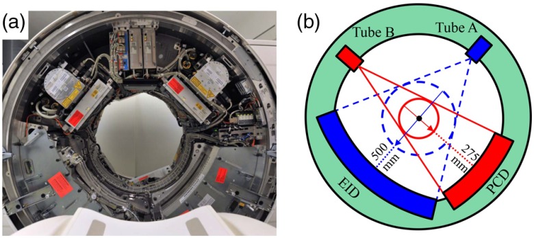
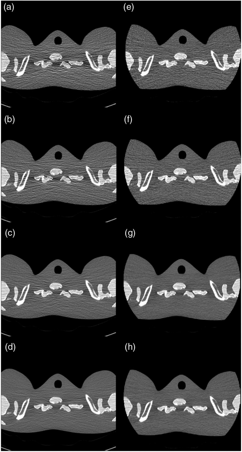
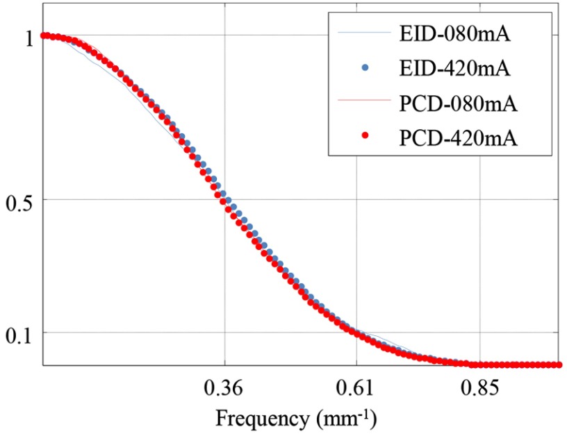
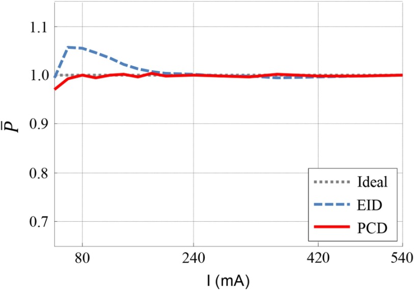
Photon counting detector (PCD)-based computed tomography (CT) is an emerging imaging technique. Compared to conventional energy integrating detector (EID)-based CT, PCD-CT is able to exclude electronic noise that may severely impair image quality at low photon counts. This work focused on comparing the noise performance at low doses between the PCD and EID subsystems of a whole-body research PCD-CT scanner, both qualitatively and quantitatively. An anthropomorphic thorax phantom was scanned, and images of the shoulder portion were reconstructed. The images were visually and quantitatively compared between the two subsystems in terms of streak artifacts, an indicator of the impact of electronic noise. Furthermore, a torso-shaped water phantom was scanned using a range of tube currents. The product of the noise and the square root of the tube current was calculated, normalized, and compared between the EID and PCD subsystems. Visual assessment of the thorax phantom showed that electronic noise had a noticeably stronger degrading impact in the EID images than in the PCD images. The quantitative results indicated that in low-dose situations, electronic noise had a noticeable impact (up to a 5.8% increase in magnitude relative to quantum noise) on the EID images, but negligible impact on the PCD images.

Keywords: computed tomography; electronic noise; low dose; photon counting.


Figures

Fig. 1
Fig. 1
Illustration of the whole-body research PCCT system. (a) Photograph. (b) Schematic drawing.
Fig. 2
Fig. 2
Phantoms used for low-dose performance assessments. (a) Anthropomorphic thorax phantom (“Lungman,” PH-1, Kyoto Kagaku, Kyoto, Japan). The shoulder portion was scanned. (b) Placement of the regions of interest on an axial image of the thorax phantom. (c) Torso-shaped water phantom with lateral width 35 cm. A tungsten wire of diameter 0.05 mm sealed in an air-filled vial was placed near the center of the water phantom for in-plane spatial resolution measurements.
Fig. 3
Fig. 3
Images of the shoulder phantom at various dose levels. (a)–(d) Images from the EID subsystem at 20, 30, 60, and 120 mAs, respectively. (e)–(h) Images from the PCD subsystem at 20, 30, 60, and 120 mAs, respectively. The PCD images had noticeably less horizontal streak artifacts and an overall more uniform appearance than the EID image. Beam hardening artifacts (dark banding) were present in both images. Display window: W/L=900/40  HU.
Fig. 4
Fig. 4
Measurement of in-plane spatial resolution. For each subsystem, there was no noticeable difference in the measured MTF curves between 80 and 420 mA, indicating consistent in-plane spatial resolution across different tube currents.
Fig. 5
Fig. 5
Normalized product of noise and square root of tube current. The normalized product for the EID subsystem was >1 at low tube currents, which is evidence of electronic noise. The normalized product for the PCD subsystem was 1 for tube currents between 80 and 540 mA.

Similar articles

Cited by

References

    1. Whiting B. R., et al. , “Properties of preprocessed sinogram data in x-ray computed tomography,” Med. Phys. 33(9), 3290–3303 (2006).MPHYA610.1118/1.2230762 - DOI - PubMed
    1. Massoumzadeh P., et al. , “Validation of CT dose-reduction simulation,” Med. Phys. 36(1), 174–189 (2009).MPHYA610.1118/1.3031114 - DOI - PMC - PubMed
    1. Duan X., et al. , “Electronic noise in CT detectors: impact on image noise and artifacts,” Am. J. Roentgenol. 201(4), W626–W632 (2013).AJROAM10.2214/AJR.12.10234 - DOI - PubMed
    1. Liu Y., et al. , “Reducing image noise in computed tomography (CT) colonography: effect of an integrated circuit CT detector,” J. Comput. Assist Tomogr. 38(3), 398–403 (2014).10.1097/RCT.0000000000000081 - DOI - PubMed
    1. Iwanczyk J. S., et al. , “Photon counting energy dispersive detector arrays for x-ray imaging,” IEEE Trans. Nucl. Sci. 56 (3), 535–542 (2009).IETNAE10.1109/TNS.2009.2013709 - DOI - PMC - PubMed