. 2016 Dec 22;1(21):e90240.
doi: 10.1172/jci.insight.90240.

Hypoxia sensing through β-adrenergic receptors

Affiliations

Hypoxia sensing through β-adrenergic receptors

Hoi I Cheong et al. JCI Insight. .

Abstract

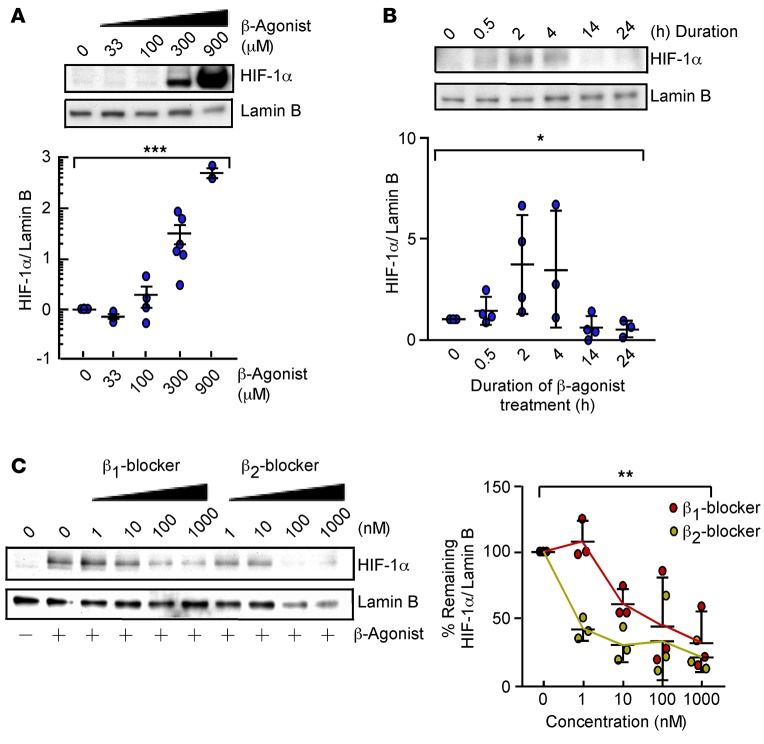
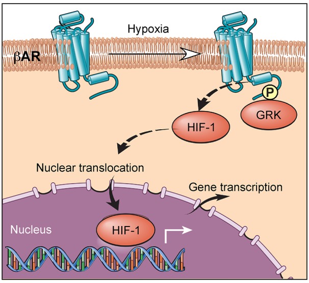
Life-sustaining responses to low oxygen, or hypoxia, depend on signal transduction by HIFs, but the underlying mechanisms by which cells sense hypoxia are not completely understood. Based on prior studies suggesting a link between the β-adrenergic receptor (β-AR) and hypoxia responses, we hypothesized that the β-AR mediates hypoxia sensing and is necessary for HIF-1α accumulation. Beta blocker treatment of mice suppressed hypoxia induction of renal HIF-1α accumulation, erythropoietin production, and erythropoiesis in vivo. Likewise, beta blocker treatment of primary human endothelial cells in vitro decreased hypoxia-mediated HIF-1α accumulation and binding to target genes and the downstream hypoxia-inducible gene expression. In mechanistic studies, cAMP-activated PKA and/or GPCR kinases (GRK), which both participate in β-AR signal transduction, were investigated. Direct activation of cAMP/PKA pathways did not induce HIF-1α accumulation, and inhibition of PKA did not blunt HIF-1α induction by hypoxia. In contrast, pharmacological inhibition of GRK, or expression of a GRK phosphorylation-deficient β-AR mutant in cells, blocked hypoxia-mediated HIF-1α accumulation. Mass spectrometry-based quantitative analyses revealed a hypoxia-mediated β-AR phosphorylation barcode that was different from the classical agonist phosphorylation barcode. These findings indicate that the β-AR is fundamental to the molecular and physiological responses to hypoxia.


Conflict of interest statement

The authors have declared that no conflict of interest exists.

Figures

Figure 1
Figure 1. Beta blocker blunts HIF-mediated erythropoiesis under hypoxia in vivo.
(A) Mice were orally administered vehicle or carvedilol (beta blocker), followed by exposure to 21% or 10% oxygen. Kidneys and serum were harvested after 2 hours, and bone marrow was collected at 24 hours (n = 5/group). (B) Expression of HIF-1α and Lamin B in nuclear extracts of kidneys, as detected by Western blot. Samples were run on the same gel but were noncontiguous. (C) HIF-1α occupancy at the erythropoietin gene Epo, detected by ChIP with control IgG or HIF-1α antibody and quantitative PCR. Fold enrichment was percentage of input (treatment) divided by average percentage of input (normoxia). (D) Expression of Epo and Actin mRNA in kidneys, as detected by reverse-transcription quantitative PCR. (E) Serum erythropoietin by ELISA. (F) Flow cytometry for erythroid progenitor populations. Proerythroblasts (I) were defined by CD44hiTER119lo and basophilic (II) and polychromatic erythroblasts (III) were defined by TER119hi as well as size and CD44 expression. Data are mean ± SD. *P < 0.05; **P < 0.005, ANOVA.
Figure 2
Figure 2. Beta blocker attenuates hypoxia responses in vitro.
(A) Expression of HIF-1α and Lamin B in HUVECs treated with escalating doses of propranolol, followed by 5-hour hypoxia (2% oxygen) (n = 2–7/condition). Data are mean ± SEM. *P < 0.05, Student’s t test. (B) HIF-1α occupancy at target genes in HUVECs treated with diluent or propranolol, followed by 12-hour hypoxia. ChIP with control IgG or HIF-1α antibody and quantification of promoter regions of CXCL12, HK2, and VEGFA by quantitative PCR. Fold enrichment was percentage of input (hypoxia with or without beta blocker) divided by average percentage of input (normoxia) (n = 3). *P < 0.05, ANOVA. (C) Heatmap showing the levels of differentially expressed transcripts of HUVECs treated with diluent (–) or propranolol (+), followed by 24-hour hypoxia. The levels of expression relative to normoxia are represented on a continuous scale from blue (lowest) to pink (highest) (n = 5 biological replicates). FDR = 0.05. (D) Kyoto Encyclopedia of Genes and Genomes pathway analysis of transcripts reversed by propranolol under hypoxia in HUVECs. Enrichment score is computed by –log (P value).
Figure 3
Figure 3. β-Agonist promotes HIF-1α accumulation under normoxia.
(A) Expression of HIF-1α and Lamin B in HUVECs treated with diluent or isoproterenol (33–900 μM) for 2 hours (n = 3–6/condition). Data are mean ± SEM. (B) Expression of HIF-1α and Lamin B in HUVECs treated with 300 μM isoproterenol for 0.5 to 24 hours (n = 3–4/condition). Data are mean ± SD. (C) Expression of HIF-1α and Lamin B in HUVECs treated with beta 1 blocker CGP-20712A or beta 2 blocker ICI-118551 (1–1,000 nM) for 45 minutes, followed by 2-hour isoproterenol (n = 3). Data are mean ± SD. *P < 0.05; **P < 0.005; ***P < 0.0005, ANOVA.
Figure 4
Figure 4. Hypoxia or β-agonist–mediated HIF-1α accumulation depends on phosphorylation of β-adrenergic receptor (β-AR) by GPCR kinase (GRK).
(A) Intracellular cAMP in HUVECs 10–15 minutes after stimulation with factors shown (n = 3–6). (B) Expression of HIF-1α and Lamin B in HUVECs treated with 300 μM β-agonist isoproterenol with or without 10 μM of the PKA inhibitor H89 or forskolin or dibutyryl cAMP (dbcAMP) for 2 hours. Cobalt chloride (CoCl2) shown as positive control (n = 3). (C) Expression of HIF-1α and Lamin B in HUVECs exposed to GRK inhibitor (1–125 μM) for 45 minutes followed by 2-hour isoproterenol. The blot is representative of 2 independent experiments. (D) Expression of HIF-1α and Lamin B in HUVECs exposed to a GRK (125 μM) or PKA inhibitor H89 (10 μM), followed by 5-hour hypoxia (2% oxygen) (n = 3). (E) Map of β-AR serine and threonine residues that are phosphorylated by PKA and GRK, mutated to alanine. (F) Expression of HIF-1α and Lamin B in human embryonic kidney cells (HEK293) overexpressing β1-AR (WTβ1-AR) or mutants lacking PKA (PKAβ1-AR) or GRK (GRKβ1-AR) sites, as shown in E, exposed to normoxia or 5-hour hypoxia (n = 3). Data are mean ± SD. *P < 0.05; **P < 0.005; ***P < 0.0005, Student’s t test.
Figure 5
Figure 5. Hypoxia induces a phosphorylation barcode in the absence of agonist binding.
Human embryonic kidney cells (HEK293) overexpressing β2-AR were exposed to the β-agonist isoproterenol or 5-hour hypoxia. β2-ARs were enriched by alprenolol for quantitative mass spectrometry analysis of phosphorylated peptides. 21% and 2% O2 (n = 3); β-agonist (n = 2). (A) Spectrum for the pS246-containing peptide. The mass difference between the y7 and y8 ions is consistent with phosphorylation at S246. (C) Spectrum for the pS261- and pS262-containing peptide. The masses of the y5, y6, and y7 ions are consistent with phosphorylation at S261 and S262. (E) Spectrum for the pS355- and pS356-containing peptide. The masses of the y16, y17, and y18 ions are consistent with phosphorylation at S355 and S356. (B, D, and F) Dot plots showing abundance of each phosphorylated peptide at 21% or 2% oxygen or with β-agonist. (G and H) Expression of phosphorylated β2-AR at S355/S356 and total β2-AR in HUVECs with 21% or 2% oxygen (vehicle or GRK inhibitor at 125 μM). Replicate samples run on parallel gels are presented (n = 2). (I) Hypoxia-specific β-AR phosphorylation barcode with increased (pink) and decreased (blue) phosphorylation at unique sites.
Figure 6
Figure 6. Working model.
Hypoxia induces a unique β-adrenergic receptor (β-AR) phosphorylation barcode that is GPCR kinase–dependent (GRK-dependent), which drives HIF-1α accumulation.

Similar articles

Cited by

References

    1. Liu Y, Cox SR, Morita T, Kourembanas S. Hypoxia regulates vascular endothelial growth factor gene expression in endothelial cells. Identification of a 5′ enhancer. Circ Res. 1995;77(3):638–643. doi: 10.1161/01.RES.77.3.638. - DOI - PubMed
    1. Semenza GL, Wang GL. A nuclear factor induced by hypoxia via de novo protein synthesis binds to the human erythropoietin gene enhancer at a site required for transcriptional activation. Mol Cell Biol. 1992;12(12):5447–5454. doi: 10.1128/MCB.12.12.5447. - DOI - PMC - PubMed
    1. Kapitsinou PP, et al. Hepatic HIF-2 regulates erythropoietic responses to hypoxia in renal anemia. Blood. 2010;116(16):3039–3048. doi: 10.1182/blood-2010-02-270322. - DOI - PMC - PubMed
    1. Beall CM, et al. Natural selection on EPAS1 (HIF2α) associated with low hemoglobin concentration in Tibetan highlanders. Proc Natl Acad Sci U S A. 2010;107(25):11459–11464. doi: 10.1073/pnas.1002443107. - DOI - PMC - PubMed
    1. Huang LE, Gu J, Schau M, Bunn HF. Regulation of hypoxia-inducible factor 1α is mediated by an O2-dependent degradation domain via the ubiquitin-proteasome pathway. Proc Natl Acad Sci U S A. 1998;95(14):7987–7992. doi: 10.1073/pnas.95.14.7987. - DOI - PMC - PubMed