. 2016 Dec 23;1(6):eaai8593.
doi: 10.1126/sciimmunol.aai8593. Epub 2016 Dec 9.

The TCF1-Bcl6 axis counteracts type I interferon to repress exhaustion and maintain T cell stemness

Affiliations

The TCF1-Bcl6 axis counteracts type I interferon to repress exhaustion and maintain T cell stemness

Tuoqi Wu et al. Sci Immunol. .

Abstract

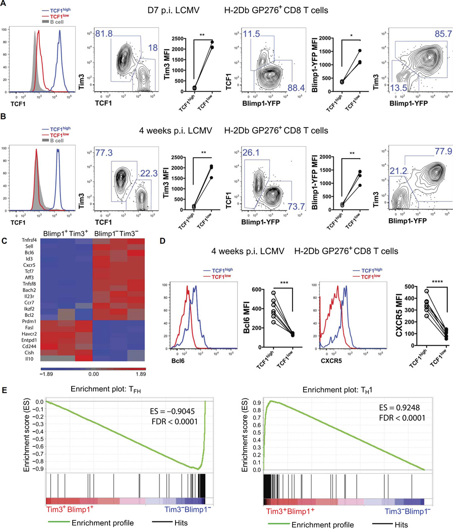
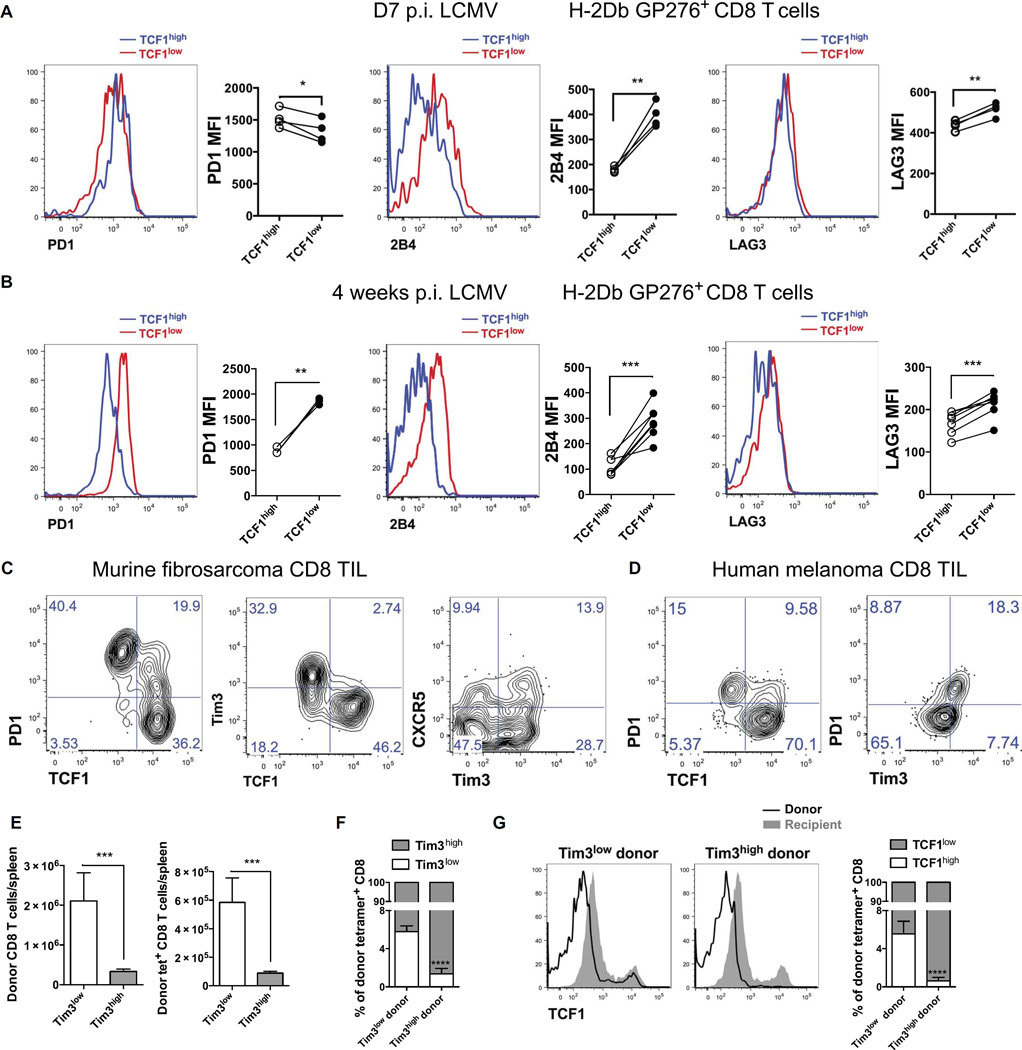
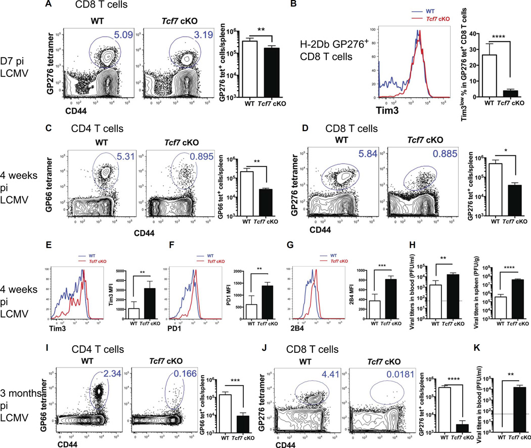
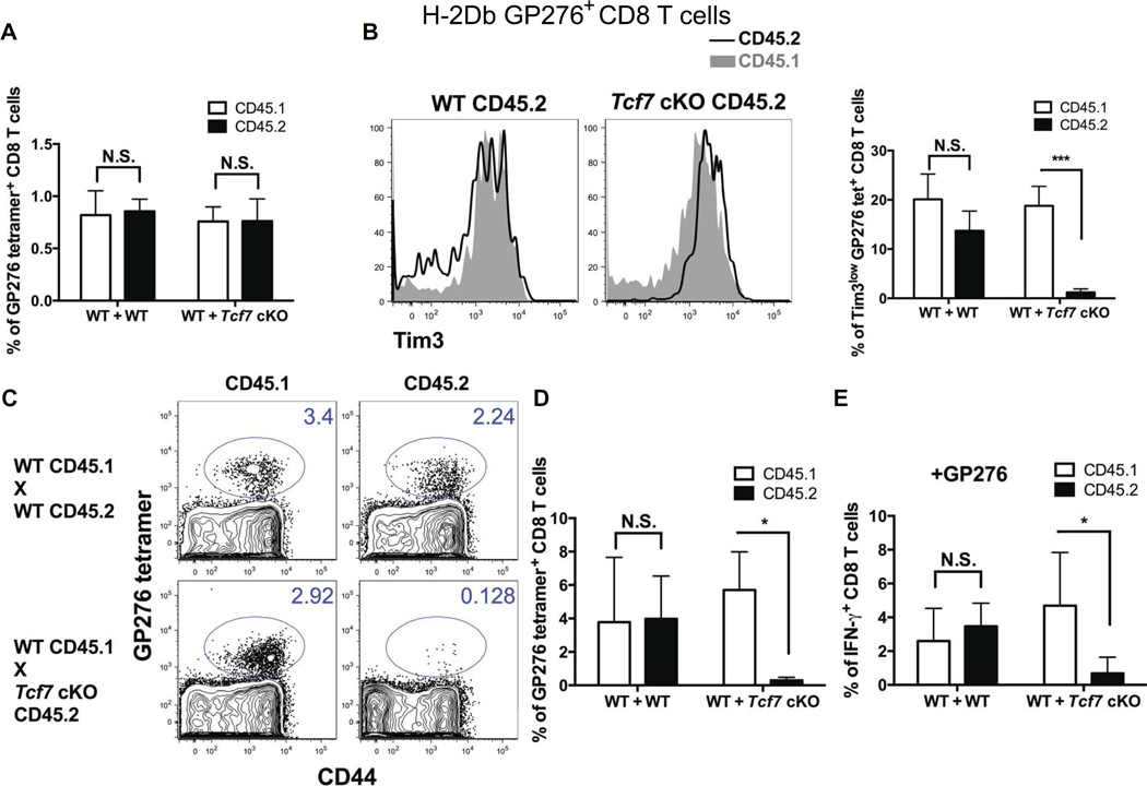
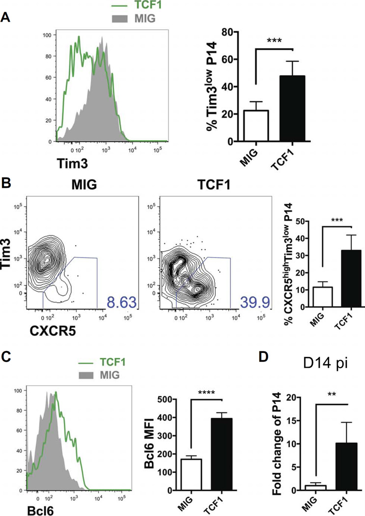
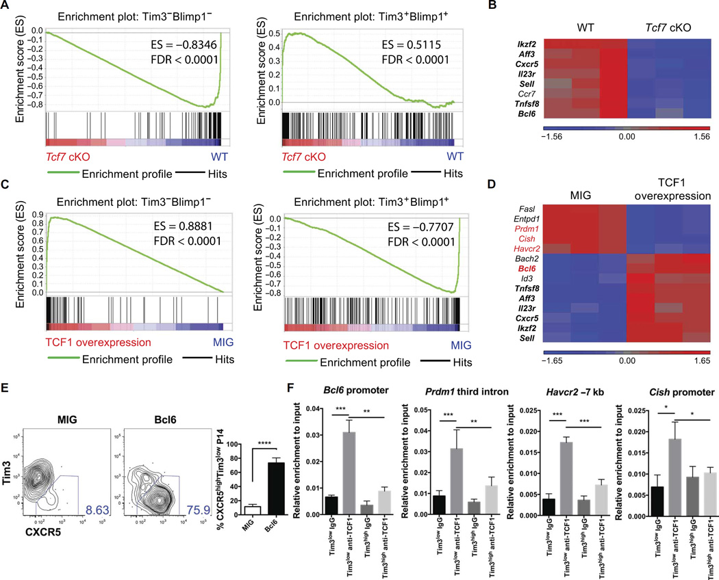
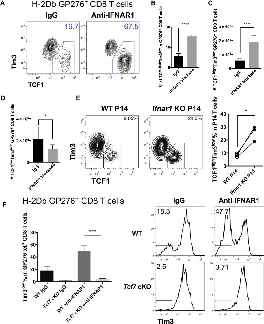
During chronic viral infections and in cancer, T cells become dysfunctional, a state known as T cell exhaustion. Although it is well recognized that memory CD8 T cells account for the persistence of CD8 T cell immunity after acute infection, how exhausted T cells persist remains less clear. Using chronic infection with lymphocytic choriomeningitis virus clone 13 and tumor samples, we demonstrate that CD8 T cells differentiate into a less exhausted TCF1high and a more exhausted TCF1low population. Virus-specific TCF1high CD8 T cells, which resemble T follicular helper (TFH) cells, persist and recall better than do TCF1low cells and act as progenitor cells to replenish TCF1low cells. We show that TCF1 is both necessary and sufficient to support this progenitor-like CD8 subset, whereas cell-intrinsic type I interferon signaling suppresses their differentiation. Accordingly, cell-intrinsic TCF1 deficiency led to a loss of these progenitor CD8 T cells, sharp contraction of virus-specific T cells, and uncontrolled viremia. Mechanistically, TCF1 repressed several pro-exhaustion factors and induced Bcl6 in CD8 T cells, which promoted the progenitor fate. We propose that the TCF1-Bcl6 axis counteracts type I interferon to repress T cell exhaustion and maintain T cell stemness, which is critical for persistent antiviral CD8 T cell responses in chronic infection. These findings provide insight into the requirements for persistence of T cell immune responses in the face of exhaustion and suggest mechanisms by which effective T cell-mediated immunity may be enhanced during chronic infections and cancer.


Figures

Fig. 1
Fig. 1. TCF1high virus-specific CD8 T cells in chronic LCMV infection resemble TFH cells
(A and B) C57BL/6 or Blimp1-YFP (yellow fluorescent protein) reporter mice were infected with LCMV clone 13. Expression of TCF1, Tim3, and Blimp1 in H-2Db GP276 tetramer+ CD8 T cells in the spleen analyzed 7 days (A) and 4weeks post-infection (p.i.) (B). MFI, mean fluorescence intensity. (C) Heat map of selected differentially expressed genes from microarrays comparing Tim3lowBlimp1low and Tim3highBlimp1high virus-specific CD8 T cells from mice 7 days after infection. (D) Expression of Bcl6 and CXCR5 on TCF1high (blue) and TCF1low (red) H-2Db GP276 tetramer+ splenic CD8 T cells analyzed 4 weeks after infection. (E) GSEA of the microarray data in (C) comparing the enrichment of TFH (left) and TH1 (right) gene signatures between Tim3lowBlimp1low and Tim3highBlimp1high virus-specific CD8 T cells. Data in (A), (B), and (D) are representative of at least two independent experiments with n ≥ 3. Statistical significance was determined by paired t tests. *P < 0.05, **P < 0.01, ***P < 0.001, ****P < 0.0001. Microarrays (C and E) were performed with three biological replicates per group. ES, enrichment score; FDR, false discovery rate.
Fig. 2
Fig. 2. TCF1 expression negatively correlates with T cell exhaustion in both chronic viral infection and solid tumors
(A and B) PD1, 2B4, and LAG3 expression on TCF1high (blue) and TCF1low (red) H-2Db GP276 tetramer+ (tet+) splenic CD8 T cells analyzed 7 days (A) and 4 weeks after infection (B). (C) C57BL/6 mice were implanted with MCA205 fibrosarcoma cells. Expression of TCF1, PD1, Tim3, and CXCR5 on tumor-infiltrating CD8 T cells analyzed 20 days after injection. (D) Expression of TCF1, PD1, and Tim3 in CD8 T cells isolated from human melanoma samples. (E to G) Tim3lowPD1+CD44high and Tim3highPD1+CD44high CD8 T cells from C57BL/6 mice 7 days after infection were transferred separately into naïve CD45.1 mice that were subsequently infected with LCMV clone 13. (E) Numbers of total and tetramer+ donor CD8 T cells in the spleen 7 days after rechallenge. (F) Frequencies of Tim3low (white) and Tim3high (gray) cells within tetramer+ progeny from Tim3low or Tim3high donors. (G) Representative histograms of TCF1 expression in donors and recipient tetramer+ CD8 T cells. Frequencies of TCF1high (white) and TCF1low (gray) cells within tetramer+ progeny from Tim3low or Tim3high donors are shown. Data in (A) to (C) and (E) to (G) are representative of at least two independent experiments with n ≥ 3. Data in (D) are representative of three melanoma patients. Statistical significance was determined by paired (A and B) and unpaired t tests (E to G). *P < 0.05, **P < 0.01, ***P < 0.001, ****P < 0.0001.
Fig. 3
Fig. 3. TCF1 deficiency compromises Tim3low virus-specific CD8 T cells and long-term persistence of T cell responses after chronic infection
Tcf7loxP/loxP; CD4-Cre (Tcf7 cKO) and littermate control (WT) mice were infected with LCMV clone 13. (A) Representative H-2Db GP276 tetramer staining and number of H-2Db GP276 tetramer+ splenic CD8 T cells 7 days after infection. (B) Representative histograms of Tim3 expression and frequencies of Tim3low cells within H-2Db GP276 tetramer+ WT and cKO CD8 T cells 7 days after infection. (C and D) Frequencies and numbers of WT and cKO I-Ab GP66 tetramer+ CD4 (C) and H-2Db GP276 tetramer+ CD8 (D) splenic T cells 4 weeks after infection. (E to G) Expression of Tim3, PD1, and 2B4 on H-2Db GP276 tetramer+ splenic CD8 T cells from WT (blue) and cKO (red) mice 4 weeks after infection. (H) Viral titers in blood and spleens 4 weeks after infection. (I and J) Frequencies and numbers of WT and cKO I-Ab GP66 tetramer+ CD4 (I) and H-2Db GP276 tetramer+ CD8 (J) splenic T cells 3 months after infection. (K) Blood viral titers 3 months after infection. Data are representative of at least two independent experiments with n ≥ 3. Statistical significance was determined by unpaired t tests. *P < 0.05, **P < 0.01, ***P < 0.001, ****P < 0.0001.
Fig. 4
Fig. 4. Cell-intrinsic requirement of TCF1 for Tim3low virus-specific CD8 T cells and persistence of T cell responses
Mixed bone marrow chimeras that received WT CD45.1 and WT CD45.2 (WT + WT) or WT CD45.1 and Tcf7 cKO CD45.2 bone marrow (WT + Tcf7 cKO) were infected with LCMV clone 13. (A) Frequencies of H-2Db GP276 tetramer+ cells within CD45.1 (white) and CD45.2 (filled) CD8 splenic T cells 7 days after infection. (B) Tim3 expression on H-2Db GP276 tetramer+ WT CD45.1 (shaded) and WT or cKO CD45.2 (solid line) CD8 T cells and frequencies of Tim3low cells within H-2Db GP276 tetramer+ CD45.1 WT (white) and CD45.2 WT or cKO (filled) splenic CD8 T cells from chimeras 7 days after infection. (C) Representative fluorescence-activated cell sorting plots of H-2Db GP276 tetramer staining on CD45.1 WT and CD45.2 WT or cKO CD8 compartments in spleens from chimeras 4 weeks after infection. (D) Frequencies of H-2Db GP276 tetramer+ cells within CD45.1 WT (white) and CD45.2 WT or cKO (filled) CD8 T cells in spleens from chimeras 4 weeks after infection. (E) Frequencies of IFN-γ+ cells within CD45.1 WT (white) and CD45.2 WT or cKO (filled) splenic CD8 T cells from chimeras 4 weeks after infection, restimulated with GP276 peptide. Data are representative of at least two independent experiments with n ≥ 4. Statistical significance was determined by paired t tests. N.S., not significant. *P < 0.05, ***P < 0.001.
Fig. 5
Fig. 5. TCF1 overexpression enhanced the differentiation of Tim3lowCXCR5high CD8 T cells and persistence of virus-specific CD8 T cells
P14 cells transduced with either control (MIG) or TCF1 overexpression (TCF1) retroviral vectors were transferred to C57BL/6 recipients that were subsequently infected with LCMV clone 13. (A to C) Mice were evaluated on day 8 after infection. (A) Tim3 expression and frequencies of Tim3low cells within transduced P14 cells. (B) Flow plot and frequencies of CXCR5highTim3low cells in transduced P14 cells. (C) Bcl6 expression in control (MIG) or TCF1-overexpressing (TCF1) P14 cells. (D) Numbers of control (MIG) and TCF1-overexpressing (TCF1) P14 cells in spleens of mice 14 days after infection. Data are representative of at least two independent experiments with n ≥ 3. Statistical significance was determined by unpaired t tests. **P < 0.01, ***P < 0.001, ****P < 0.0001.
Fig. 6
Fig. 6. TCF1 regulates the differentiation of Tim3lowBlimp1low virus-specific CD8 T cells and the expression of Bcl6 and pro-exhaustion genes
(A and B) Microarray experiments with tetramer+ WT and Tcf7 cKO CD8 T cells on day 7 after infection. (A) GSEA comparing the enrichment of Tim3lowBlimp1low (left) and Tim3highBlimp1high (right) gene signatures between WT and cKO virus-specific CD8 T cells. (B) Heat map of selected up-regulated or down-regulated genes in cKO cells relative to WT cells. (C and D) Microarrays of TCF1-overexpressing and control (MIG) P14 T cells on day 8 after infection. (C) GSEA comparing the enrichment of Tim3lowBlimp1low (left) and Tim3highBlimp1high (right) gene signatures between TCF1-overexpressing and control P14 cells. (D) Heat map of selected up-regulated or down-regulated genes in TCF1-overexpressing relative to control P14 cells. Genes marked in bold were reciprocally expressed in Tcf7 cKO cells. Genes marked in red were evaluated by ChIP. (E) Tim3 and CXCR5 expression, as well as frequencies of CXCR5highTim3low cells in control (MIG), or Bcl6-transduced P14 cells on day 8 after infection. (F) ChIP assays performed on day 7 after infection. Tim3lowPD1+CD44high and Tim3highPD1+CD44high CD8 T cells. Binding of TCF1 to indicated loci determined by ChIP and quantitative reverse transcription polymerase chain reaction. Data in (E) and (F) are representative of two independent experiments with n ≥ 3. Statistical significance in (E) and (F) was determined by unpaired t tests. IgG, immunoglobulin G. *P < 0.05, **P < 0.01, ***P < 0.001, ****P < 0.0001.
Fig. 7
Fig. 7. Type I IFN signaling suppresses the generation of TCF1highTim3low virus-specific CD8 T cells, upstream of TCF1
(A to D) C57BL/6 mice were treated with anti-IFNAR1 or IgG before LCMV clone 13 infection. Splenocytes analyzed 7 days after infection. (A) Representative TCF1 and Tim3 expression inH-2DbGP276 tetramer+ CD8 T cells. Frequencies (B) and numbers (C) of TCF1highTim3low GP276 tetramer+ CD8 T cells. (D) Numbers of TCF1lowTim3high GP276 tetramer+ CD8 T cells. (E) Ten thousand Ifnar1 KO and WT P14 cells were mixed in a 1:1 ratio and transferred into NK-depleted WT hosts that were subsequently infected with LCMV clone 13. TCF1highTim3low CD8 T cells 7 days after infection. (F) WT and Tcf7 cKO mice were treated with 1 mg of anti-IFNAR1 or isotype control IgG before LCMV clone 13 infection. Splenocytes analyzed 7 days after infection. Representative Tim3 expression and frequencies of Tim3low cells within GP276 tetramer+ CD8 T cells in each group. Data are representative of two independent experiments with n ≥ 3. Statistical significance in (B) to (D) and (F) was determined by unpaired t tests. Statistical significance in (E) was determined by paired t tests. *P < 0.05, ***P < 0.001, ****P < 0.0001.

References

    1. Ahmed R, Gray D. Immunological memory and protective immunity: Understanding their relation. Science. 1996;272:54–60. - PubMed
    1. Kaech SM, Wherry EJ. Heterogeneity and cell-fate decisions in effector and memory CD8+ T cell differentiation during viral infection. Immunity. 2007;27:393–405. - PMC - PubMed
    1. Wherry EJ, Kurachi M. Molecular and cellular insights into T cell exhaustion. Nat. Rev. Immunol. 2015;15:486–499. - PMC - PubMed
    1. Wherry EJ. T cell exhaustion. Nat. Immunol. 2011;12:492–499. - PubMed
    1. Angelosanto JM, Blackburn SD, Crawford A, Wherry EJ. Progressive loss of memory T cell potential and commitment to exhaustion during chronic viral infection. J Virol. 2012;86:8161–8170. - PMC - PubMed