. 2017 Mar;81(3):395-406.
doi: 10.1002/ana.24852. Epub 2017 Jan 23.

The melanoma-linked "redhead" MC1R influences dopaminergic neuron survival

Affiliations

The melanoma-linked "redhead" MC1R influences dopaminergic neuron survival

Xiqun Chen et al. Ann Neurol. 2017 Mar.

Abstract

Objective: Individuals with Parkinson disease are more likely to develop melanoma, and melanoma patients are reciprocally at higher risk of developing Parkinson disease. Melanoma is strongly tied to red hair/fair skin, a phenotype of loss-of-function polymorphisms in the MC1R (melanocortin 1 receptor) gene. Loss-of-function variants of MC1R have also been linked to increased risk of Parkinson disease. The present study is to investigate the role of MC1R in dopaminergic neurons in vivo.

Methods: Genetic and pharmacological approaches were employed to manipulate MC1R, and nigrostriatal dopaminergic integrity was determined by comprehensive behavioral, neurochemical, and neuropathological measures.

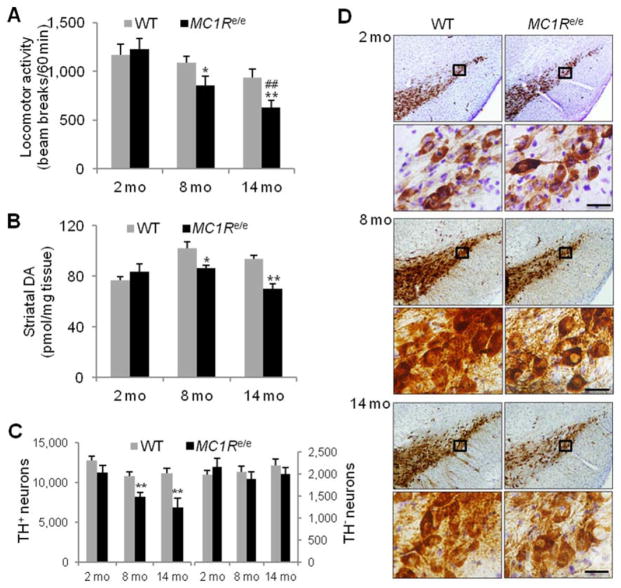
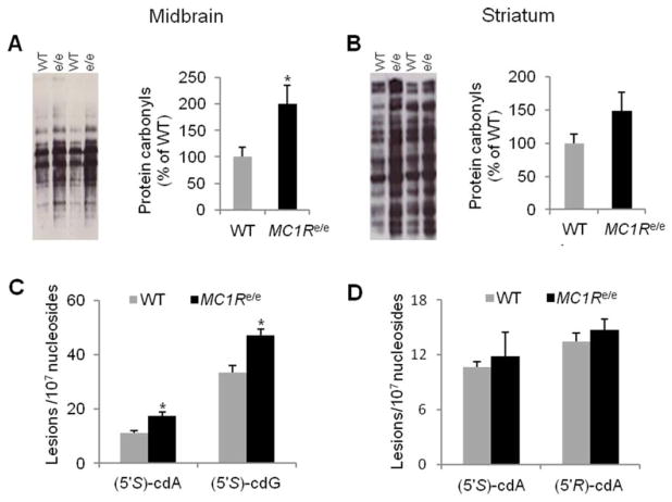
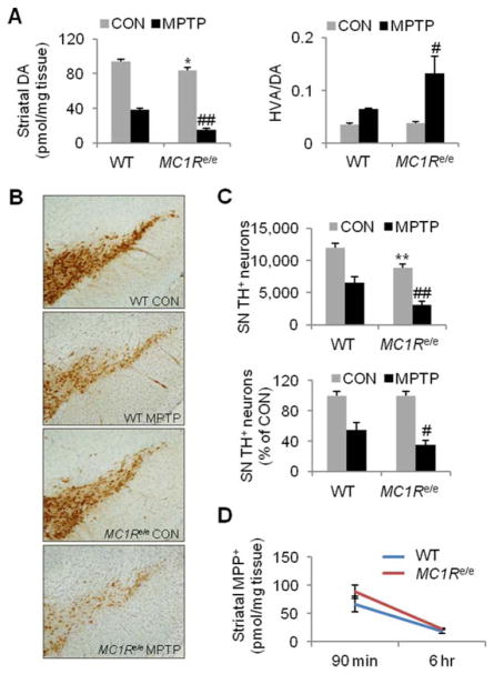
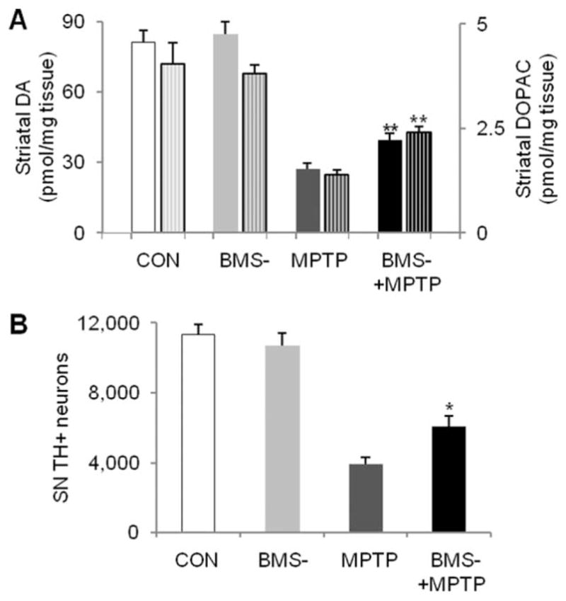
Results: MC1Re/e mice, which carry an inactivating mutation of MC1R and mimic the human redhead phenotype, have compromised nigrostriatal dopaminergic neuronal integrity, and they are more susceptible to dopaminergic neuron toxins 6-hydroxydopamine and 1-methyl-4-phenyl-1,2,3,6-tetrahydropyridine (MPTP). Furthermore, a selective MC1R agonist protects against MPTP-induced dopaminergic neurotoxicity.

Interpretation: Our findings reveal a protective role of MC1R in the nigrostriatal dopaminergic system, and they provide a rationale for MC1R as a potential therapeutic target for Parkinson disease. Together with its established role in melanoma, MC1R may represent a common pathogenic pathway for melanoma and Parkinson disease. Ann Neurol 2017;81:395-406.


Conflict of interest statement

Potential Conflicts of Interest

Nothing to report.

Figures

FIGURE 1
FIGURE 1
Expression of MC1R in the substantia nigra (SN) in adult naive C57BL/6J mouse brain. (A) Western blot using anti- MC1R antibody with total tissue lysate (left panel), membrane (m) or cytosolic fraction (c) (middle panel) isolated from ventral midbrain (MB) and the striatum (Str), or B16 mouse melanoma cells treated with shRNA targeting mouse MC1R or scrambled construct (right panel). Adrenal (Adr), where MC1R is known to be expressed, serves as a control. β-Tubulin and Na+/K+-adenosine triphosphatase (ATPase) are loading controls for total and cytosolic proteins, and membrane proteins, respectively. (B) Immunostaining of MC1R and Nissl counterstaining in a ventral midbrain coronal section (top panel). Cells in the box in the SN pars compacta are shown at higher magnification on the right. Scale bar=25 μm. Bottom panels show ventral midbrain coronal sections incubated with blocking peptide (left) or no primary antibody (right). (C) Fluorescence double labeling for tyrosine hydroxylase (TH; red) and MC1R (green) in the SN. Scale bars=10 μm. (D) Laser capture microdissection of TH-positive neurons from the SN and reverse transcriptase polymerase chain reaction analysis of MC1R mRNA. Glyceraldehyde-3- phosphate dehydrogenase (GAPDH) serves as internal control.
FIGURE 2
FIGURE 2
Compromised nigrostriatal dopaminergic integrity in MC1Re/e mice. (A) Locomotor activity in MC1Re/e mice and littermate wild-type (WT) controls assessed by adjacent beam breaks during the first hour of the dark cycle in an open field chamber (n=15, WT and MC1Re/e 2- and 8-month-old mice; n=14 and 13, WT and MC1Re/e 14-month-old mice). *p=0.05, **p=0.01 versus WT littermates; ##p=0.01 versus 2-month-old self-control MC1Re/e mice by 2-way analysis of variance (ANOVA) followed by Tukey post hoc test. p=0.038 for genotype effect; p=0.0002 for time effect; p=0.11 for interaction. (B) Striatal dopamine (DA) content in MC1Re/e mice and WT littermates determined by high-performance liquid chromatography coupled with electrochemical detection (n=5, WT and MC1Re/e at all 3 age points). *p < 0.05, **p < 0.01 versus WT littermates, 1-way ANOVA followed by Tukey post hoc test. (C) Stereological quantification of tyrosine hydroxylase (TH)-positive and TH-negative (Nissl-positive) neurons in substantia nigra pars compacta in MC1Re/e mice and littermate WT controls (n=5, WT and MC1Re/e 2- and 14-month-old mice; n=6, WT and MC1Re/e 8-month-old mice). **p=0.01 versus WT littermates, 1- way ANOVA followed by Tukey post hoc test. (D) Representative sets of nigral sections from MC1Re/e mice and littermate WT controls stained for TH. Cells in the black boxes at indicated age points are shown at higher magnification in the lower panels. Scale bars=20 μm.
FIGURE 3
FIGURE 3
MC1Re/e mice have greater oxidative damage in the ventral midbrain. (A, B) Protein carbonyls assessed by Oxyblot in (A) the ventral midbrain and (B) the striatum in adult (3–6-month-old) MC1Re/e mice and littermate wild-type (WT) controls (n=4, WT and MC1Re/e ventral midbrain; n=5, WT and MC1Re/e striatum). *p< 0.05 versus WT by Student t test. (C, D) DNA damage markers 8,5′-cyclo-2′-deoxyadenosine (cdA) and 8,5′-cyclo-2′-deoxyguanosine (cdG) assessed by liquid chromatography–tandem mass spectrometry in (C) the ventral midbrain and (D) the striatum in adult MC1Re/e mice (5–8 months old) and littermate WT controls (n=4, WT and MC1Re/e mice, pooled from 12 animals of each genotype). *p< 0.05 versus WT by Student t test. [Color figure can be viewed at wileyonlinelibrary.com]
FIGURE 4
FIGURE 4
MC1Re/e mice are more susceptible to dopaminergic toxin 6-hydroxydopamine (6-OHDA). (A) Adult (6–8-month-old) MC1Re/e and littermate wild-type (WT) mice were infused with 10μg 6-OHDA into the left striatum. Rotational behavior was assessed at 3 weeks, and animals were sacrificed at 4 weeks after 6-OHDA lesion. (B) Spontaneous and 5mg/kg amphetamine-induced net ipsilateral rotations in MC1Re/e mice and littermate WT controls (n=12, WT and MC1Re/e). *p< 0.05 versus WT by Student t test. (C) Striatal dopamine (DA) measured by high-performance liquid chromatography (HPLC) and DA turnover rate (homovanillic acid [HVA]/DA ratio, HVA determined by HPLC) on contralateral (Contra) unlesioned side and ipsilateral (Ipsi) lesioned side in MC1Re/e mice and WT littermates (n=12, WT and MC1Re/e). **p < 0.01 versus WT Contra side; ##p< 0.01 versus WT Ipsi side, 1-way analysis of variance (ANOVA) followed by Tukey post hoc test. (D) Disruption of substantia nigra (SN) dopaminergic neurons (stained positive for tyrosine hydroxylase [TH]) on the Ipsi lesioned side of an MC1Re/e mouse after 6-OHDA lesion. (E) Stereological quantification of nigral TH-positive neurons expressed as absolute counts and percentage of Contra side in MC1Re/e mice and WT littermates (n=12,WT and MC1Re/e). **p< 0.01 versus WT Contra side; ##p< 0.01 versus WT Ipsi side, 1-way ANOVA followed by Tukey post hoc test for absolute counts and Student t test for normalized counts. [Color figure can be viewed at wileyonlinelibrary.com]
FIGURE 5
FIGURE 5
MC1Re/e mice are more susceptible to dopaminergic toxin 1-methyl-4-phenyl-1,2,3,6-tetrahydropyridine (MPTP). Adult (6–8-month-old) MC1Re/e and littermate wildtype (WT) mice were injected intraperitoneally (i.p.) with 20mg/kg MPTP-HCl or control vehicle (saline) once daily for 4 days and sacrificed 7 days after the last MPTP administration. (A) Dopamine (DA) in the striatum measured by high-performance liquid chromatography (HPLC) and DA turnover rate (homovanillic acid [HVA]/DA ratio, HVA determined by HPLC) in MPTP or vehicle control (CON) MC1Re/e mice and WT littermates (n=6, WT and MC1Re/e CON; n=8 and 7, WT and MC1Re/e MPTP). *p< 0.05 versus WT CON; #p < 0.05, ##p< 0.01 versus WT MPTP, 1-way analysis of variance (ANOVA) followed by Tukey post hoc test. (B) Substantial loss of TH-positive neurons in the substantia nigra (SN) of an MC1Re/e mouse after MPTP subacute treatment. (C) Stereological quantification of nigral tyrosine hydroxylase (TH)- positive neurons expressed as absolute counts and percentage of CON group in MC1Re/e mice and WT littermates (n=6, WT and MC1Re/e CON; n=8 and 7, WT and MC1Re/e MPTP). **p< 0.01versus WT CON; #p< 0.05, ##p< 0.01 versus WT MPTP, 1-way ANOVA followed by Tukey post hoc test for absolute counts and Student t test for normalized counts. (D) Striatal 1 methyl-4-phenylpyridinium (MPP+) detected by HPLC in MC1Re/e and littermate WT mice following 20mg/kg MPTP-HCl intraperitoneal administration (n=6 and 8, WT and MC1Re/e, both 90 minutes and 6 hours). [Color figure can be viewed at wileyonlinelibrary.com]
FIGURE 6
FIGURE 6
MC1R agonist protects against 1-methyl-4- phenyl-1,2,3,6-tetrahydropyridine (MPTP)-induced dopaminergic neurotoxicity. Adult (3-month-old) C57Bl/6 mice were administered with a single dose of MPTP-HCl at 40mg/kg or vehicle (saline) intraperitoneally, and BMS-470539 (BMS-) 100mg/kg or vehicle (water) was injected subcutaneously 10 minutes before and 60 minutes after MPTP. Mice were sacrificed 7 days later. (A) Striatal dopamine (DA) and metabolite 3,4-dihydroxyphenylacetic acid (DOPAC; patterned columns, the right y-axis) were analyzed by high-performance liquid chromatography. (B) Numbers of nigral tyrosine hydroxylase (TH)-positive neurons were counted by stereological method. *p < 0.05, **p < 0.01 versus MPTP group, 1-way analysis of variance followed by Tukey post hoc test (n=5, 4, 6, 7 for control [CON], BMS-, MPTP, and BMS-+MPTP groups, respectively). SN=substantia nigra.

References

    1. Liu R, Gao X, Lu Y, Chen H. Meta-analysis of the relationship between Parkinson disease and melanoma. Neurology. 2011;76:2002–2009. - PMC - PubMed
    1. Devine MJ, Plun-Favreau H, Wood NW. Parkinson’s disease and cancer: two wars, one front. Nat Rev Cancer. 2011;11:812–823. - PubMed
    1. Gao X, Simon KC, Han J, et al. Family history of melanoma and Parkinson disease risk. Neurology. 2009;73:1286–1291. - PMC - PubMed
    1. Kareus SA, Figueroa KP, Cannon-Albright LA, Pulst SM. Shared predispositions of parkinsonism and cancer: a population-based pedigree-linked study. Arch Neurol. 2012;69:1572–1577. - PubMed
    1. Flaherty KT, Hodi FS, Fisher DE. From genes to drugs: targeted strategies for melanoma. Nat Rev Cancer. 2012;12:349–361. - PubMed

MeSH terms

Substances