Transforming Growth Factor-β1 Modulates the Expression of Syndecan-4 in Cultured Vascular Endothelial Cells in a Biphasic Manner
- PMID: 28019669
- PMCID: PMC5485002
- DOI: 10.1002/jcb.25861
Transforming Growth Factor-β1 Modulates the Expression of Syndecan-4 in Cultured Vascular Endothelial Cells in a Biphasic Manner
Abstract
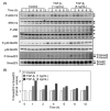
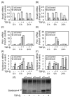
Proteoglycans are macromolecules that consist of a core protein and one or more glycosaminoglycan side chains. Previously, we reported that transforming growth factor-β1 (TGF-β1 ) regulates the synthesis of a large heparan sulfate proteoglycan, perlecan, and a small leucine-rich dermatan sulfate proteoglycan, biglycan, in vascular endothelial cells depending on cell density. Recently, we found that TGF-β1 first upregulates and then downregulates the expression of syndecan-4, a transmembrane heparan sulfate proteoglycan, via the TGF-β receptor ALK5 in the cells. In order to identify the intracellular signal transduction pathway that mediates this modulation, bovine aortic endothelial cells were cultured and treated with TGF-β1 . Involvement of the downstream signaling pathways of ALK5-the Smad and MAPK pathways-in syndecan-4 expression was examined using specific siRNAs and inhibitors. The data indicate that the Smad3-p38 MAPK pathway mediates the early upregulation of syndecan-4 by TGF-β1 , whereas the late downregulation is mediated by the Smad2/3 pathway. Multiple modulations of proteoglycan synthesis may be involved in the regulation of vascular endothelial cell functions by TGF-β1 . J. Cell. Biochem. 118: 2009-2017,2017. © 2016 The Authors. Journal of Cellular Biochemistry Published by Wiley Periodicals, Inc.
Keywords: ENDOTHELIAL CELL; PROTEOGLYCAN; SYNDECAN-4; TGF-β.
© 2016 The Authors. Journal of Cellular Biochemistry Published by Wiley Periodicals, Inc.
Figures


Similar articles
-
Biglycan Intensifies ALK5-Smad2/3 Signaling by TGF-β1 and Downregulates Syndecan-4 in Cultured Vascular Endothelial Cells.J Cell Biochem. 2017 May;118(5):1087-1096. doi: 10.1002/jcb.25721. Epub 2017 Jan 10. J Cell Biochem. 2017. PMID: 27585241 Free PMC article.
-
TGF-β1/ALK5-induced monocyte migration involves PI3K and p38 pathways and is not negatively affected by diabetes mellitus.Cardiovasc Res. 2011 Aug 1;91(3):510-8. doi: 10.1093/cvr/cvr100. Epub 2011 Apr 8. Cardiovasc Res. 2011. PMID: 21478266
-
Activation of extracellular signal-regulated kinase by TGF-beta1 via TbetaRII and Smad7 dependent mechanisms in human bronchial epithelial BEP2D cells.Cell Biol Toxicol. 2007 Mar;23(2):113-28. doi: 10.1007/s10565-006-0097-x. Epub 2006 Nov 9. Cell Biol Toxicol. 2007. PMID: 17096210
-
Syndecan-4 signaling at a glance.J Cell Sci. 2013 Sep 1;126(Pt 17):3799-804. doi: 10.1242/jcs.124636. Epub 2013 Aug 22. J Cell Sci. 2013. PMID: 23970415 Free PMC article. Review.
-
[Design, synthesis, and structure-activity relationship study of organoantimony compounds that induce the expression of perlecan in vascular endothelial cells].Yakugaku Zasshi. 2014;134(7):789-91. doi: 10.1248/yakushi.14-00017-2. Yakugaku Zasshi. 2014. PMID: 24989466 Review. Japanese.
Cited by
-
Amelioration of Cisplatin-Induced Ototoxicity in Rats by L-arginine: The Role of Nitric Oxide, Transforming Growth Factor Beta 1 and Nrf2/HO-1 Pathway.Asian Pac J Cancer Prev. 2020 Jul 1;21(7):2155-2162. doi: 10.31557/APJCP.2020.21.7.2155. Asian Pac J Cancer Prev. 2020. PMID: 32711445 Free PMC article.
-
TGF-β1 suppresses syndecan-2 expression through the ERK signaling pathway in nucleus pulposus cells.Int J Clin Exp Pathol. 2018 Apr 1;11(4):2017-2024. eCollection 2018. Int J Clin Exp Pathol. 2018. PMID: 31938308 Free PMC article.
-
TGF-β1 Potentiates the Cytotoxicity of Cadmium by Induction of a Metal Transporter, ZIP8, Mediated by the ALK5-Smad2/3 and ALK5-Smad3-p38 MAPK Signal Pathways in Cultured Vascular Endothelial Cells.Int J Mol Sci. 2021 Dec 31;23(1):448. doi: 10.3390/ijms23010448. Int J Mol Sci. 2021. PMID: 35008873 Free PMC article.
-
Association of serum transforming growth factor β1 with left ventricular hypertrophy in children with primary hypertension.Eur J Pediatr. 2023 Dec;182(12):5439-5446. doi: 10.1007/s00431-023-05219-2. Epub 2023 Sep 27. Eur J Pediatr. 2023. PMID: 37755472 Free PMC article.
-
Epigenetic Regulation of the Biosynthesis & Enzymatic Modification of Heparan Sulfate Proteoglycans: Implications for Tumorigenesis and Cancer Biomarkers.Int J Mol Sci. 2017 Jun 26;18(7):1361. doi: 10.3390/ijms18071361. Int J Mol Sci. 2017. PMID: 28672878 Free PMC article. Review.
References
-
- Berenson GS, Radhakrishnamurthy B, Srinivasan SR, Vijayagopal P, Dalferes ER, Jr. , Sharma C. 1984. Recent advances in molecular pathology. Carbohydrate‐protein macromolecules and arterial wall integrity‐a role in atherogenesis. Exp Mol Pathol 41:267–287. - PubMed
-
- Brooks AR, Lelkes PI, Rubanyi GM. 2002. Gene expression profiling of human aortic endothelial cells exposed to disturbed flow and steady laminar flow. Physiol Genomics 9:27–41. - PubMed
-
- Camejo G. 1981. The interaction of lipids and lipoproteins with the intercellular matrix of arterial tissue: Its possible role in atherogenesis. Adv Lipid Res 19:1–53. - PubMed
MeSH terms
Substances
LinkOut - more resources
Full Text Sources
Other Literature Sources
Molecular Biology Databases

