. 2017 Feb;18(2):236-245.
doi: 10.1038/ni.3654. Epub 2016 Dec 26.

Ubiquitination of hnRNPA1 by TRAF6 links chronic innate immune signaling with myelodysplasia

Affiliations

Ubiquitination of hnRNPA1 by TRAF6 links chronic innate immune signaling with myelodysplasia

Jing Fang et al. Nat Immunol. 2017 Feb.

Erratum in

Abstract

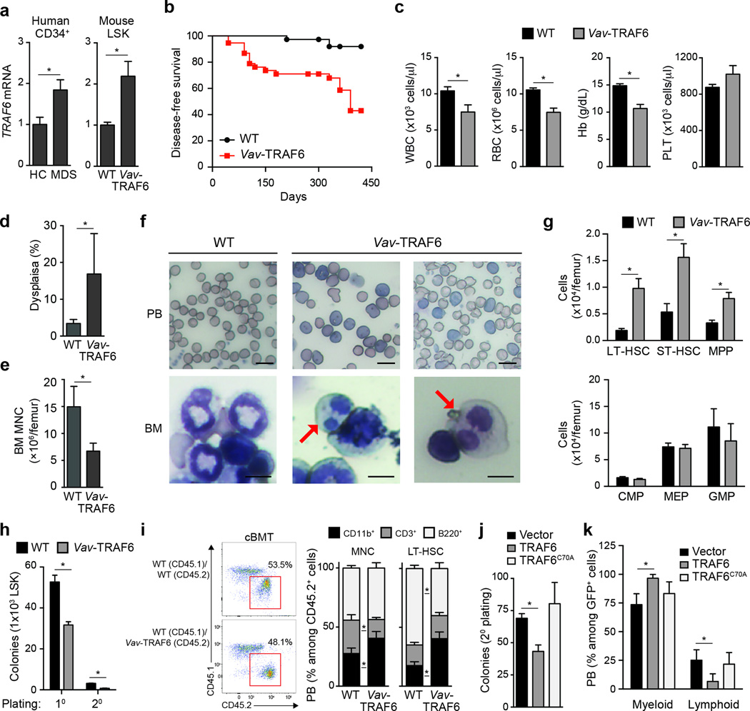
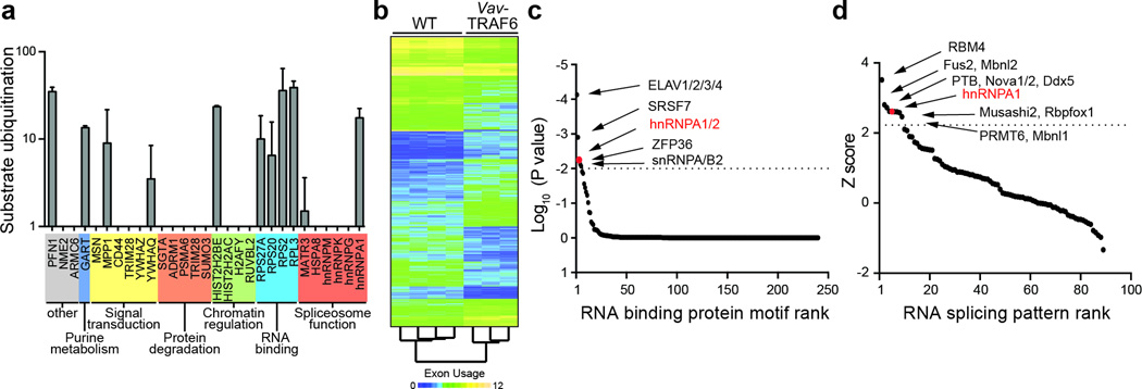
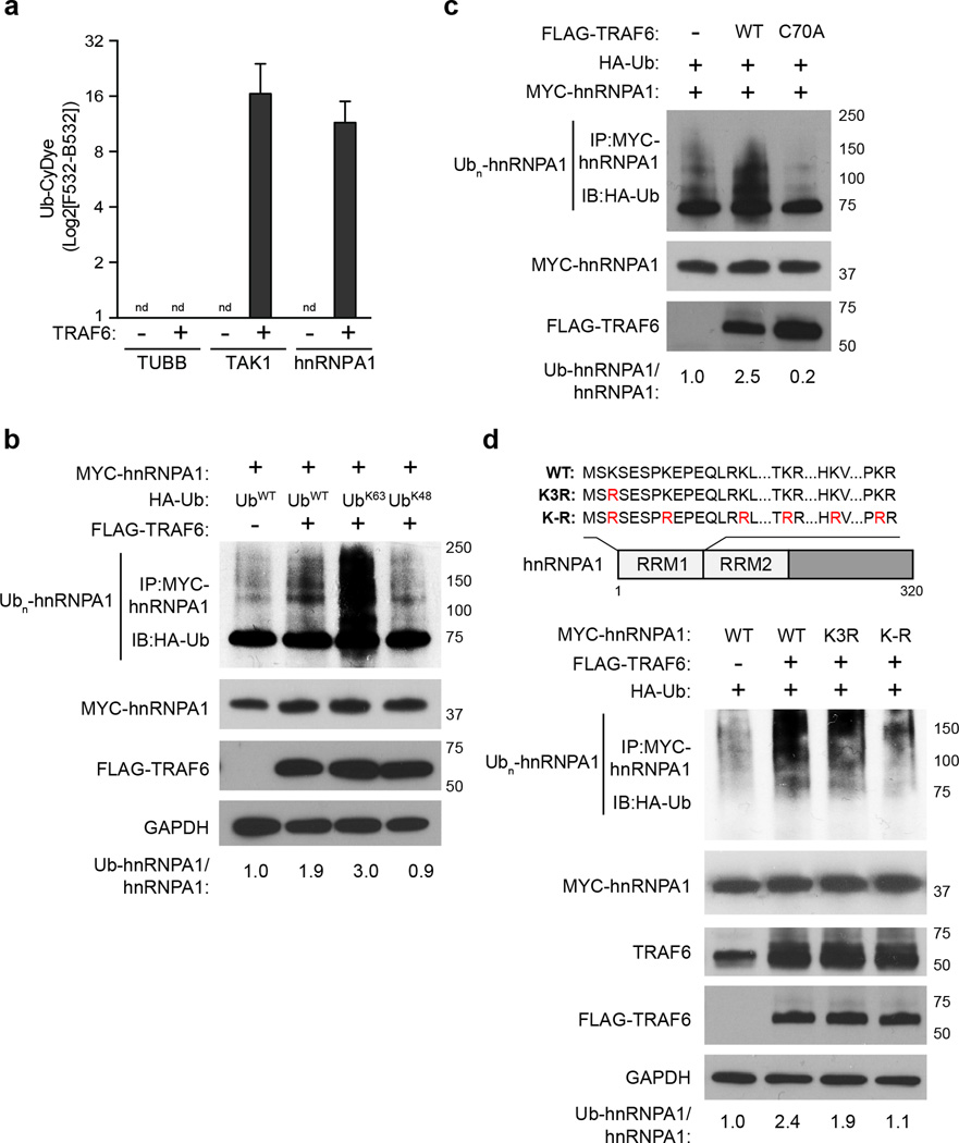
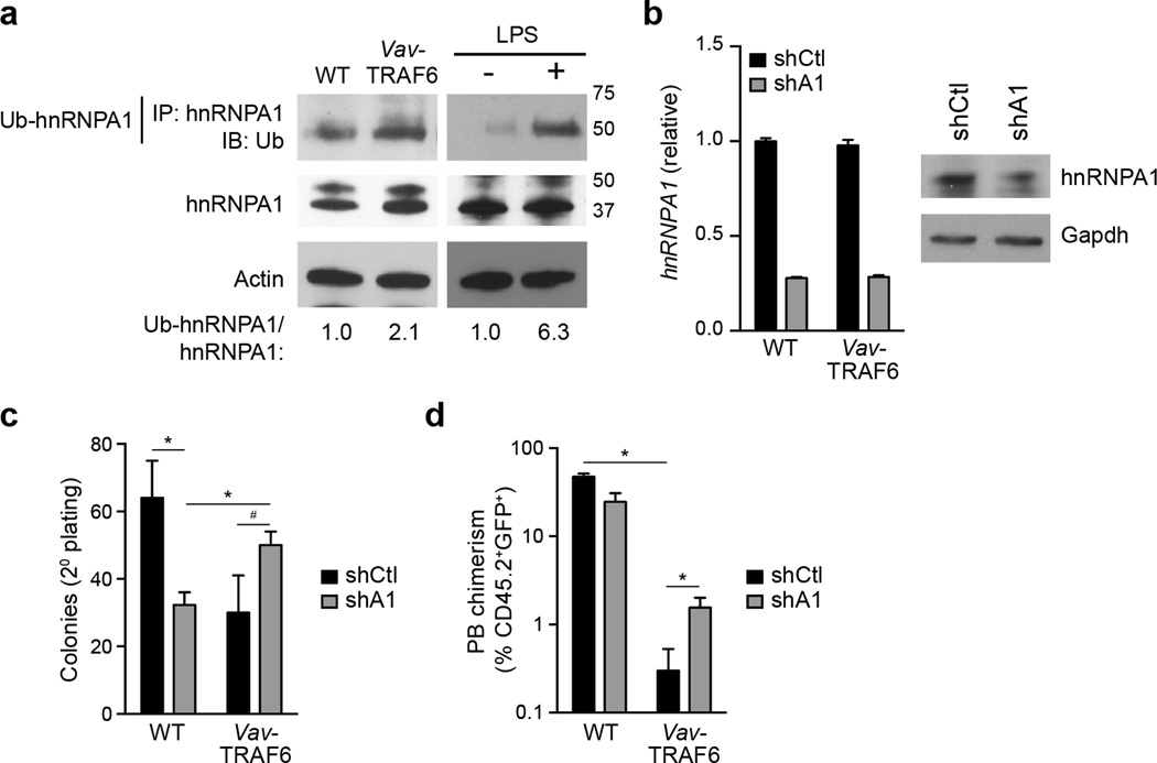
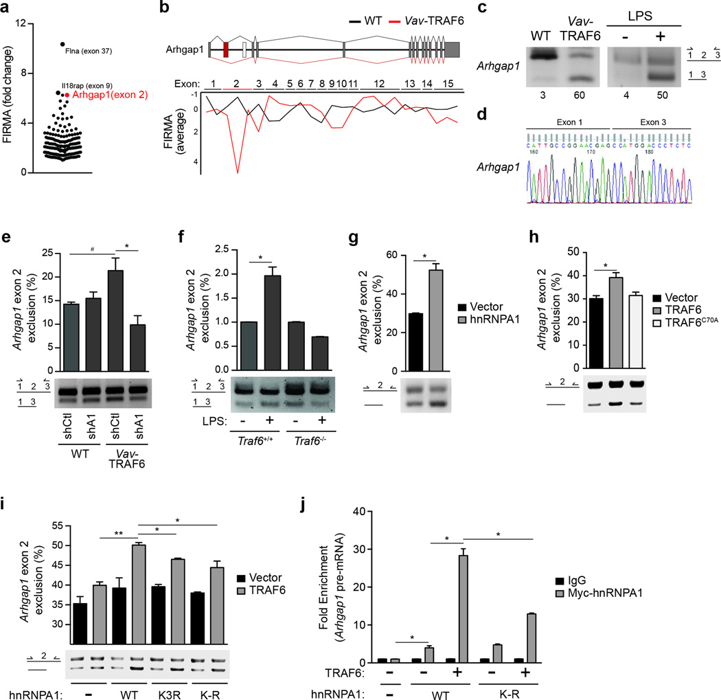
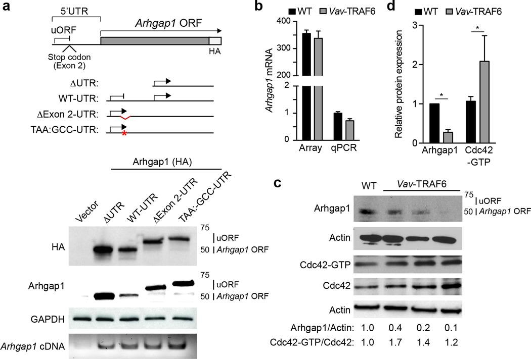
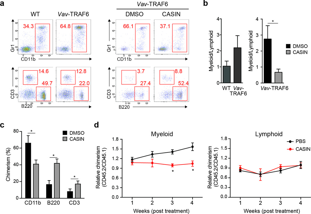
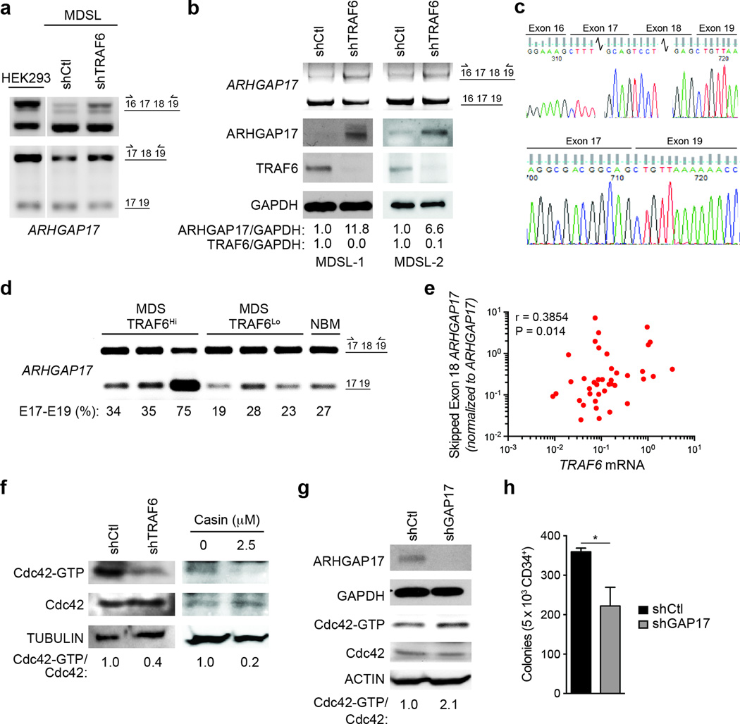
Toll-like receptor (TLR) activation contributes to premalignant hematologic conditions, such as myelodysplastic syndromes (MDS). TRAF6, a TLR effector with ubiquitin (Ub) ligase activity, is overexpressed in MDS hematopoietic stem/progenitor cells (HSPCs). We found that TRAF6 overexpression in mouse HSPC results in impaired hematopoiesis and bone marrow failure. Using a global Ub screen, we identified hnRNPA1, an RNA-binding protein and auxiliary splicing factor, as a substrate of TRAF6. TRAF6 ubiquitination of hnRNPA1 regulated alternative splicing of Arhgap1, which resulted in activation of the GTP-binding Rho family protein Cdc42 and accounted for hematopoietic defects in TRAF6-expressing HSPCs. These results implicate Ub signaling in coordinating RNA processing by TLR pathways during an immune response and in premalignant hematologic diseases, such as MDS.


Figures

Figure 1
Figure 1. Overexpression of TRAF6 results in hematopoietic stem/progenitor cell defects
(a) TRAF6 mRNA in CD34+ BM cells from low-risk MDS patients (n = 36) and age-matched healthy controls (HC; n = 7) (left), and LSK cells from Vav-TRAF6 (n = 6) and WT FVB/NJ (n = 4) mice (primers are designed to measure both endogenous mouse Traf6 and ectopic human TRAF6). P = 0.02. (b) Disease-free survival of Vav-TRAF6 (n = 60) and WT FVB/NJ mice (n = 40) *, P = 0.009. (c) Complete blood counts for WT FVB/NJ (n = 33) and moribund Vav-TRAF6 (n = 18) mice. *. P < 0.01. (d) BM neutrophil dysplasia in age-matched WT (n = 3) and Vav-TRAF6 (n = 5) FVB/NJ mice. At least 100 cells were examined for each mouse. *, P = 0.04. (e) BM mononuclear cell counts from WT (n = 3) and Vav-TRAF6 FVB/NJ mice (n = 4). *, P = 0.004. (f) Representative Wright-Giemsa-stained peripheral blood smears (PB) and bone marrow cytospins (BM) from WT (FVB/NJ) and 2 moribund Vav-TRAF6 mice. The red arrow indicates dysplastic myeloid cells with Pseudo-Pelger Hüet anomaly. Scale bars, 10 µm (top) and 7.5 µm (bottom) (g) Number of long-term hematopoietic stem cells (LT-HSC, P = 0.0056), short-term HSC (ST-HSC, P = 0.0011), multipotent progenitors (MPP, P = 0.014), common myeloid progenitors (CMP), megakaryocyte-erythroid progenitor (MEP), and granulocyte-monocytic progenitor (GMP) in the BM of WT (C57Bl/6) (n = 4) and Vav-TRAF6 (n = 4) ~6 month old mice. (h) Colony numbers were determined after primary and secondary plating LSK BM cells from WT (FVB/NJ; n = 3) and Vav-TRAF6 (n = 3) mice in methylcellulose. *, P < 0.001. (i) FACS analysis of WT (C57Bl/6) or Vav-TRAF6 donor-derived (CD45.2+) and competitor-derived (CD45.1+) mononuclear cells from peripheral blood 12 weeks post competitive transplantation (left panel). Proportion of myeloid (CD11b+) and lymphoid (CD3+ and B220+) cells within the donor-derived compartment (gated on CD45.2+) after competitive transplantation with either BM mononuclear cells (MNC, left) or LT-HSC (right). (n = 8 mice per genotype) (right panel). *, P < 0.05. (j) Colony formation in methylcellulose after serial replating of GFP-sorted Lin BM cells (C57Bl/6) transduced with retroviral vectors (MSCV-IRES-GFP) encoding WT (n = 3) or an E3 ligase-mutant TRAF6 (TRAF6C70A, n = 3). *, P < 0.01. (k) Proportion of donor-derived (gated on CD45.2+;GFP+) peripheral blood myeloid (Cd11b+) and lymphoid (CD3+ and B220+) cells were examined by FACS for competitively transplanted GFP-sorted Lin BM cells (C57Bl/6) transduced with retroviral vectors encoding WT (n = 5) or an E3 ligase-mutant TRAF6 (TRAF6C70A, n = 10) co-expressing GFP (MSCV-IRES-GFP). *, P < 0.05. Data are from three experiments (a, c–g; means and s.e.m.) or from two experiments (h–k; means and s.e.m.).
Figure 2
Figure 2. TRAF6 regulates ubiquitination of RNA binding proteins and RNA splicing
(a) In vitro Ub reconstitution assays were performed on the identified candidates from the cell-based screen with purified TRAF6 (n = 29). Shown is the substrate ubiquitination in the presence of TRAF6 (F532 CyDye) as compared to without TRAF6 (B532 CyDye) for 2 independent replicates. (b) Exon-level microarray expression analysis of LSK from 6 month WT (FVB/NJ, n = 4) and Vav-TRAF6 mice (n = 3). (c) Motif enrichment in differentially spliced exons from Vav-TRAF6 LSK as compared to WT LSK in the form of position frequency matrix motifs from the CisBP-RNA database. Each point represents one motif. Motifs are sorted left to right in decreasing order of significance. Motifs enriched in Vav-TRAF6 LSK are above the horizontal dotted line. hnRNPA1, P = 9.7x10⁻⁵. (d) Enrichment scores for exon usage patterns for Vav-TRAF6 LSK as compared to 46 known splicing factor exon usage patterns. Each point represents the enrichment Z-score for an individual splicing pattern of the splicing factor patterns examined. Exon usage patterns enriched in Vav-TRAF6 LSK are above the horizontal dotted line. hnRNPA1, P = 0.017. Data are from two experiments (a; means and s.d.).
Figure 3
Figure 3. TRAF6 ubiquitinates hnRNPA1
(a) In vitro ubiquitin reconstitution assay performed in duplicate and analyzed by CyeDye fluorescence (F532 subtracted from background, B532) for hnRNPA1, TAK1, and Tubulin (TUBB; a negative control). Individual assays contained recombinant Ub, UBE1, Ubc13/UBE2V1, ATP, and the indicated substrates. nd, not detectable. (b) HEK293 cells transfected with MYC-hnRNPA1, FLAG-TRAF6, and either WT HA-Ub or mutant Ub that form only K63- (UbK63) or K48- (UbK48) linkages were immunoblotted for HA (Ub) on IP MYC-hnRNPA1. (c) HEK293 cells transfected with MYC-hnRNPA1, and either WT or E3 ligase defective (C70A) TRAF6 were IB for IP MYC-hnRNPA1 (anti HA-Ub). (d) Schematic representation of hnRNPA1 protein, and corresponding lysine-to-arginine mutations within the RNA recognition motif (RRM) (above image). HEK293 cells transfected with WT, Lys3-mutant hnRNPA1 (K3R), or hnRNPA1 with mutation of the lysines within the first RRM domain (K–R) were immunoprecipitated (IP) for MYC-hnRNPA1 and then immunoblotted for HA (Ub). Data are from two experiments (a; means and s.d.).
Figure 4
Figure 4. hnRNPA1 contributes to the hematopoietic phenotype in Vav TRAF6 mice
(a) Immunoprecipitation and immunobloting of ubiquitinated hnRNPA1 from WT (FVB/NJ) and Vav-TRAF6 Lin BM cells and LPS-treated WT (C57Bl/6) (Traf6+/+) and TRAF6-deficient (Traf6−/−) Lin BM cells. (b) hnRNPA1 mRNA and protein expression was determined following expression of shRNAs targeting hnRNPA1 (shA1) or non-targeting shRNA (shCtl). (c) Colony formation in methylcellulose after serial replating of GFP-sorted WT (FVB/NJ, n = 3) and Vav-TRAF6 (n = 3) Lin BM cells transduced with shRNAs targeting hnRNPA1 (shA1) or control shRNA (shCtl) co-expressing GFP. *, P < 0.05; #, P = 0.08. (d) Chimerism of donor-derived (CD45.2+GFP+) peripheral blood cells from lethally-irradiated recipient mice receiving Lin BM cells transduced with shRNAs targeting hnRNPA1 (shA1) or control shRNA (shCtl) co-expressing GFP competitively transplanted with wild-type (CD45.1+) total BM cells (n = 8 per genotype). *, P < 0.05. Data are from two experiments (b–d; means and s.e.m.).
Figure 5
Figure 5. TRAF6 regulates RNA splicing of Arhgap1
(a) Significantly excluded or included exons as determined by FIRMA index (−4.0 ≥ a ≤ 4.0) in Vav-TRAF6 versus WT (FVB/NJ) LSK (MIDAS P < 0.05). Each point represents a differentially spliced exon. Highlighted are the top 3 differentially spliced exons in Vav-TRAF6 LSK. (b) Schematic representation of the mouse Arhgap1 gene structure with the approximate position of exons and introns. Overview of Arhgap1 exon usage is shown for WT (FVB/NJ) (black lines) and Vav-TRAF6 LSK (red lines). The average of FIRMA index values for individual exons of Arhgap1 in WT (n = 4) and Vav-TRAF6 LSK (n = 3) are shown below. FIRMA index scores indicate exon exclusion (negative values) or exon retention (~0). (c) RT-PCR analysis (using primers flanking exon 2) of the Arhgap1 cassette exon in Vav-TRAF6 and WT (FVB/NJ) LSK (left), and in LPS-treated WT Lin BM cells (right). Values under the plot represent the short isoform as a percentage of the short and long isoforms. (d) Sequence analysis of the Arhgap1 cassette exon using primers flanking skipped exon 2 isolated from Vav-TRAF6 LSK. (e) RT-PCR analysis (using primers flanking exon 2, as depicted in the schematic) of the Arhgap1 cassette exon in WT (FVB/NJ) or Vav-TRAF6 Lin BM cells were transduced with non-targeting shRNA (shCtl) or shRNA targeting mouse hnRNPA1 (shA1). Summary of Arhgap1 exon 2 exclusion calculated based on the short isoform as a percentage of the short and long isoforms is shown above. n = 3 per group; #, P = 0.06; *, P = 0.04. (f) RT-PCR analysis (using primers flanking exon 2, as depicted in the schematic) of the Arhgap1 cassette exon in WT (C57Bl/6) (Traf6+/+;Mx1Cre) and TRAF6-deficient Lin BM cells (Traf6fl/fl;Mx1Cre mice treated with PolyIC) stimulated with 10 ng/mL of LPS for 2 hrs. Summary of Arhgap1 exon 2 exclusion calculated based on the short isoform as a percentage of the short and long isoforms is shown above. n = 3 per group; *, P < 0.05. (g) RT-PCR analysis of a minigene plasmid containing the Arhgap1 cassette exon 2 with 50 bp of flanking intron sequences (using primers flanking exon 2, as depicted in the schematic on the left) in HEK293 cells (HEK293-Arhgap1exon2) transfected with hnRNPA1. Summary of Arhgap1 exon 2 exclusion from the minigene calculated based on the short isoform as a percentage of the short and long isoforms is shown above from 2 independent experiments. *, P = 0.02. (h) RT-PCR analysis of a minigene plasmid containing the Arhgap1 cassette exon 2 (using primers flanking exon 2, as depicted in the schematic on the left) in HEK293-Arhgap1exon2 cells transfected with WT (n = 7) and an E3 ligase-mutant (C70A; n = 5) TRAF6. Summary of Arhgap1 exon 2 exclusion from the minigene was calculated based on the short isoform as a percentage of the short and long isoforms is shown above from 2 independent experiments. *, P = 0.03. (i) RT-PCR analysis of a minigene plasmid containing the Arhgap1 cassette exon 2 (using primers flanking exon 2, as depicted in the schematic on the left) in HEK293-Arhgap1exon2 cells transfected with TRAF6 and hnRNPA1 (WT and lysine mutants). Summary of Arhgap1 exon 2 exclusion from the minigene was calculated based on the short isoform as a percentage of the short and long isoforms is shown above from 2 independent experiments (n = 2 per group). Vec, vector; A1WT, hnRNPA1 WT; A1K3R, hnRNPA1 with K3R mutation; A1K-R, hnRNPA1 with mutation of all lysines within the first RNA recognition motif. *, P < 0.01; **, P = 0.003. (j) qRT-PCR of Arhgap1 cassette exon 2 expressed from the minigene plasmid following RNA immunoprecipitation of Myc-hnRNPA1 in HEK293-Arhgap1exon2 cells transfected with Myc-hnRNPA1 (WT and K-R) and TRAF6. Shown is the average of 2 independent experiments (n = 2 per group). *, P < 0.01. Data are from two experiments (e–j; means and s.e.m.).
Figure 6
Figure 6. Exclusion of exon 2 results in reduced Arhgap1 protein and Cdc42 activation
(a) Schematic of the Arhgap1 5’-UTR and coding region. uORF, upstream open reading frame; HA, hemagglutinin. The indicated Arhgap1 5’-UTR cDNA fusions were transfected (pCMV-GFP) into HEK293 cells and evaluated by Arhgap1 and HA immunoblotting. Transfected Arhgap1 5’-UTR cDNA were measured by RT-PCR using primers aligned to Arhgap1 coding sequences. (b) Microarray and qRT-PCR analysis of Arhgap1 total RNA from LSK of WT (FVB/NJ) and Vav-TRAF6 mice. (c) Immunoblotting of Arhgap1 in Lin BM cells from WT (FVB/NJ) and Vav-TRAF6 mice. Cdc42 activity (Cdc42-GTP) in WT (FVB/NJ) and Vav-TRAF6 Lin BM cells was determined by a pull-down/immunoblot assay. Shown is a representative blot from one WT (FVB/NJ) and three Vav-TRAF6 mice. (d) Densitometric analysis of Arhgap1 protein and Cdc42-GTP (WT, n = 3; Vav-TRAF6, n = 5). *, P < 0.05. Data are from three experiments (b,d; means and s.e.m.).
Figure 7
Figure 7. Cdc42 contributes to HSPC defects in Vav TRAF6 mice
(a) FACS analysis of donor-derived (gated on CD45.2+) myeloid (CD11b+Gr1+) and lymphoid (B220+ and CD3+) proportions 8 weeks post transplantation for competitively transplanted WT (C57Bl/6) and Vav-TRAF6 LT-HSC, and for Vav-TRAF6 LT-HSC treated in vitro with CASIN (or DMSO) for 24 hr. *, P < 0.05. (b) Summary of independent experiments of myeloid and lymphoid proportions of donor-derived (gated on CD45.2+) peripheral blood shown as a ratio (CD11b/[B220 + CD3]). (c) Peripheral blood myeloid (CD11b+) and lymphoid (B220+ and CD3+) chimerism (CD45.2+) of donor-derived cells determined 8 weeks after competitive transplantation with Vav-TRAF6 LT-HSC treated with CASIN. (n = 6 per group). *, P < 0.005. (d) Donor-derived LT-HSC cells (CD45.2+) from Vav-TRAF6 mice and competitor BM cells (CD45.1+) were transplanted into lethally-irradiated recipient mice (CD45.1+). Three month post secondary transplantation, the recipients were treated with 30 mg/kg CASIN and vehicle control (PBS) for 16 days. Ratio of donor-derived (gated on CD45.2+) and competitor-derived (gated on CD45.1+) myeloid (CD11b) and lymphoid (CD3 and B220) cells was determined by flow cytometry 4 weeks post-CASIN treatment in vivo. Donor-derived chimerism is normalized (1.0) for each mouse at week 1 (n = 5 per group). *, P = 0.007. Data are from two experiments (b–d; means and s.e.m.).
Figure 8
Figure 8. Cdc42 activation is associated with primary human MDS
(a) RT-PCR using primers flanking endogenous ARHGAP17 exon 18 or exons 17 and 18 (as depicted in the schematic on the left) in HEK293 or MDSL cells expressing shRNAs targeting TRAF6 (shTRAF6) or a non-targeting control (shCtl). (b) Immunoblotting of ARHGAP17 in independently transduced MDSL cells (MDSL-1-2) expressing a non-targeting shRNA (shCtl) or shRNA targeting TRAF6 (shTRAF6). (c) Sequence analysis of the ARHGAP17 exons 16, 17, 18, and 19 in MDSL cells. (d) RT-PCR using primers flanking endogenous ARHGAP17 exon 18 (as depicted in the schematic on the left) in MDS patient BM mononuclear cells with high (>2-fold TRAF6, n = 3) or low (< 1.0-fold TRAF6, n =3) TRAF6 mRNA, and 1 normal BM sample. (e) qRT-PCR of ARHGAP17 exon 18 using primers to the exon 17–19 junction and normalized to total ARHGAP17 in MDS/AML BM cells (n = 35). Spearman correlation: r = 0.3854, P = 0.014. (f) Cdc42-GTP pulldown assays in MDSL cells expressing non-targeting shRNA (shCtl) or shRNA targeting TRAF6 (shTRAF6), or in MDSL treated with 2.5 µM CASIN. (g) Cdc42-GTP pulldown assays in MDSL expressing non-targeting shRNA (shCtl) or shRNA targeting ARHGAP17 (shGAP17). (h) Colony formation in methylcellulose of human CD34+ cells expressing a non-targeting shRNA (shCtl) or shRNA targeting ARHGAP17 (shGAP17) (n = 3). *, P = 0.047. Data are from three experiments (h; means and s.e.m.).

Comment in

References

    1. Esplin BL, et al. Chronic exposure to a TLR ligand injures hematopoietic stem cells. J Immunol. 2011;186:5367–5375. - PMC - PubMed
    1. Wong JJ, et al. Orchestrated intron retention regulates normal granulocyte differentiation. Cell. 2013;154:583–595. - PubMed
    1. Shalek AK, et al. Single-cell transcriptomics reveals bimodality in expression and splicing in immune cells. Nature. 2013;498:236–240. - PMC - PubMed
    1. Nagai Y, et al. Toll-like receptors on hematopoietic progenitor cells stimulate innate immune system replenishment. Immunity. 2006;24:801–812. - PMC - PubMed
    1. Starczynowski DT, et al. Identification of miR-145 and miR-146a as mediators of the 5q- syndrome phenotype. Nat Med. 2010;16:49–58. - PubMed

Publication types

MeSH terms

Substances