. 2017 Apr;16(4):489-496.
doi: 10.1038/nmat4822. Epub 2016 Dec 26.

Designer vaccine nanodiscs for personalized cancer immunotherapy

Affiliations

Designer vaccine nanodiscs for personalized cancer immunotherapy

Rui Kuai et al. Nat Mater. 2017 Apr.

Abstract

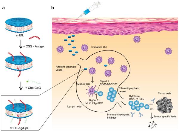
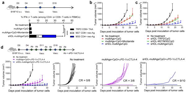
Despite the tremendous potential of peptide-based cancer vaccines, their efficacy has been limited in humans. Recent innovations in tumour exome sequencing have signalled the new era of personalized immunotherapy with patient-specific neoantigens, but a general methodology for stimulating strong CD8α+ cytotoxic T-lymphocyte (CTL) responses remains lacking. Here we demonstrate that high-density lipoprotein-mimicking nanodiscs coupled with antigen (Ag) peptides and adjuvants can markedly improve Ag/adjuvant co-delivery to lymphoid organs and sustain Ag presentation on dendritic cells. Strikingly, nanodiscs elicited up to 47-fold greater frequencies of neoantigen-specific CTLs than soluble vaccines and even 31-fold greater than perhaps the strongest adjuvant in clinical trials (that is, CpG in Montanide). Moreover, multi-epitope vaccination generated broad-spectrum T-cell responses that potently inhibited tumour growth. Nanodiscs eliminated established MC-38 and B16F10 tumours when combined with anti-PD-1 and anti-CTLA-4 therapy. These findings represent a new powerful approach for cancer immunotherapy and suggest a general strategy for personalized nanomedicine.


Figures

Figure 1
Figure 1. Design of sHDL nanodisc platform for personalized cancer vaccines
a, sHDL nanodiscs, composed of phospholipids and apolipoprotein-1 mimetic peptides (22A), are engineered for co-delivery of antigen (Ag) peptides and adjuvants. Pre-formed sHDL nanodiscs displaying 4 mol% DOPE-PDP are mixed with cysteine-modified Ag peptides, including tumor-specific mutated neo-antigens identified via tumor exome DNA sequencing, and subsequent incubation with cholesterol-modified immunostimulatory molecules (Cho-CpG) leads to formation of sHDL nanodiscs co-loaded with Ag and CpG (sHDL-Ag/CpG). b, Upon administration, sHDL nanodiscs efficiently co-deliver Ag and CpG to draining lymph nodes, promote strong and durable Ag presentation by dendritic cells (DCs) (Signal 1), and induce DC maturation (Signal 2), resulting in elicitation of robust Ag-specific CD8α+ cytotoxic T lymphocyte (CTL) responses. Activated CTLs recognize and kill their target cancer cells in peripheral tissues and exert strong anti-tumor efficacy. Combination immunotherapy with immune checkpoint blockade further amplifies the potency of nanodisc vaccination, leading to elimination of established tumors.
Figure 2
Figure 2. Strong and durable Ag presentation mediated by sHDL nanodiscs
a, Dynamic light scattering analysis and b, transmission electron microscopy imaging showed uniform sHDL-Ag/CpG (10.5 nm ± 0.5 average diameter) with nanodisc-like morphology. Scale bar = 100 nm. Scale bar in the inset = 20 nm. c, Homogeneity of nanodiscs was maintained after sterile-filtration (0.22 μm), and long-term storage (8 weeks) at −20°C, followed by thawing at 37°C. d-e, BMDCs were incubated with vaccine formulations for d, 24 h or e, indicated lengths of time, and Ag presentation was quantified by flow-cytometry analysis of DCs stained with 25-D1.16 mAb that recognizes SIINFEKL-H-2Kb complex. f-g, Confocal microscopy images of JAWSII cells (immature DCs). f, JAWSII cells were incubated with free Ag+CpG or sHDL-Ag/CpG for 24 h and stained with 25-D1.16 mAb. Scale bars = 20 μm. g, JAWSII cells were incubated with free CSSSIINFEK(FITC)L + CpG or sHDL-CSSSIINFEK(FITC)L/CpG for 6, 24, or 48 h, followed by staining with Hochest and Lysotracker. Scale bars = 10 μm. h, BMDCs were incubated with different concentrations of indicated formulations: low dose = 20 nM SIINFEKL and 3 nM CpG; medium dose = 100 nM SIINFEKL and 15 nM CpG; and high dose = 500 nM SIINFEKL and 75 nM CpG. After incubation for 24 h or 48 h, BMDCs were co-cultured with SIINFEKL-specific B3Z T-cell hybridoma for another 24 h, followed by assessment of T cell activation. The data show mean ± SD from a representative experiment (n = 3) from 2-4 independent experiments. * p < 0.05, ** p < 0.01, *** p < 0.001, and **** p < 0.0001, analyzed by (d) one-way or (e,h) two-way ANOVA with Bonferroni multiple comparisons post-test.
Figure 3
Figure 3. Vaccine nanodiscs for LN-targeting of Ag and adjuvants and elicitation of CTL responses
a-b, C57BL/6 mice were administered subcutaneously at tail base with a, 31 nmol FITC-tagged Ag (CSSSIINFEK(FITC)L) or b, 2.3 nmol Cho-CpG (20% labeled by Cy5) in free soluble or sHDL form, and fluorescence signal in the draining inguinal LNs were quantified with IVIS after 24 h. c-e, C57BL/6 mice were immunized with the indicated formulations (15.5 nmol Ag peptide and 2.3 nmol CpG) on days 0, 21, and 42. c, Shown are their representative scatter plots on day 49 and d the frequency of SIINFEKL-specific CD8α+ T-cells in peripheral blood measured 7 days post each immunization by flow-cytometry analysis of tetramer+ CD8α+ T-cells. e, On day 50, pre-vaccinated animals were challenged with subcutaneous flank injection of 2×10⁵ B16OVA cells, and tumor growth was measured over time. f-h, C57BL/6 mice were immunized in a biweekly interval. Shown are f, percent of SIINFEKL-specific CD8α+ T-cells in peripheral blood; g, ELISPOT analysis of IFN-γ spot-forming cells among splenocytes after ex vivo restimulation with SIINFEKL on day 35; and h, Ag-specific CD8α+ T-cell responses measured over 12 weeks post vaccination (black arrows indicate days of immunizations). i, Vaccinated mice in (h) were intravenously challenged with 5×10⁴ B16OVA cells two months after the third vaccination. Shown are pictures of the lungs and numbers of lung metastatic nodules counted on day 20 after the B16OVA challenge. The data show mean ± SD from a representative experiment (n = 4-5) from 2-3 independent experiments. * p < 0.05, ** p < 0.01, *** p < 0.001, and **** p < 0.0001, analyzed by (a-b) two-tailed unpaired Student’s t test, (d,e,f,h) two-way ANOVA, or (i) one-way ANOVA with Bonferroni multiple comparisons post-test. Asterisks in panel e indicate statistically significant differences between sHDL-Ag/CpG and SIINFEKL+CpG+Montanide.
Figure 4
Figure 4. Nanodisc-based neo-antigen vaccination for personalized immunotherapy
a, Mutation of Adpgk in MC-38 murine colon adenocarcinoma cells was confirmed by sequencing cDNA of Adpgk. b-d, C57BL/6 mice were vaccinated three times with the indicated formulations (equivalent to 15.5 nmol mutated Adpgk peptide and 2.3 nmol CpG) in a bi-weekly interval, and the frequency of Adpgk-specific CD8α+ T-cells in peripheral blood was measured . Shown are b, the representative scatter plots, and c, the frequency of Adpgk-specific CTLs on day 35. d, Clonal contraction of Ag-specific CD8α+ T-cell responses elicited by sHDL-Adpgk/CpG and sHDL-SIINFEKL/CpG vaccines was monitored for eight weeks after the last vaccination. e, C57BL/6 mice were inoculated subcutaneously with 105 MC-38 tumor cells and vaccinated with the indicated formulations (equivalent to 15.5 nmol mutated Adpgk peptide and 2.3 nmol CpG) on days 10, 17, and 24. Shown are the percent of intracellular IFN-γ+, TNF-α+, and IFN-γ+TNF-α+ CD8α+ T-cells in peripheral blood on day 30 after ex vivo restimulation with the mutated Adpgk Ag. Average and individual MC-38 tumor growth curves are shown with fraction of complete tumor regression (CR). f, C57BL/6 mice were inoculated subcutaneously with 105 MC-38 tumor cells and vaccinated with the indicated formulations (equivalent to 15.5 nmol mutated Adpgk peptide and 2.3 nmol CpG) on days 10 and 17. On days 1 and 4 after each vaccination, mice were administered intraperitoneally with αPD-1 (100 μg/mouse). Average and individual MC-38 tumor growth curves are shown. The data show mean ± SD from a representative experiment (n = 5-10) from 2-3 independent experiments. * p < 0.05, ** p < 0.01, *** p < 0.001, and **** p < 0.0001, analyzed by (c) one-way ANOVA or (e,f) two-way ANOVA with Bonferroni multiple comparisons post-test. Asterisks in (e,f) indicate statistically significant differences between sHDL-Ag/CpG and all other treatment groups.
Figure 5
Figure 5. Tumor eradication by combination immunotherapy with multi-epitope vaccine nanodiscs and immune checkpoint blockade
a-d, C57BL/6 mice were inoculated subcutaneously with 105 melanoma B16F10 cells and vaccinated on days 4, 11, and 18 with indicated formulations (10 nmol of each antigen peptide and 2.3 nmol of CpG). For the combination immunotherapy, on days 1 and 4 after each vaccination, αPD-1 and αCTLA-4 (100 μg/mouse each) were administered intraperitoneally. Shown are a, the percent of IFN-γ+ CD8α+ or CD4+ T cells in peripheral blood measured by intracellular cytokine staining, and b-d, average and individual B16F10 tumor growth curves. The data show mean ± SD from a representative experiment (n = 5-10) from 2-3 independent experiments. * p < 0.05, ** p < 0.01, *** p < 0.001, and **** p < 0.0001, analyzed by (a) one-way ANOVA or (b-d) two-way ANOVA with Bonferroni multiple comparisons post-test. Asterisks in (b-d) indicate statistically significant differences between sHDL-Ag/CpG and all other treatment groups.

References

    1. Melief CJ, van der Burg SH. Immunotherapy of established (pre)malignant disease by synthetic long peptide vaccines. Nat Rev Cancer. 2008;8:351–360. - PubMed
    1. Speiser DE, et al. Rapid and strong human CD8+ T cell responses to vaccination with peptide, IFA, and CpG oligodeoxynucleotide 7909. J Clin Invest. 2005;115:739–746. - PMC - PubMed
    1. Fourcade J, et al. Immunization with analog peptide in combination with CpG and montanide expands tumor antigen-specific CD8+ T cells in melanoma patients. J Immunother. 2008;31:781–791. - PMC - PubMed
    1. Hailemichael Y, et al. Persistent antigen at vaccination sites induces tumor-specific CD8+ T cell sequestration, dysfunction and deletion. Nat Med. 2013;19:465–472. - PMC - PubMed
    1. Reddy ST, et al. Exploiting lymphatic transport and complement activation in nanoparticle vaccines. Nat Biotechnol. 2007;25:1159–1164. - PubMed

Publication types

MeSH terms