Review
. 2017 Jan:57-58:178-189.
doi: 10.1016/j.matbio.2016.12.005. Epub 2016 Dec 23.

Force-dependent breaching of the basement membrane

Affiliations
Review

Force-dependent breaching of the basement membrane

Tammy T Chang et al. Matrix Biol. 2017 Jan.

Abstract

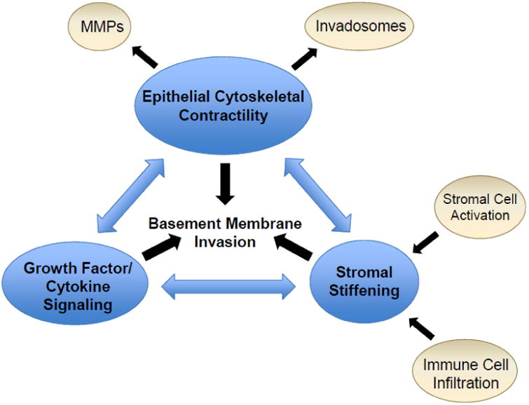
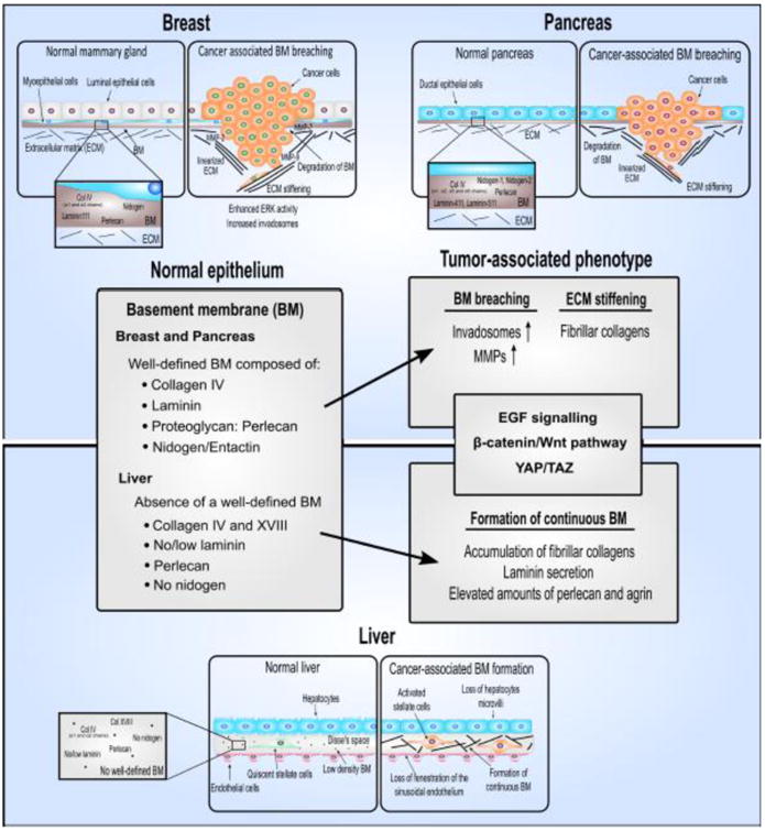
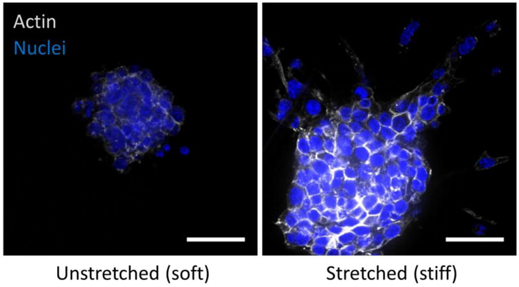
Clinically, non-invasive carcinomas are confined to the epithelial side of the basement membrane and are classified as benign, whereas invasive cancers invade through the basement membrane and thereby acquire the potential to metastasize. Recent findings suggest that, in addition to protease-mediated degradation and chemotaxis-stimulated migration, basement membrane invasion by malignant cells is significantly influenced by the stiffness of the associated interstitial extracellular matrix and the contractility of the tumor cells that is dictated in part by their oncogenic genotype. In this review, we highlight recent findings that illustrate unifying molecular mechanisms whereby these physical cues contribute to tissue fibrosis and malignancy in three epithelial organs: breast, pancreas, and liver. We also discuss the clinical implications of these findings and the biological properties and clinical challenges linked to the unique biology of each of these organs.

Keywords: Breast cancer; Hepatocellular carcinoma; Matrix rigidity; Pancreatic cancer; Tumor invasion.


Figures

Figure 1
Figure 1
Inter-related and cross-regulating factors that promote basement membrane invasion in cancer progression. MMPs, matrix metalloproteinases.
Figure 2
Figure 2
Primary components of the basement membrane in healthy breast, pancreas, and liver tissue and the changes that occur in the basement membrane during development of fibrosis and cancer.
Figure 3
Figure 3
Malignant mammary epithelial cells cultured in reconstituted basement membrane as spherical multicellular organoids and then embedded in collagen hydrogels placed under either 0 (unstretched - soft) or 10% strain (stretched - stiff) in a tension bioreactor. After three days, cultures were fixed and stained for actin cytoskeleton (phalloidin) and nuclei (DAPI) to assess the extent of tumor cell invasion and migration (scale bar = 20 μm).

References

    1. Allred DC. Ductal carcinoma in situ: terminology, classification, and natural history. J Natl Cancer Inst Monogr. 2010;2010:134–138. doi: 10.1093/jncimonographs/lgq035. - DOI - PMC - PubMed
    1. Lopez-Garcia MA, Geyer FC, Lacroix-Triki M, Marchio C, Reis-Filho JS. Breast cancer precursors revisited: molecular features and progression pathways. Histopathology. 2010;57:171–192. doi: 10.1111/j.1365-2559.2010.03568.x. - DOI - PubMed
    1. Cowell CF, Weigelt B, Sakr RA, Ng CK, Hicks J, King TA, Reis-Filho JS. Progression from ductal carcinoma in situ to invasive breast cancer: revisited. Mol Oncol. 2013;7:859–869. doi: 10.1016/j.molonc.2013.07.005. - DOI - PMC - PubMed
    1. Allred DC, Wu Y, Mao S, Nagtegaal ID, Lee S, Perou CM, Mohsin SK, O'Connell P, Tsimelzon A, Medina D. Ductal carcinoma in situ and the emergence of diversity during breast cancer evolution. Clin Cancer Res. 2008;14:370–378. doi: 10.1158/1078-0432.CCR-07-1127. - DOI - PubMed
    1. Evans AJ, Pinder SE, Snead DR, Wilson AR, Ellis IO, Elston CW. The detection of ductal carcinoma in situ at mammographic screening enables the diagnosis of small, grade 3 invasive tumours. Br J Cancer. 1997;75:542–544. - PMC - PubMed

Publication types

MeSH terms