. 2017 Feb 1;26(3):599-610.
doi: 10.1093/hmg/ddw419.

Mutations in BOREALIN cause thyroid dysgenesis

Affiliations

Mutations in BOREALIN cause thyroid dysgenesis

Aurore Carré et al. Hum Mol Genet. .

Abstract

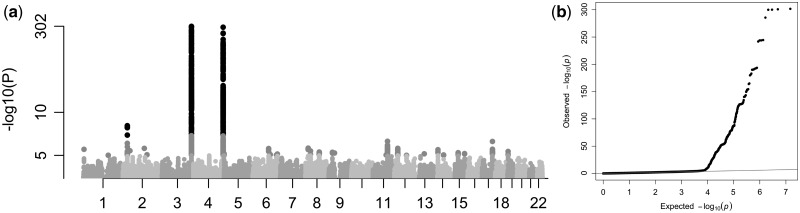
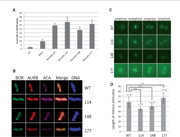
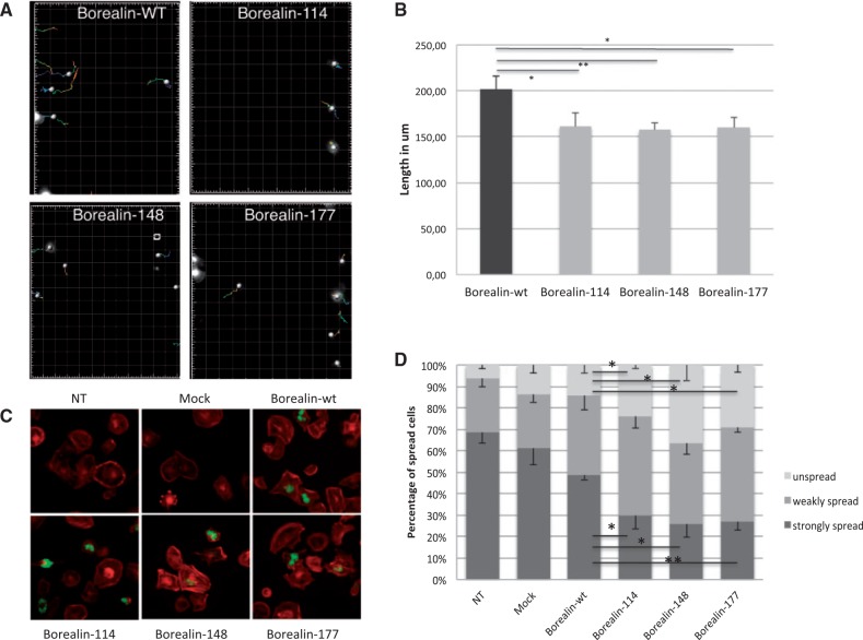
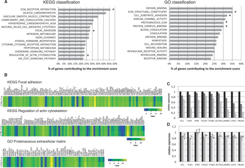
Congenital hypothyroidism is the most common neonatal endocrine disorder and is primarily caused by developmental abnormalities otherwise known as thyroid dysgenesis (TD). We performed whole exome sequencing (WES) in a consanguineous family with TD and subsequently sequenced a cohort of 134 probands with TD to identify genetic factors predisposing to the disease. We identified the novel missense mutations p.S148F, p.R114Q and p.L177W in the BOREALIN gene in TD-affected families. Borealin is a major component of the Chromosomal Passenger Complex (CPC) with well-known functions in mitosis. Further analysis of the missense mutations showed no apparent effects on mitosis. In contrast, expression of the mutants in human thyrocytes resulted in defects in adhesion and migration with corresponding changes in gene expression suggesting others functions for this mitotic protein. These results were well correlated with the same gene expression pattern analysed in the thyroid tissue of the patient with BOREALIN-p.R114W. These studies open new avenues in the genetics of TD in humans.

Trial registration: ClinicalTrials.gov NCT01916018.


Figures

Figure 1.
Figure 1.
Molecular genetics. (A) Pedigrees of families with Borealin mutations. Family F1. Characterization of a homozygous missense mutation by WES, c.443C>T, p.S148F, in a consanguineous family F1. Familial pedigree with four children including two siblings with TD and the homozygous mutation (m/m). Parents are heterozygous (m/+) for the mutation. The mother has an asymmetric thyroid lobes and the father has nodules. Mutation and familial segregation are confirmed by Sanger sequencing. Family F2. Characterization of a heterozygous missense mutation, c.530T>G, p.R114Q, identified by Sanger sequencing in the mother and her daughter. The daughter has CH with ectopy and the mother had an asymmetric thyroid and developed later a papillary thyroid cancer. Family F3. Characterization of a heterozygous missense mutation, c.341G>A, p.L177W, found by Sanger sequencing in a girl with CH due to athyreosis and congenital heart defect and in the mother. The mother has nodules (in light gray). Affected individuals are indicated by solid black/gray symbols (black for thyroid ectopy, thyroid hemiagenesis, athyreosis, asymmetric thyroid and light gray for nodules). (B) Schematic representation of the Borealin protein with domain structure. (C). Multiple sequence alignment of Borealin proteins. For the three missense mutations (in gray) conservation across evolution of altered amino acid residues is shown.
Figure 2.
Figure 2.
Borealin expression during thyroid development. (A) Gene expression by quantitative PCR of Borealin in thyroid tissue at 8GW, 12GW and in adult thyroid, reported to one thyroid tissue at 8GW and Peptidylpropyl isomerase A. Experiments with three tissues by stage. (B) Immunohistofluorescence of the Borealin (A, E, in red), Ecadherin (B, in green) and TG (F, in green) in thyroid tissue at 8 and 12GW. In C, co-staining of Borealin and E-cadherin. In G, co-staining of Borealin and TG. Boxes indicating enlarged areas (D, H).
Figure 3.
Figure 3.
Effects on mitosis of Borealin mutants. (A). G2/M phases in Nthy transfected with Borealin-wt, Borealin-114, Borealin-148 and Borealin-177. Borealin transfection in cells lead to increase the mitosis. The percentage of cells was determined by flow cytometry. Values are represented as mean ± SEM from three independent measurements. NT, non-transfected; Mock, cells transfected with empty vector; Borealin-wt, Borealin-114, Borealin-148 and Borealin-177 were cells transfected with sh-vector and with vector containing Borealin-wt or mutated. (B) Localization of Borealin mutants during metaphase. HeLa M cells were transiently transfected with GFP-tagged wild-type and mutant Borealin. Single z-planes of representative transfected cells are shown. BOR, Borealin, AURB, Aurora B kinase, ACA, anti-centromere autoantibodies. (C) Time lapse. Examples of cells progressing through mitosis after transfection with wild-type and mutant Borealin-GFP along with Borealin shRNA. (D) Length of mitosis. GFP-positive cells as described in C were tracked from prophase to telophase. Average + SEM are shown.
Figure 4.
Figure 4.
Modifications in migration and spreading of cells by Borealin mutants. (A) Tracking of GFP-Nthy transfected with Borealin-wt, Borealin-114, Borealin-148 and Borealin-177 during 8 h after wound healing assay on time-lapse video microscopy as described in "Materials and Methods." (B) The length of migration in um was measured by quantifying the total distance that the positively transfected cells (GFP) moved from the edge of the wound toward the center of the wound in 8 h. Mean of the length of the migration from four independent experiments. *P = 0.04, 0.0036, 0.0202 respectively for Nthy with Borealin-114, Borealin-148 and Borealin-148 in comparison with Nthy with Borealin-wt, calculated by t test. (C) Analysis of spreading by immunofluorescence. Staining of Borealin-Flag (in green) and filamentous actin (in red) showed decreased spreading of Borealin-114, Borealin-148, Borealin-177 Nthy compared to Borealin-wt Nthy (NT, no transfected; mock, empty vectors). (D) The percentage of spread and unspread Nthy was estimated by counting Nthy for each cell line (light gray, unspread; medium gray, weakly spread; dark gray, strongly spread). The graph represents the mean five independent experiments. *P< 0.05, P < 0.01 calculated by t test.
Figure 5.
Figure 5.
Transcriptome analysis of thyroid tissue with Borealin-114 and quantitative PCR in transfected HPT. (A) Genes with decreased expression in transcriptome analysis of the thyroid tissue with Borealin-114 compared to thyroid controls. On the left, KEGG Pathway analysis of expressed mRNAs. Top fifteen enriched down-regulated pathways. On the right, GO enrichment analysis of expressed mRNAs. Top fifteen enriched down-regulated pathways. The bar plot represents the percentage of genes contributing to the enrichment score. *Pathways involved in adhesion and/or migration. (B) Examples of pathways of interest with decreased expression genes in thyroid tissue with Borealin-114 (T114) compared to thyroid tissues control (CTRL): KEGG Focal adhesion, KEGG Regulation of actin cytoskeleton, GO Proteinaceous extracellular matrix. Colour scale for each pathway is shown. From left to right, gene expression difference between control tissues and T114 is increased. *Genes involved in adhesion and/or migration and validated in HPT. (C) Gene expression of Borealin-114 tissue compared to controls in trancriptome analysis and validated by quantitative PCR (black, thyroid controls; medium gray, transcriptome results of Borealin-114 tissue; light gray, validation by quantitative PCR in Borealin-114 tissue). (D) Genes involved in adhesion and/or migration in transfected HPT analysed by quantitative PCR 48 h post-transfection (black, Borealin-wt HPT, dark gray, Borealin-114 HPT; medium gray, Borealin-148 HPT; light gray, Borealin-177 HPT). The graph represents the mean of four independent experiments. *P < 0.05, **P<0.01 calculated by t-test.

References

    1. Stoupa A., Kariyawasam D., Carré A., Polak M. (2016) Update of Thyroid Developmental Genes. Endocrinol. Metab. Clin. North. Am., 45, 243–254. - PubMed
    1. Léger J., Marinovic D., Garel C., Bonaıïti-Pellié C., Polak M., Czernichow P. (2002) Thyroid developmental anomalies in first degree relatives of children with congenital hypothyroidism. J. Clin. Endocrinol. Metab., 87, 575–580. - PubMed
    1. Damante G., Tell G., Di Lauro R. (2001) A unique combination of transcription factors controls differentiation of thyroid cells. Prog. Nucleic. Acid. Res. Mol. Biol., 66, 307–356. - PubMed
    1. Fagman H., Nilsson M. (2010) Morphogenesis of the thyroid gland. Mol. Cell. Endocrinol., 35–54. - PubMed
    1. Johansson E., Andersson L., Örnros J., Carlsson T., Ingeson-Carlsson C., Liang S., Dahlberg J., Jansson S., Parrillo L., Zoppoli P., et al. (2015) Revising the embryonic origin of thyroid C cells in mice and humans. Development, 142, 3519–3528. - PMC - PubMed

Publication types

Associated data