

CD11c⁺ CD8⁺ T Cells Reduce Renal Fibrosis Following Ureteric Obstruction by Inducing Fibroblast Apoptosis
- PMID: 28025478
- PMCID: PMC5297636
- DOI: 10.3390/ijms18010001
CD11c⁺ CD8⁺ T Cells Reduce Renal Fibrosis Following Ureteric Obstruction by Inducing Fibroblast Apoptosis
Abstract
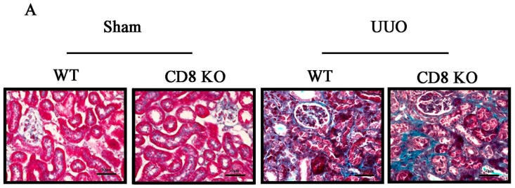
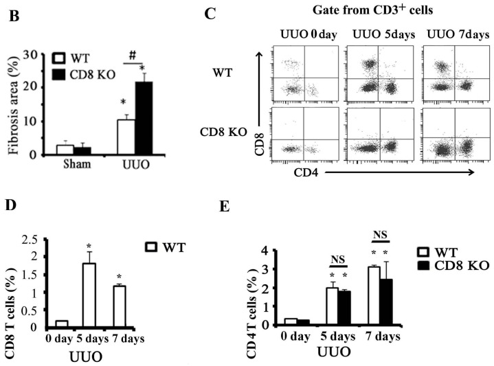
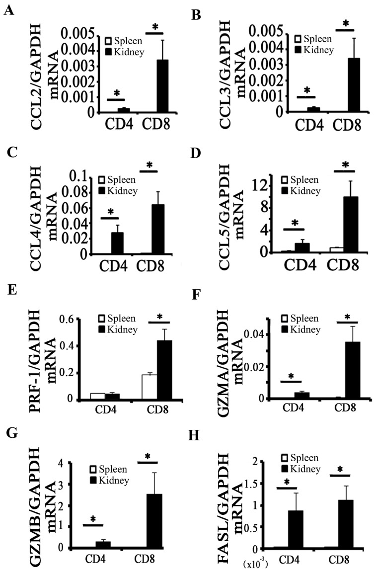
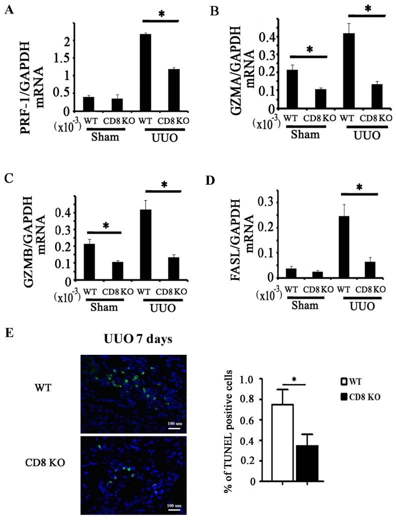
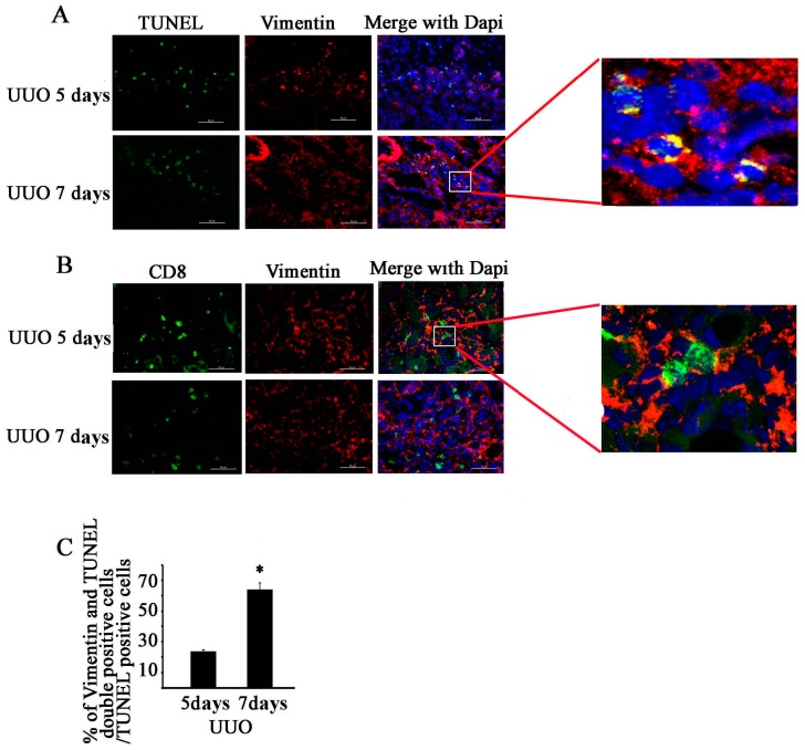
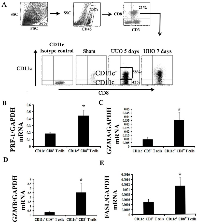
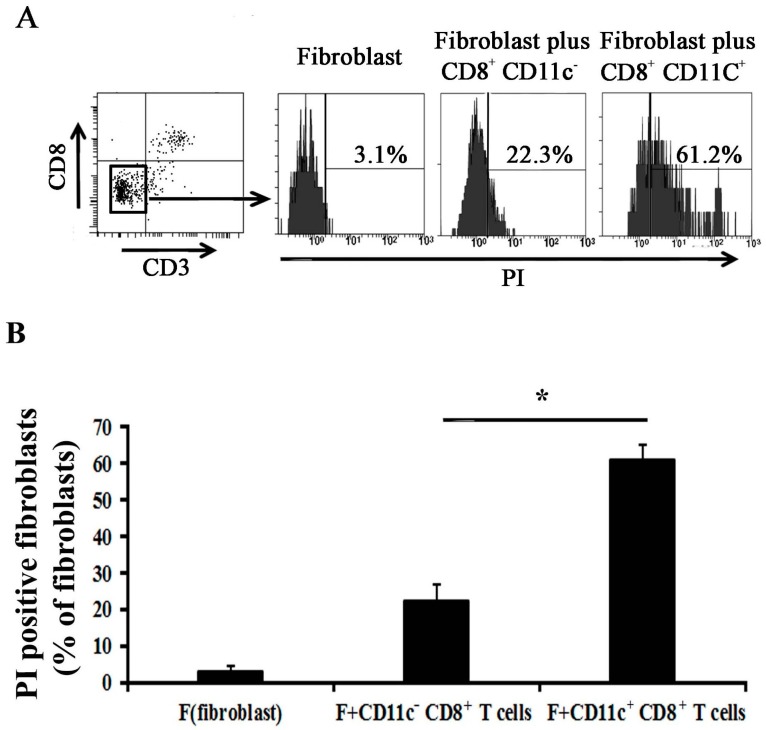
Tubulointerstitial fibrosis is a common consequence of various kidney diseases that lead to end-stage renal failure, and lymphocyte infiltration plays an important role in renal fibrosis. We previously found that depletion of cluster of differentiation 8⁺ (CD8⁺) T cells increases renal fibrosis following ureteric obstruction, and interferon-γ (IFN-γ)-expressing CD8⁺ T cells contribute to this process. CD8⁺ T cells are cytotoxic T cells; however, whether their cytotoxic effect reduces fibrosis remains unknown. This study showed that CD8⁺ T cells isolated from obstructed kidney showed mRNA expression of the cytotoxicity-related genes perforin 1, granzyme A, granzyme B, and FAS ligand; additionally, CD8 knockout significantly reduced the expression levels of these genes in obstructed kidney. Infiltrated CD8⁺ T cells were distributed around fibroblasts, and they are associated with fibroblast apoptosis in obstructed kidney. Moreover, CD11c⁺ CD8⁺ T cells expressed higher levels of the cytotoxicity-related genes than CD11c- CD8⁺ T cells, and infiltrated CD11c⁺ CD8⁺ T cells in obstructed kidney could induce fibroblast death in vitro. Results indicated that induction of fibroblast apoptosis partly contributed to the effect of CD8⁺ T cells on reduction of renal fibrosis. Given that inflammatory cells are involved in fibrosis, our results suggest that kidney fibrosis is a multifactorial process involving different arms of the immune system.
Keywords: T cells; fibroblasts; fibrosis; inflammation; kidney.
Conflict of interest statement
The authors declare no conflict of interest.
Figures
Similar articles
-
The Mechanism of CD8+ T Cells for Reducing Myofibroblasts Accumulation during Renal Fibrosis.Biomolecules. 2021 Jul 5;11(7):990. doi: 10.3390/biom11070990. Biomolecules. 2021. PMID: 34356613 Free PMC article. Review.
-
Depletion of CD8+ T Cells Exacerbates CD4+ T Cell-Induced Monocyte-to-Fibroblast Transition in Renal Fibrosis.J Immunol. 2016 Feb 15;196(4):1874-81. doi: 10.4049/jimmunol.1501232. Epub 2016 Jan 15. J Immunol. 2016. PMID: 26773152
-
CD11c(high)CD8+ regulatory T cell feedback inhibits CD4 T cell immune response via Fas ligand-Fas pathway.J Immunol. 2013 Jun 15;190(12):6145-54. doi: 10.4049/jimmunol.1300060. Epub 2013 May 15. J Immunol. 2013. PMID: 23677464
-
Single-cell perforin and granzyme expression reveals the anatomical localization of effector CD8+ T cells in influenza virus-infected mice.Proc Natl Acad Sci U S A. 2003 Mar 4;100(5):2657-62. doi: 10.1073/pnas.0538056100. Epub 2003 Feb 24. Proc Natl Acad Sci U S A. 2003. PMID: 12601154 Free PMC article.
-
Perforin-independent CD8(+) T-cell-mediated cytotoxicity of alveolar epithelial cells is preferentially mediated by tumor necrosis factor-alpha: relative insensitivity to Fas ligand.Am J Respir Cell Mol Biol. 1999 May;20(5):849-58. doi: 10.1165/ajrcmb.20.5.3585. Am J Respir Cell Mol Biol. 1999. PMID: 10226053
Cited by
-
Hematuria as a risk factor for progression of chronic kidney disease and death: findings from the Chronic Renal Insufficiency Cohort (CRIC) Study.BMC Nephrol. 2018 Jun 26;19(1):150. doi: 10.1186/s12882-018-0951-0. BMC Nephrol. 2018. PMID: 29940877 Free PMC article.
-
Critical Role of Cysteine-Rich Protein 61 in Mediating the Activation of Renal Fibroblasts.Front Physiol. 2019 May 3;10:464. doi: 10.3389/fphys.2019.00464. eCollection 2019. Front Physiol. 2019. PMID: 31130867 Free PMC article.
-
Identification of the key immune-related genes and immune cell infiltration changes in renal interstitial fibrosis.Front Endocrinol (Lausanne). 2023 Nov 8;14:1207444. doi: 10.3389/fendo.2023.1207444. eCollection 2023. Front Endocrinol (Lausanne). 2023. PMID: 38027143 Free PMC article.
-
Redefining the Immunobiology of Organ Transplantation for New Clinical Horizons.Scand J Immunol. 2025 Aug;102(2):e70045. doi: 10.1111/sji.70045. Scand J Immunol. 2025. PMID: 40717391 Free PMC article. Review.
-
The Mechanism of CD8+ T Cells for Reducing Myofibroblasts Accumulation during Renal Fibrosis.Biomolecules. 2021 Jul 5;11(7):990. doi: 10.3390/biom11070990. Biomolecules. 2021. PMID: 34356613 Free PMC article. Review.
References
-
- Anders H.J., Vielhauer V., Frink M., Linde Y., Cohen C.D., Blattner S.M., Kretzler M., Strutz F., Mack M., Gröne H.J., et al. A chemokine receptor CCR-1 antagonist reduces renal fibrosis after unilateral ureter ligation. J. Clin. Investig. 2002;109:251–259. doi: 10.1172/JCI0214040. - DOI - PMC - PubMed
-
- Eis V., Luckow B., Vielhauer V., Siveke J.T., Linde Y., Segerer S., Lema G.P., Cohen C.D., Kretzler M., Mack M., et al. Chemokine receptor CCR1 but not CCR5 mediates leukocyte recruitment and subsequent renal fibrosis after unilateral ureteral obstruction. J. Am. Soc. Nephrol. 2004;15:337–347. doi: 10.1097/01.ASN.0000111246.87175.32. - DOI - PubMed
MeSH terms
Substances
LinkOut - more resources
Full Text Sources
Other Literature Sources
Research Materials
Miscellaneous
