. 2016 Dec 22;18(1):15.
doi: 10.3390/ijms18010015.

CHARMM Force Field Parameterization of Peroxisome Proliferator-Activated Receptor γ Ligands

Affiliations

CHARMM Force Field Parameterization of Peroxisome Proliferator-Activated Receptor γ Ligands

Melina Mottin et al. Int J Mol Sci. .

Abstract

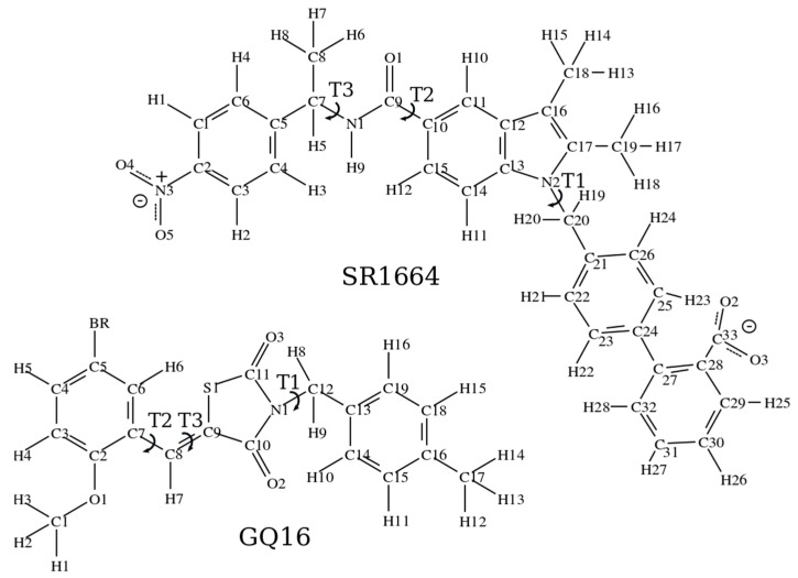
The peroxisome proliferator-activated receptor γ (PPARγ) ligands are important therapeutic drugs for the treatment of type 2 diabetes, obesity and cardiovascular diseases. In particular, partial agonists and non-agonists are interesting targets to reduce glucose levels, presenting few side effects in comparison to full agonists. In this work, we present a set of CHARMM-based parameters of a molecular mechanics force field for two PPARγ ligands, GQ16 and SR1664. GQ16 belongs to the thiazolidinedione class of drugs and it is a PPARγ partial agonist that has been shown to promote the "browning" of white adipose tissue. SR1664 is the precursor of the PPARγ non-agonist class of ligands that activates PPARγ in a non-classical manner. Here, we use quantum chemical calculations consistent with the CHARMM protocol to obtain bonded and non-bonded parameters, including partial atomic charges and effective torsion potentials for both molecules. The newly parameterized models were evaluated by examining the behavior of GQ16 and SR1664 free in water and bound to the ligand binding pocket of PPARγ using molecular dynamics simulations. The potential parameters derived here are readily transferable to a variety of pharmaceutical compounds and similar PPARγ ligands.

Keywords: CHARMM parameters; GQ16; PPARγ ligands; SR1664; molecular dynamics; nuclear receptor.


Conflict of interest statement

The authors declare no conflict of interest.

Figures

Figure 1
Figure 1
Chemical structures of the PPARγ ligands SR1664 and GQ16, indicating atom numbering and the parameterized torsions. The arc-shaped arrows indicate the torsion (T) bonds for which full revolution is possible.
Figure 2
Figure 2
Potential energy curves generated for the torsions T1, T2, and T3 of the SR1664 molecule. The values of the potential energy computed by density functional theory are represented by the blue line (QM), whereas the corresponding values generated from the classical potential are represented by the green line (MM). The QM and MM energy differences are depicted by the black dotted line; the solid lines are the fitted dihedral potentials; the blue dotted lines are the sum MM and fitted dihedral potential. The structures on the right side of each graph depict the fragments used to calculate the potential energy curves (the calculated dihedrals are colored).
Figure 3
Figure 3
Potential energy curves generated for the torsions T1, T2, and T3 of the GQ16 molecule. The values of the potential energy computed by density functional theory are represented by the blue line (QM), whereas the corresponding values generated from the classical potential are represented by the green line (MM). The QM and MM energy differences are depicted by the black dotted line; the solid lines are the fitted dihedral potentials; the blue dotted lines are the sum MM and fitted dihedral potential. The structures on the right side of each graph depict the fragments used to calculate the potential energy curves (the calculated dihedrals are colored).
Figure 4
Figure 4
SR1664 fragment structures minimized at the MP2 level (carbon atoms in gray) and classically minimized (carbon atoms in purple), superposed with snapshots extracted from the MD simulations in vacuum (thin gray lines): (A) T1 fragment; (B) T2 fragment and (C) T3 fragment.
Figure 5
Figure 5
(A) SR1664 structures minimized at the MP2 level (carbon atoms in gray) and classically minimized (carbon atoms in purple), superposed with molecular snapshots of MD simulations in vacuum (thin gray lines); (B) molecular snapshots of MD simulations in water (thin light blue lines) and (C) molecular snapshots of MD simulations of PPARγ LBD (thin magenta lines) superposed with the crystallographic structure (carbon atoms in lime).
Figure 6
Figure 6
(A) Minimized GQ16 structures: QM (carbon atoms in gray) and MM (carbon atoms in purple) superposed with molecular snapshots from the GQ16 MD trajectory in vacuum (thin gray lines), aligning the central ring; (B) molecular snapshots of GQ16 MD simulations in water (thin light blue lines) and (C) molecular snapshots of GQ16 MD simulations in PPARγ LBD (thin magenta lines) superposed with crystallographic structure (carbon atoms in lime).

Similar articles

References

    1. Lehrke M., Lazar M.A. The many faces of PPARγ. Cell. 2005;123:993–999. doi: 10.1016/j.cell.2005.11.026. - DOI - PubMed
    1. Willson T.M., Brown P.J., Sternbach D.D., Henke B.R. The PPARs: From orphan receptors to drug discovery. J. Med. Chem. 2000;43:527–550. doi: 10.1021/jm990554g. - DOI - PubMed
    1. Ahmadian M., Suh J.M., Hah N., Liddle C., Atkins A.R., Downes M., Evans R.M. PPARγ signaling and metabolism: The good, the bad and the future. Nat. Med. 2013;19:557–566. doi: 10.1038/nm.3159. - DOI - PMC - PubMed
    1. Francis G.A., Fayard E., Picard F., Auwerx J. Nuclear receptors and the control of metabolism. Annu. Rev. Physiol. 2003;65:261–311. doi: 10.1146/annurev.physiol.65.092101.142528. - DOI - PubMed
    1. Desvergne B., Wahli W. Peroxisome proliferator-activated receptors: Nuclear control of metabolism. Endocr. Rev. 1999;20:649–688. doi: 10.1210/er.20.5.649. - DOI - PubMed

Substances

LinkOut - more resources