. 2017 May;74(10):1923-1936.
doi: 10.1007/s00018-016-2445-1. Epub 2016 Dec 26.

Retinoic acid maintains human skeletal muscle progenitor cells in an immature state

Affiliations

Retinoic acid maintains human skeletal muscle progenitor cells in an immature state

Marina El Haddad et al. Cell Mol Life Sci. 2017 May.

Abstract

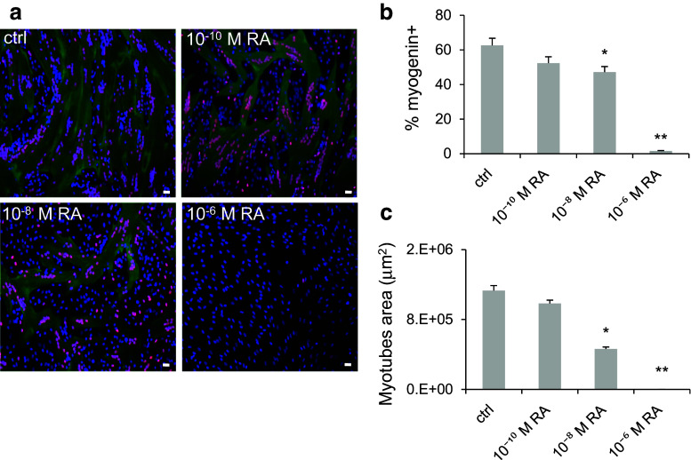
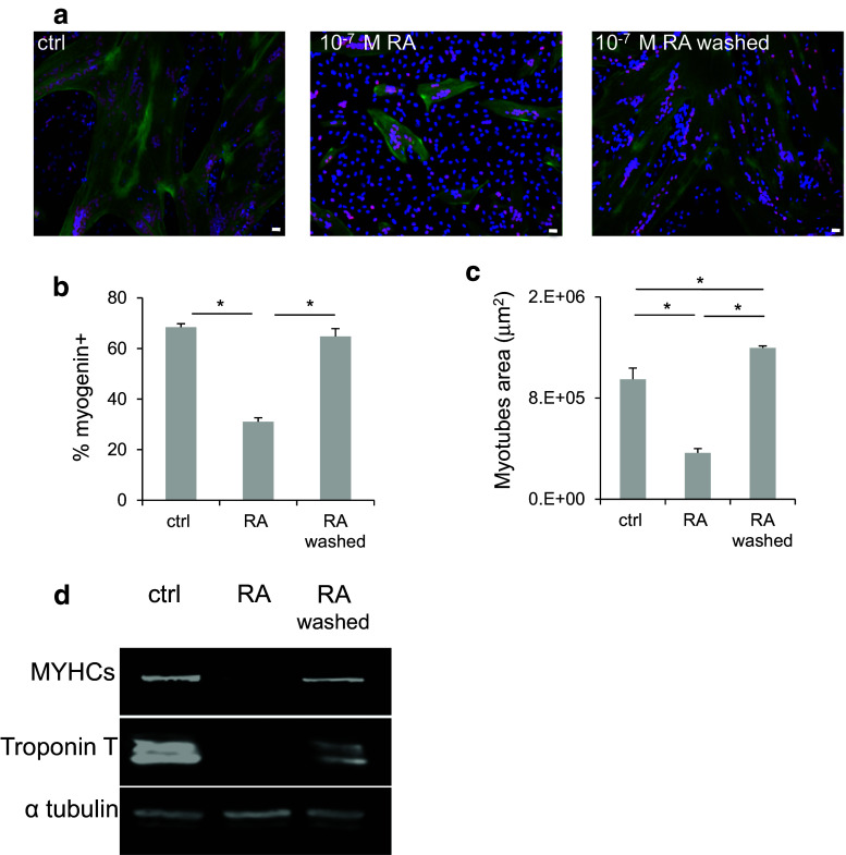
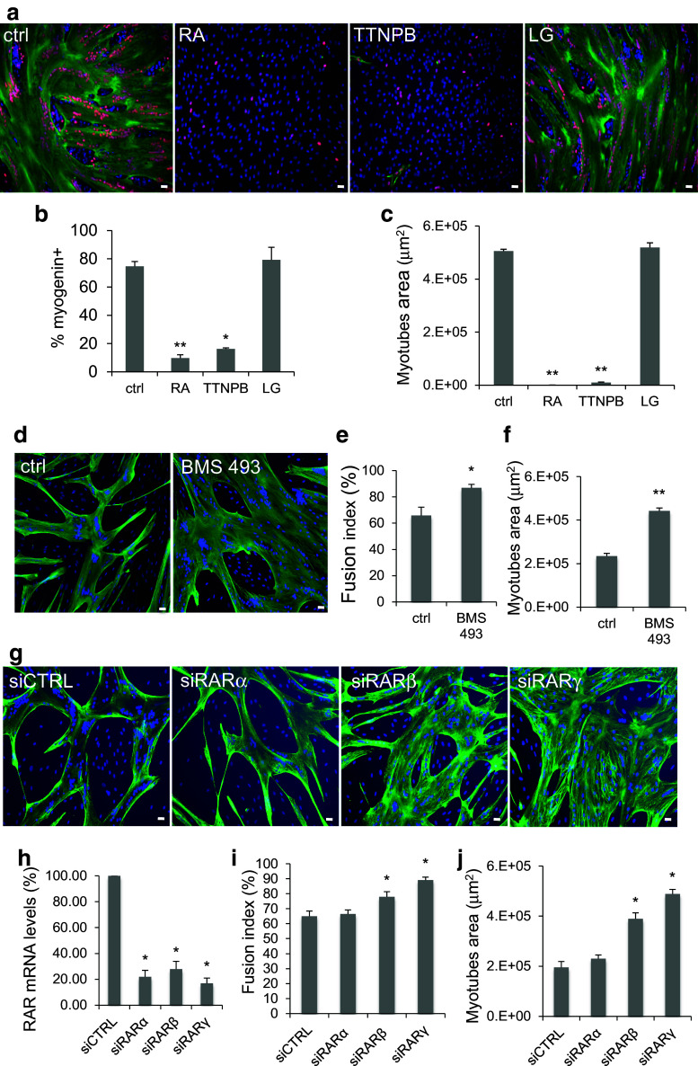
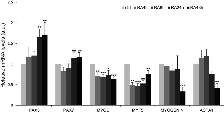
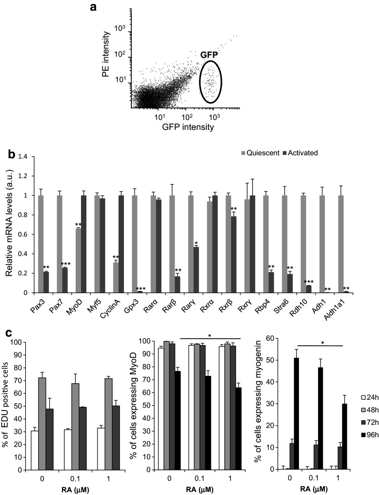
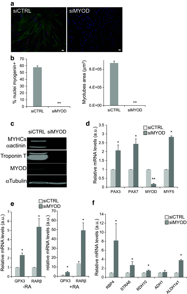
Muscle satellite cells are resistant to cytotoxic agents, and they express several genes that confer resistance to stress, thus allowing efficient dystrophic muscle regeneration after transplantation. However, once they are activated, this capacity to resist to aggressive agents is diminished resulting in massive death of transplanted cells. Although cell immaturity represents a survival advantage, the signalling pathways involved in the control of the immature state remain to be explored. Here, we show that incubation of human myoblasts with retinoic acid impairs skeletal muscle differentiation through activation of the retinoic-acid receptor family of nuclear receptor. Conversely, pharmacologic or genetic inactivation of endogenous retinoic-acid receptors improved myoblast differentiation. Retinoic acid inhibits the expression of early and late muscle differentiation markers and enhances the expression of myogenic specification genes, such as PAX7 and PAX3. These results suggest that the retinoic-acid-signalling pathway might maintain myoblasts in an undifferentiated/immature stage. To determine the relevance of these observations, we characterised the retinoic-acid-signalling pathways in freshly isolated satellite cells in mice and in siMYOD immature human myoblasts. Our analysis reveals that the immature state of muscle progenitors is correlated with high expression of several genes of the retinoic-acid-signalling pathway both in mice and in human. Taken together, our data provide evidences for an important role of the retinoic-acid-signalling pathway in the regulation of the immature state of muscle progenitors.

Keywords: Differentiation; MyoD; Myoblasts; RAR; Satellite cells.


Conflict of interest statement

The authors have declared no conflict of interest

Figures

Fig. 1
Fig. 1
Retinoic acid inhibits human myoblast differentiation. Human myoblasts were induced to differentiate in differentiation medium for 4 days in the presence of increasing concentrations of RA or not (ctrl) for the first 48 h (refreshed after 24 h). a Representative images (six fields for each condition) showing myogenin (red) and Troponin T (green) expression. DNA was stained with DAPI (blue). b Quantification of the percentage of myogenin-positive nuclei relative to all cell nuclei and c quantification of the average area of myotubes (in μm2) in the cultures, as shown in a. p ≤ 0.05 (*) and p ≤ 0.01 (**). Scale bars 10 μM
Fig. 2
Fig. 2
Retinoic acid exerts a reversible inhibition on human myoblast differentiation. Human myoblasts were treated or not (ctrl) with 10−7 M RA for 2 days. RA was then washed off (or not), and cells were allowed to differentiate for 4 days. a Representative images (six fields for each condition) showing immunofluorescence analysis of myogenin (red) and Troponin T (green) expression. DNA was stained with DAPI (blue). b Quantification of the percentage of myogenin-positive nuclei relative to all cell nuclei and c quantification of the average area of myotubes (in μm2) in the cultures shown in a. d Western blotting was performed using antibodies against the muscle differentiation markers myosin heavy chain slow (MYHCs) and Troponin T. The expression of alpha tubulin (α-tubulin) was used as loading control. p ≤ 0.05 (*). Scale bars 10 μM
Fig. 3
Fig. 3
RAR activation prevents human myoblast differentiation. Human myoblasts were induced to differentiate in differentiation medium for 4 days in the presence of 10−6 M RA, 10−6 M TTNPB (RAR agonist), 10−6 M LG100268 (LG; RXR agonist), 10−7 M BMS493 (RAR agonist inverse), or not (ctrl) for the first 48 h (refreshed after 24 h). Because differentiation is already optimal in our culture conditions, we changed the culture parameters to be able to measure the BMS493 effect on the size of myotubes by plating myoblasts at lower density to reduce the average myotube area. a Images (six fields for each condition) showing immunofluorescence analysis of myogenin (red) and Troponin T (green) expression in RA, TTNPB, and LG-treated myoblasts. DNA was stained with DAPI (blue). b Quantification of the percentage of myogenin-positive nuclei relative to all nuclei and c quantification of the average area of myotubes (in μm2) in control cultures and in cells treated with RA, TTNPB, or LG, as shown in a. d Images (six fields for each condition) showing immunofluorescence analysis of Troponin T (green) expression in ctrl and BMS 493 treated myoblasts. DNA was stained with DAPI (blue). e, f Quantification of the immunofluorescence data for the cultures shown in d: e myotube fusion index (number of nuclei in myotubes relative to the total number of nuclei) and f myotube average area (in μm2), calculated using the Image J software. gj Myoblast were transfected with siCTRL, siRARα, β, or γ and induced to differentiate for 3–4 days. RARα, RARβ, or RARγ mRNA levels (h). Troponin T immunofluorescence pictures (g; Troponin T green, DAPI blue) were quantified to determine the fusion index (i) and the myotube average area (j). p ≤ 0.05 (*); p ≤ 0.01 (**). Scale bars 10 μM
Fig. 4
Fig. 4
Retinoic acid inhibits the expression of members of the MYOD gene family, but preserves the expression of the determination genes PAX7 and PAX3. RT-qPCR analysis of the expression of myogenic determination and differentiation genes in human myoblasts incubated or not (ctrl) with 10−7M RA for 48 h. p < 0.05 (*), p < 0.01 (**), and p < 0.001 (***). Data are shown as mRNA levels relative to the expression in Ctrl/non-treated cells at each timepoint, which was set at 1
Fig. 5
Fig. 5
RA-signalling pathway is activated in quiescent mouse satellite cells. a Graph showing GFP-positive satellite cells isolated by FACS from adult Tg: Pax7-nGFP mice (n = 3). b RT-qPCR analysis of the expression of myogenic determination, differentiation and proliferation genes, some RA-target genes, RA receptors, and genes encoding proteins involved in RA biogenesis in quiescent and activated mouse satellite cells. c Percentage of EdU, MyoD, and myogenin-positive nuclei relative to all cell nuclei in activated mouse satellite cells treated (RA) or not (ctrl) with 10−7M RA or 10−6M RA for indicated days. p < 0.05 (*), p < 0.01 (**), and p < 0.001 (***)
Fig. 6
Fig. 6
MYOD silencing blocks human myoblast differentiation and activates the retinoic-acid-signalling pathway. a Representative images (six fields from each condition) showing immunofluorescence analysis of the expression of myogenin (red) and Troponin T (green) in human myoblasts transfected with siCTRL (control) or siMYOD. DNA was stained with DAPI (blue). b Quantification of the percentage of myogenin-positive nuclei relative to all nuclei (left panel) and of the average area of myotubes in the cultures shown in a. c Western blot analysis of the expression of MyoD and the muscle differentiation markers MYHC slow (MYHCs), Troponin T, and alpha-actinin (α-actinin) in human myoblasts transfected with siCTRL or siMYOD. Alpha tubulin (α-tubulin) was used as loading control. d RT-qPCR analysis of the expression of myogenic determination genes in human myoblasts transfected with siMYOD relative to myoblasts transfected with sictrl (set to 1). e RT-qPCR evaluation of the expression of the RA-target genes RARβ and GPX3 in human myoblasts transfected with siCTRL or siMYOD incubated (+RA) or not (−RA, 0.1% DMSO) with RA. f RT-qPCR analysis of the expression of genes involved in the RA-signalling pathway in myoblasts transfected with siMYOD relative to control myoblasts (siCTRL). p ≤ 0.05 (*), p < 0.01 (**). Scale bars 10 μM

References

    1. Dumont NA, Wang YX, Rudnicki MA. Intrinsic and extrinsic mechanisms regulating satellite cell function. Development. 2015;142:1572–1581. doi: 10.1242/dev.114223. - DOI - PMC - PubMed
    1. Sambasivan R, Yao R, Kissenpfennig A, Van Wittenberghe L, Paldi A, Gayraud-Morel B, Guenou H, Malissen B, Tajbakhsh S, Galy A. Pax7-expressing satellite cells are indispensable for adult skeletal muscle regeneration. Development. 2011;138:3647–3656. doi: 10.1242/dev.067587. - DOI - PubMed
    1. Lepper C, Partridge TA, Fan CM. An absolute requirement for Pax7-positive satellite cells in acute injury-induced skeletal muscle regeneration. Development. 2011;138:3639–3646. doi: 10.1242/dev.067595. - DOI - PMC - PubMed
    1. Chang NC, Rudnicki MA. Satellite cells: the architects of skeletal muscle. Curr Top Dev Biol. 2014;107:161–181. doi: 10.1016/B978-0-12-416022-4.00006-8. - DOI - PubMed
    1. Chang NC, Chevalier FP, Rudnicki MA. Satellite cells in muscular dystrophy—lost in polarity. Trends Mol Med. 2016;22:479–496. doi: 10.1016/j.molmed.2016.04.002. - DOI - PMC - PubMed

Publication types