Review
. 2017 Feb;133(2):155-175.
doi: 10.1007/s00401-016-1662-x. Epub 2016 Dec 26.

Alzheimer's disease: experimental models and reality

Affiliations
Review

Alzheimer's disease: experimental models and reality

Eleanor Drummond et al. Acta Neuropathol. 2017 Feb.

Abstract

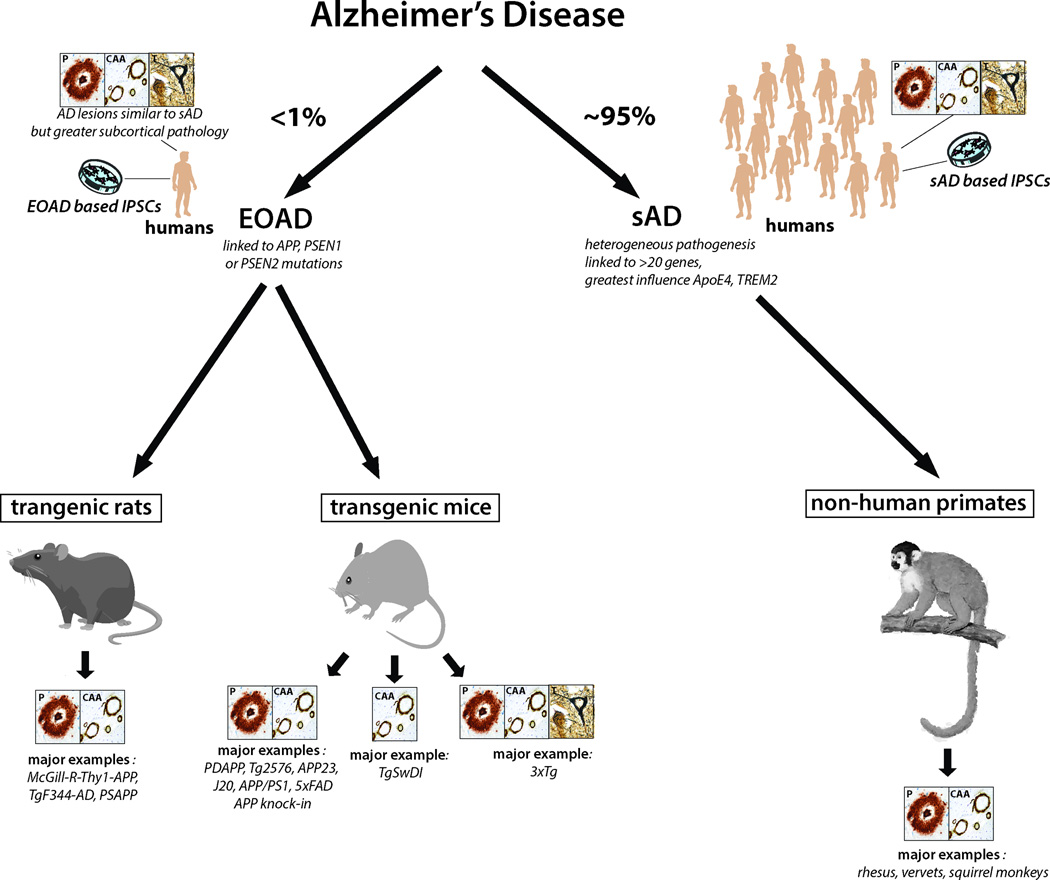
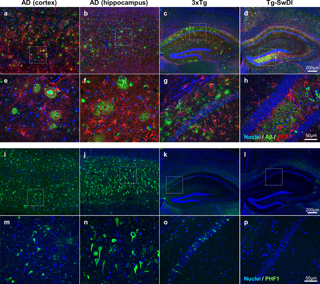
Experimental models of Alzheimer's disease (AD) are critical to gaining a better understanding of pathogenesis and to assess the potential of novel therapeutic approaches. The most commonly used experimental animal models are transgenic mice that overexpress human genes associated with familial AD (FAD) that result in the formation of amyloid plaques. However, AD is defined by the presence and interplay of both amyloid plaques and neurofibrillary tangle pathology. The track record of success in AD clinical trials thus far has been very poor. In part, this high failure rate has been related to the premature translation of highly successful results in animal models that mirror only limited aspects of AD pathology to humans. A greater understanding of the strengths and weakness of each of the various models and the use of more than one model to evaluate potential therapies would help enhance the success of therapy translation from preclinical studies to patients. In this review, we summarize the pathological features and limitations of the major experimental models of AD, including transgenic mice, transgenic rats, various physiological models of sporadic AD and in vitro human cell culture models.


Figures

Figure 1
Figure 1. Schematic of the major animal models of Alzheimer’s disease
Less than 1% of AD cases are early onset familial Alzheimer’s disease (EOAD) cases that are caused by autosomal dominant mutations in APP, PSEN1 or PSEN2. However, all major transgenic rodent models express these mutated forms of APP and PS1. The best animal models available of sAD are non-human primates. The consistent presence of the types of neuropathology present in each model is shown in the boxes; P: plaques; CAA; congophilic amyloid angiopathy; T: neurofibrillary tangles. We did not consider the presence of pre-tangle pathology in these animal models sufficient to indicate the presence of neurofibrillary tangle pathology. As such, only 3xTg mice express all 3 pathological hallmarks of AD. The specific types animal models included in each category are examples of the most common animal models currently used in AD research.
Figure 2
Figure 2. Neuropathological differences between humans with AD and transgenic mouse models of AD
Fluorescent immunohistochemistry was performed on formalin-fixed paraffin embedded brain sections using the same conditions for human and mouse tissue to highlight species differences. Immunostaining in the human AD cortex (a, e, I, m), human AD hippocampus (b, f, j, n), 3xTg mouse (28 months old) hippocampus and cortex (c, g, k, o), and Tg-SwDI mouse(16 months old) hippocampus and cortex (d, h, l, p) is shown. a–h: shows immunohistochemistry for Aβ (green; labelled using a combination of 4G8 and 6E10 antibodies) and astrocytes (red; labelled using GFAP). i–p: shows immunohistochemistry for phosphorylated tau (green; labelled using PHF1). All sections were counterstained with Hoechst to label nuclei. a–d and i–l show the differences in distribution of Aβ, astrocytes and phosphorylated tau at low magnification throughout the hippocampus and cortex (scale bar for all = 200 µm). e–h and m–p show the differences in morphology of plaques and neurofibrillary tangles at higher magnification (scale bar for all = 50 µm) of the areas outlined by a box in a–d and i–l. The most obvious species differences include the preferential presence of plaques in the cortex in humans (a) in comparison to 3xTg (c) or TgSwDI mice (d), the presence of both extensive numbers of cored and diffuse plaques in humans (e), but not in mice (g, h), and the greater density of neurofibrillary tangles in humans (i, j) in comparison to 3xTg mice (k). As expected, there were no neurofibrillary tangles present in Tg-SwDI mice (l, p).

References

    1. Alexander AG, Marfil V, Li C. Use of Caenorhabditis elegans as a model to study Alzheimer's disease and other neurodegenerative diseases. Front Genet. 2014;5:279. - PMC - PubMed
    1. Andorfer C, Kress Y, Espinoza M, de SR, Tucker KL, Barde YA, Duff K, Davies P. Hyperphosphorylation and aggregation of tau in mice expressing normal human tau isoforms. J Neurochem. 2003;86:582–590. - PubMed
    1. Bales KR, Liu F, Wu S, Lin S, Koger D, DeLong C, Hansen JC, Sullivan PM, Paul SM. Human APOE isoform-dependent effects on brain beta-amyloid levels in PDAPP transgenic mice. J Neurosci. 2009;29:6771–6779. - PMC - PubMed
    1. Bales KR, Verina T, Cummins DJ, Du Y, Dodel RC, Saura J, Fishman CE, DeLong CA, Piccardo P, Petegnief V, et al. Apolipoprotein E is essential for amyloid deposition in the APPV717F transgenic mouse model of Alzheimer's disease. PNAS. 1999;96:15233–15238. - PMC - PubMed
    1. Bales KR, Verina T, Dodel RC, Du YS, Altstiel L, Bender M, Hyslop P, Johnstone EM, Little SP, Cummins DJ, et al. Lack of apolipoprotein E dramatically reduces amyloid β-peptide deposition. Nature Gen. 1997;17:263–264. - PubMed

Publication types