. 2016 Nov;38(5):1359-1366.
doi: 10.3892/ijmm.2016.2759. Epub 2016 Sep 29.

Charge-based precipitation of extracellular vesicles

Affiliations

Charge-based precipitation of extracellular vesicles

Maria Chiara Deregibus et al. Int J Mol Med. 2016 Nov.

Abstract

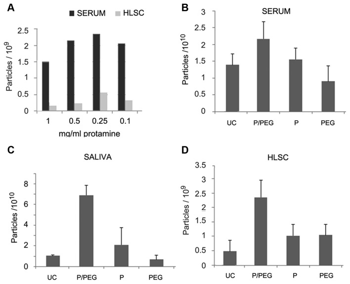
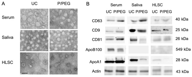
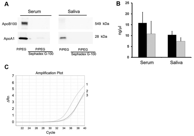
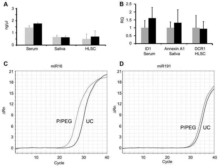
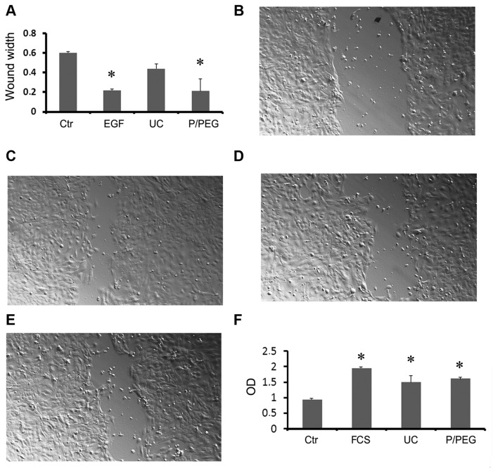
Vesicular-mediated communication between cells appears critical in many biological processes. Extracellular vesicles (EVs) released from healthy and diseased cells are involved in a network of exchange of biologically active molecules. Since EVs present in biological fluids carry the signature of the cell of origin, they are potential biomarkers for ongoing physiological or pathological processes. Despite the knowledge on EV biology accrued in recent years, techniques of EV purification remain a challenge and all the described methods have some advantages and disadvantages. In the present study, we described a method based on charge precipitation of EVs from biological fluids and from cell supernatants in comparison with the differential ultracentrifugation, which is considered the gold standard for EV purification. The analysis of ζ‑potential revealed that EVs have a negative charge that allows the interaction with a positively charged molecule, such as protamine. Protamine was shown to induce EV precipitation from serum and saliva and from cell culture media without the need for ultracentrifugation. EV resuspension was facilitated when protamine (P) precipitation was performed in the presence of PEG 35,000 Da (P/PEG precipitation). The recovery of precipitated EVs evaluated by NanoSight analysis was more efficient than that obtained by ultracentrifugation. By electron microscopy the size of EVs was similar after both methods were used, and the expression of CD63, CD9 and CD81 exosomal markers in the P/PEG‑precipitated EVs indicated an enrichment in exosomes. The RNA recovery of P/PEG‑precipitated EVs was similar to that of EVs isolated by ultracentrifugation. In addition, P/PEG‑precipitated EVs retained the biological activity in vitro as observed by the induction of wound closure by keratinocytes and of proliferation of tubular epithelial cells. In conclusion, charge-based precipitation of EVs has the merit of simplicity and avoids the requirement of expensive equipments and may be used for the efficient isolation of EVs from small biological samples.


Figures

Figure 1
Figure 1
Nanoparticle tracking analysis (NTA) of extracellular vesicles (EVs) isolated from serum, saliva and culture medium of human liver stem cells (HLSCs). (A) Number of particles precipitated from 250 µl of serum and 1.5 ml of HLSC culture medium by the addition of different doses of protamine (1, 0.5, 0.25 and 0.1 mg/ml). (B–D) Number of particles isolated from 750 µl of serum (B), 2.5 ml saliva (C) and 1.5 ml culture medium of HLSCs (2×10⁶ cells; (D) by ultracentrifugation (UC) or P/PEG, P alone and PEG alone. Data are mean ± 1SD of three independent experiments evaluated in triplicate. ANOVA with Dunnet's multicomparison test was performed all samples vs. UC; *P<0.05.
Figure 2
Figure 2
Comparison of extracellular vesicles (EVs) purified by ultracentrifugation and P/PEG precipitation. (A) Representative transmission electron microscopy of EVs isolated by ultracentrifugation (UC) or by P/PEG precipitation and negatively stained with NanoVan. EVs were viewed using a JEOL Jem 1010 electron microscope (black line, 100 nm). Three experiments were performed with similar results. (B) Representative western blot analysis of CD63, CD9, CD81 and Actin expression by EVs isolated with UC or by P/PEG precipitation from serum, saliva and human liver stem cell (HLSC) (four experiments were performed with similar results) and of apolipoprotein B100 (ApoB100) and apolipoprotein A1 (ApoA1) associated with EVs (five experiments were performed with similar results).
Figure 3
Figure 3
Apolipoprotein is associated with extracellular vesicles (EVs) purified by ultracentrifugation and P/PEG precipitation from serum, saliva and human liver stem cell (HLSC). (A) Representative western blot analysis of apolipoprotein B100 (ApoB100) and apolipoprotein A1 (ApoA1) associated with EVs isolated by P/PEG precipitation from serum and saliva after gel-filtration with Sephadex G-100 spin columns to remove lipoproteins. Two experiments were performed with similar results. (B) Total RNA extraction from EVs separated from serum and saliva before and after gel-filtration with Sephadex G-100 spin columns to remove lipoproteins. Data are mean ± 1SD of three independent experiments. (C) Representative PCR analysis for ID1 mRNA expressed by serum EVs before 1) and after gel-filtration 2,3) with Sephadex G-100 spin columns to remove lipoproteins. Two experiments were performed with similar results.
Figure 4
Figure 4
RNA quantification of extracellular vesicles (EVs) purified by ultracentrifugation and P/PEG precipitation from serum, saliva and human liver stem cells (HLSCs) and PCR analysis of mRNA and miRNA. (A) Quantification of total RNA extracted from EVs separated by ultracentrifugation (UC) (gray columns) and P/PEG precipitation (dark columns) from serum, saliva and HLSCs. Data are mean ± 1SD of three experiments. (B) RT-qPCR analysis of representative mRNA expressed by serum (ID1), saliva (Annexin A1) and HLSC (DCR1) EVs isolated with UC (grey columns) or P/PEG precipitation (black columns). 18S was used to normalize RNA input and data are expressed as the relative quantification level (RQ) (mean ± 1SD of three experiments). (C and D) Representative detection by PCR of (C) miR-16 and (D) miR-191 in EVs isolated from serum by UC or P/PEG precipitation. Similar results were obtained with other miRNAs expressed by serum EVs (miR-29a, -99b and -223; data not shown).
Figure 5
Figure 5
Biological activity of extracellular vesicles (EVs) purified by ultracentrifugation and P/PEG precipitation from saliva and human liver stem cells (HLSCs). (A–E) Evaluation of wound healing on normal dermal keratinocytes (HaCaT) by scratch test. Quantitative evaluation of wound size reduction after 36-h incubation with the vehicle alone (Ctr), 10 ng/ml EGF as a positive control, and EVs isolated by ultracentrifugation (UC) or P/PEG precipitation (50,000 EVs/cell). Data are mean ± 1SD of three independent experiments evaluated in triplicate. ANOVA with Dunnet's multicomparison test was performed in all the samples vs. UC; *P<0.05. Representative micrographs of Ctr (B), EGF (C), UC EVs (D) and P/PEG EVs (E) induced wound healing. Original magnification, ×100. (F) Proliferation of TEC evaluated by BrdU incorporation after 12-h incubation with EVs isolated by UC or P/PEG precipitation (10,000 EVs/cell). As negative control, TEC was incubated with the vehicle alone in the absence of fetal calf serum (FCS); as positive control, cells were incubated with 10% FCS. Data are mean ± 1SD of three independent experiments evaluated in triplicate. ANOVA with Dunnet's multicomparison test was performed in all the samples versus Ctr; *P<0.05.

References

    1. Ratajczak J, Wysoczynski M, Hayek F, Janowska-Wieczorek A, Ratajczak MZ. Membrane-derived microvesicles: Important and underappreciated mediators of cell-to-cell communication. Leukemia. 2006;20:1487–1495. doi: 10.1038/sj.leu.2404296. - DOI - PubMed
    1. Cocucci E, Racchetti G, Meldolesi J. Shedding microvesicles: Artefacts no more. Trends Cell Biol. 2009;19:43–51. doi: 10.1016/j.tcb.2008.11.003. - DOI - PubMed
    1. Quesenberry PJ, Aliotta J, Deregibus MC, Camussi G. Role of extracellular RNA-carrying vesicles in cell differentiation and reprogramming. Stem Cell Res Ther. 2015;6:153. doi: 10.1186/s13287-015-0150-x. - DOI - PMC - PubMed
    1. Shelke GV, Lässer C, Gho YS, Lötvall J. Importance of exosome depletion protocols to eliminate functional and RNA-containing extracellular vesicles from fetal bovine serum. J Extracell Vesicles. 2014 Sep 30; doi: 10.3402/jev.v3.24783. Epub ahead of print. - DOI - PMC - PubMed
    1. Szatanek R, Baran J, Siedlar M, Baj-Krzyworzeka M. Isolation of extracellular vesicles: Determining the correct approach (Review) Int J Mol Med. 2015;36:11–17. - PMC - PubMed