. 2016 Nov;38(5):1490-1498.
doi: 10.3892/ijmm.2016.2762. Epub 2016 Sep 30.

A novel Chk1-binding peptide that enhances genotoxic sensitivity through the cellular redistribution of nuclear Chk1

Affiliations

A novel Chk1-binding peptide that enhances genotoxic sensitivity through the cellular redistribution of nuclear Chk1

Kwang Seok Kim et al. Int J Mol Med. 2016 Nov.

Abstract

Since checkpoint kinase 1 (Chk1) is an essential factor for cell viability following DNA damage, the inhibition of Chk1 has been a major focus of pharmaceutical development to enhance the sensitivity of tumor cells to chemo- and radiotherapy that damage DNA. However, due to the off-target effects of conventional Chk1-targeting strategies and the toxicity of Chk1 inhibitors, alternative strategies are required to target Chk1. To facilitate such efforts, in this study, we identified a specific Chk1-binding 12-mer peptide from the screening of a phage display library and characterized the peptide in terms of cellular cytotoxicity, and in terms of its effect on Chk1 activity and sensitivity to genotoxic agents. This peptide, named N-terminal Chk1-binding peptide (Chk1‑NP), bound the kinase domain of Chk1. Simulation of the binding revealed that the very N-terminus of the Chk1 kinase domain is the potential peptide binding site. Of note, the polyarginine-mediated internalization of Chk1‑NP redistributed nuclear Chk1 with a prominent decrease in the nucleus in the absence of DNA damage. Treatment with Chk1‑NP peptide alone decreased the viability of p53-defective HeLa cells, but not that of p53-functional NCI-H460 cells under normal conditions. The treatment of HeLa or NCI-H460 cells with the peptide significantly enhanced radiation sensitivity following ionizing radiation (IR) with a greater enhancement observed in HeLa cells. Moreover, the IR-induced destabilization of Chk1 was aggravated by treatment with Chk1‑NP. Therefore, the decreased nuclear localization and protein levels of Chk1 seem to be responsible for the enhanced cancer cell killing following combined treatment with IR and Chk1‑NP. The approach using the specific Chk1-binding peptide may facilitate the mechanistic understanding and potential modulation of Chk1 activities and may provide a novel rationale for the development of specific Chk1-targeting agents.


Figures

Figure 1
Figure 1
Isolation of the recombinant N-terminal fragment of checkpoint kinase 1 (Chk1) (aa 2-270). Hexahistidine-tagged Chk1 fragment (amino acid 2-270) was isolated from bacterial extracts subjected to IPTG induction. The arrow indicates the isolated Chk1 N-terminal fragment.
Figure 2
Figure 2
Identification of a peptide that can specifically bind to the N-terminus of checkpoint kinase 1 (Chk1). (A) Domain structure of Chk1 (human full length, amino acid 1-476). The kinase domain (amino acid 8-265) and SQ motif are indicated. The SQ motif is a cluster of serine and glutamine residues that is phosphorylated following DNA damage. The SQ motif encompasses serine 317 and 345 residues that are phosphorylated by ATR. Dotted line indicates the N-terminal fragment of Chk1 (amino acid 2-270) which is expressed as a recombinant protein and used as a bait in the screening of a phage display library. (B) Amino acid sequence of the screened 12-mer Chk1 N-terminal binding peptide (APNKTLSVNKMV) and potential sites of the peptide interactions on the Chk1 N-terminal fragment (PDB ID: 2E9N). Candidate sites of peptide interactions on the Chk1 fragment are presented in blue, which are the very N-terminal β-sheets in the Chk1 fragment. PEP-Site Finder program was used to find potential sites of peptide interactions on Chk1. (C) Biotin-labeled peptides for experiments. R9 has biotin-lebeled 9-mer polyarginine peptide and serves as a control. N-terminal Chk1-binding peptide (Chk1-NP) is a fusion of biotin-labeled 9-mer polyarginine and 12-mer peptide that can bind the Chk1 N-terminal fragment. 9-mer polyarginine is used for internalization of the peptides. (D) Interaction of Chk1-NP with Chk1. Pull-down assay of the biotinylated peptides with streptavidin beads and subsequent western blot analysis revealed a specific interaction between Chk1-NP and Chk1.
Figure 3
Figure 3
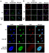
Peptide internalization and its effect on the cellular distribution of checkpoint kinase 1 (Chk1). (A) HeLa cells were treated with 5 µM R9 or N-terminal Chk1-binding peptide (Chk1-NP) for 2 h and analyzed by immunofluorescence microscopy. FITC-conjugated streptavidin was used to detect biotinylated peptides. C is a no-peptide treatment control. Nuclear staining by DAPI is also shown. (B) Effect of the peptides on Chk1 localization. HeLa cells were treated as described in (A). A prominent decrease in the nuclear pool of Chk1 is observed in Chk1-NP treated cells. (C) Decreased nuclear localization of Chk1 by Chk1-NP treatment. Nuclear localization of endogenous Chk1 is significantly decreased only in Chk1-NP treated cells. HJURP, a nuclear resident protein, is unaffected by the peptides. The image in the inset was enlarged in the panel below.
Figure 4
Figure 4
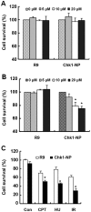
Effect of the R9 or N-terminal Chk1-binding peptide (Chk1-NP) on cell viability. (A) Viability of NCI-H460 cells was measured by MTT assay following treatment with 0, 5, 10 or 20 µM of R9 or Chk1-NP. Peptide-treated cells were incubated up to 48 h. (B) Viability of HeLa cells was measured by MTT assay following the same treatment as (A). (C) Effect of R9 or Chk1-NP on viability of HeLa cells following genotoxic treatments. Cells were treated with 5 µM of R9 or Chk1-NP for 1 h prior to treatment with genotoxic agents such as 500 nM camptothecin (CPT), 5 mM hydroxyurea (HU) or 10 Gy ionizing radiation (IR). Cell survival was measured by MTT assay after 24 h. Con stands for control. Error bars represent the means ± tandard deviation. The means of 3 independent experiments were graphed. *P<0.05 compared to R9-treated group.
Figure 5
Figure 5
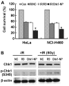
Effect of R9 or N-terminal Chk1-binding peptide (Chk1-NP) on the radiosensitivity of HeLa and NCI-H460 cells. (A) HeLa or NCI-H460 cells were treated with 5 µM of R9 or Chk1-NP peptide for 1 h prior to treatment with 5 Gy IR. MTT assay was used to measure cell viability after 24 h. Con stands for control. The means of 3 independent experiments were graphed. (B) Effect of R9 or Chk1-NP on Chk1 activation following exposure to IR. HeLa cells were treated as described in (A). Western blot analysis of Chk1 and phosphorylated (p-)Chk1 (S345) was carried out. β-actin was used as a loading control. *P<0.05 compared to only IR or IR/R9-treated group.
Figure 6
Figure 6
Proposed model for conformational regulation of checkpoint kinase 1 (Chk1) by N-terminal Chk1-binding peptide (Chk1-NP). Autoinhibitory closed conformation of Chk1 is maintained by the intramolecular interaction between the N-terminal kinase domain and the C-terminal domain (CD). Binding of Chk1-NP to the very N-terminal part of the Chk1 kinase domain disrupts the closed conformation and releases the C-terminal domain. Serine (S)345 residue is phosphorylated and the pChk1 is exported to the cytoplasm. Open conformation of Chk1 upon Chk1-NP binding may facilitate the phosphorylation of Chk1 at S345, which leads to the activation of Chk1. pChk1 may follow the following fates: transduction of Chk1 signaling to downstream targets, export to the cytoplasm and its degradation. Therefore Chk1-NP may enhance radiation sensitivity by changing conformation, localization, activity and the protein stability of Chk1.

Similar articles

References

    1. Sancar A, Lindsey-Boltz LA, Unsal-Kaçmaz K, Linn S. Molecular mechanisms of mammalian DNA repair and the DNA damage checkpoints. Annu Rev Biochem. 2004;73:39–85. doi: 10.1146/annurev.biochem.73.011303.073723. - DOI - PubMed
    1. Ciccia A, Elledge SJ. The DNA damage response: Making it safe to play with knives. Mol Cell. 2010;40:179–204. doi: 10.1016/j.molcel.2010.09.019. - DOI - PMC - PubMed
    1. Smith J, Tho LM, Xu N, Gillespie DA. The ATM-Chk2 and ATR-Chk1 pathways in DNA damage signaling and cancer. Adv Cancer Res. 2010;108:73–112. doi: 10.1016/B978-0-12-380888-2.00003-0. - DOI - PubMed
    1. Sørensen CS, Syljuåsen RG, Falck J, Schroeder T, Rönnstrand L, Khanna KK, Zhou BB, Bartek J, Lukas J. Chk1 regulates the S phase checkpoint by coupling the physiological turnover and ionizing radiation-induced accelerated proteolysis of Cdc25A. Cancer Cell. 2003;3:247–258. doi: 10.1016/S1535-6108(03)00048-5. - DOI - PubMed
    1. Falck J, Mailand N, Syljuåsen RG, Bartek J, Lukas J. The ATM-Chk2-Cdc25A checkpoint pathway guards against radio-resistant DNA synthesis. Nature. 2001;410:842–847. doi: 10.1038/35071124. - DOI - PubMed