. 2016 Dec 27;14(12):e2000631.
doi: 10.1371/journal.pbio.2000631. eCollection 2016 Dec.

Collective Resistance in Microbial Communities by Intracellular Antibiotic Deactivation

Affiliations

Collective Resistance in Microbial Communities by Intracellular Antibiotic Deactivation

Robin A Sorg et al. PLoS Biol. .

Abstract

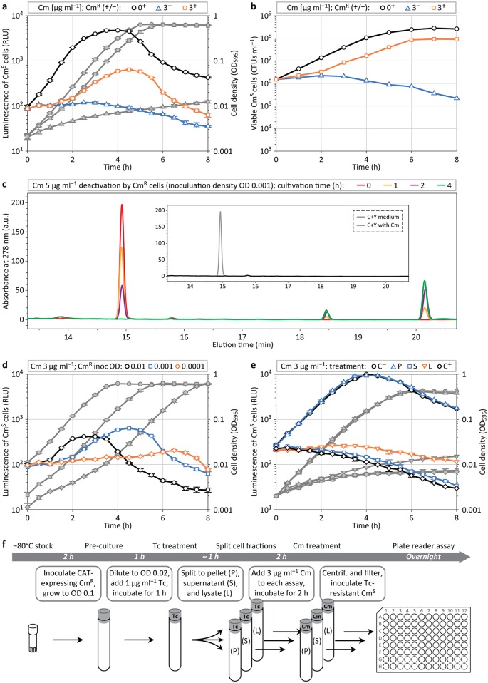
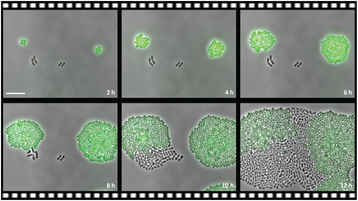
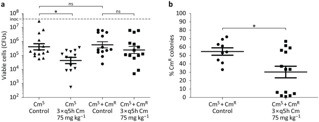
The structure and composition of bacterial communities can compromise antibiotic efficacy. For example, the secretion of β-lactamase by individual bacteria provides passive resistance for all residents within a polymicrobial environment. Here, we uncover that collective resistance can also develop via intracellular antibiotic deactivation. Real-time luminescence measurements and single-cell analysis demonstrate that the opportunistic human pathogen Streptococcus pneumoniae grows in medium supplemented with chloramphenicol (Cm) when resistant bacteria expressing Cm acetyltransferase (CAT) are present. We show that CAT processes Cm intracellularly but not extracellularly. In a mouse pneumonia model, more susceptible pneumococci survive Cm treatment when coinfected with a CAT-expressing strain. Mathematical modeling predicts that stable coexistence is only possible when antibiotic resistance comes at a fitness cost. Strikingly, CAT-expressing pneumococci in mouse lungs were outcompeted by susceptible cells even during Cm treatment. Our results highlight the importance of the microbial context during infectious disease as a potential complicating factor to antibiotic therapy.


Conflict of interest statement

The authors have declared that no competing interests exist.

Figures

Fig 1
Fig 1. Experimental setup to determine passive resistance.
Antibiotic-susceptible cells (AbS) constitutively expressing luc are grown together with antibiotic-resistant cells (AbR, which do not express luc). Only when the concentration of the antibiotic in the medium is reduced by enzymatic deactivation of resistant cells will the genetically antibiotic-susceptible cells be able to grow and produce light.
Fig 2
Fig 2. Cm deactivation during mixed population assays.
(a) Plate reader assay sets in quadruplicate (average and standard error of the mean [s.e.m.]) measuring luminescence (symbols with color outline) and cell density (corresponding grey symbols) of S. pneumoniae CmS growing in the presence of 3 μg ml−1 Cm, in presence (+) or absence (−) of CmR cells. (b) Development of the count of viable CmS cells (colony-forming units per ml [CFUs ml−1]) during the cultivation assay presented in a, determined via plating in the presence of kanamycin; average values of duplicates are shown. (c) Culture supernatant (S) samples after 0, 1, 2, and 4 h of CmR cultivation (inoculation at optical density OD 0.001) in the presence of 5 μg ml−1 Cm, analyzed for Cm content by high-performance liquid chromatography (HPLC) separation and ultraviolet (UV) detection at 278 nm. (d) Luminescence and cell density profiles of CmS cells treated with 3 μg ml−1 Cm (inoculation at OD 0.001) in dependency of the inoculum size of CmR cells. (e, f), CmS luminescence and growth analysis (e) in Cm-supplemented medium (3 μg ml−1) that was pretreated with CmR cell pellet (P), S, and culture lysate (L), and controls without (C) and with Cm (C+); (f) schematic overview of the assay (see also Methods and S1 Data).
Fig 3
Fig 3. Interspecies collective resistance.
Still images (overlay of phase contrast and fluorescence microscopy) of a time-lapse experiment of S. pneumoniae CmS, cocultivated with a strain of the pneumococcal niche competitor S. aureus (strain LAC pCM29) that expresses CAT and GFP, growing on a semi-solid surface supplemented with 3 μg ml−1 Cm. Scale bar 10 μm.
Fig 4
Fig 4. Population dynamics of bacterial communities.
(a) Simulated growth trajectories for CmR and CmS populations subject to antibiotic stress and resource competition. (b) Dynamic of intracellular Cm (yr and ys) and growth-limiting resource (z). Simulation time is scaled relative to the mean residence time of cells in a chemostat, which is equal to the generation time at steady state. At low population densities, the CmR strain can grow, whereas CmS cannot, due to a high concentration of Cm. However, the invasion of CmR lowers antibiotic stress, generating permissive conditions for the growth of CmS cells. The chemostat is then rapidly colonized by both strains (shortly after t = 180) until the resource becomes limiting. From that moment onwards, total cell density changes little, while the relative frequencies of the two strains continue to shift. Eventually, a stable equilibrium is reached, at which the cost and benefit of CAT expression (i.e., reduced growth rate efficiency for CmR cells versus their lower intracellular Cm concentration) balance out. Inset (c), The dark-red dot pinpoints the parameter set used in the simulation shown in a and b: r = 20.0, η = 0.9, kz = 4.0, c = 1.0, p = 50.0, hY = 0.25/Y0, kY = 2.5/Y0, d = 30.0/Y0 and Y0 = 0.8. These parameters were selected to lie in a restricted area of parameter space (highlighted in red) where stable coexistence between CmS and CmR cells is observed Alternative model outcomes, which were identified by a numerical bifurcation analysis (see S1 Text and S4 Fig), include establishment of CmS only (area S), establishment of CmR only (area R), no bacterial growth (area N), and competition-induced extinction (area E, where CmS bacteria first outcompete CmR bacteria and subsequently are cleared by the antibiotic; see S5 Fig).
Fig 5
Fig 5. Cross-protection in a mouse pneumonia model.
(a) Eight-wk-old female CD1 mice were infected intratracheally with CmS pneumococci or an equivalent amount of CmS + CmR pneumococci in a one-to-one ratio. One h post infection, mice were treated with one intraperitoneal injection of Cm 75 mg kg−1 followed by two additional doses spaced 5 h apart. Control mice received an injection of the vehicle alone. n = 14 for CmS control; 13 for CmS Cm-treated; 13 for CmS + CmR control; and 14 for CmS + CmR Cm-treated. Data plotted as average and s.e.m. of two independent experiments combined. Dashed line ‘inoc’ denotes the initial inoculum. *p < 0.05; one-way ANOVA with Tukey's multiple comparison post-test. (b) Bacterial colonies recovered from the CmS + CmR control and CmS + CmR Cm-treated mice were individually picked and used to inoculate THY media in 96-well plates. These 96-well plates were then used to inoculate 96-well plates with THY media containing either 15 μg ml−1 Cm or 100 μg ml−1 kanamycin to determine whether or not the original bacterial colony was CmS or CmR. n = 9 for CmS + CmR control and 14 for CmS + CmR Cm-treated. Data plotted as average and s.e.m. of two independent experiments combined. *p = 0.04; Mann–Whitney U test (see S1 Data).

References

    1. World Health Organization. Antimicrobial resistance: global report on surveillance. 2014 http://www.who.int/drugresistance/documents/surveillancereport/en.
    1. Vega NM, Gore J. Collective antibiotic resistance: mechanisms and implications. Curr Opin Microbiol. 2014. October;21C:28–34. - PMC - PubMed
    1. Levin BR, Rozen DE. Non-inherited antibiotic resistance. Nat Rev Microbiol. 2006. July;4(7):556–62. 10.1038/nrmicro1445 - DOI - PubMed
    1. Chi DH, Hendley JO, French P, Arango P, Hayden FG, Winther B. Nasopharyngeal reservoir of bacterial otitis media and sinusitis pathogens in adults during wellness and viral respiratory illness. Am J Rhinol. 2003. August;17(4):209–14. - PubMed
    1. Allen EK, Koeppel AF, Hendley JO, Turner SD, Winther B, Sale MM. Characterization of the nasopharyngeal microbiota in health and during rhinovirus challenge. Microbiome. 2014. June 25;2(1):22. - PMC - PubMed

MeSH terms

Substances