. 2017 Feb:502:97-105.
doi: 10.1016/j.virol.2016.12.018. Epub 2016 Dec 25.

Characterization of founder viruses in very early SIV rectal transmission

Affiliations

Characterization of founder viruses in very early SIV rectal transmission

Zhe Yuan et al. Virology. 2017 Feb.

Abstract

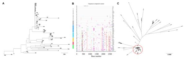
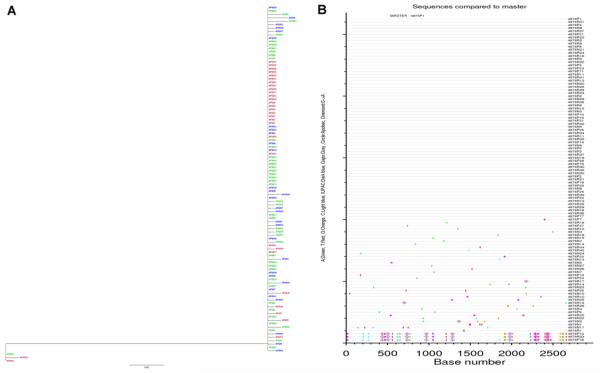
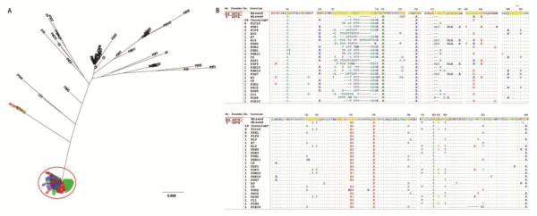
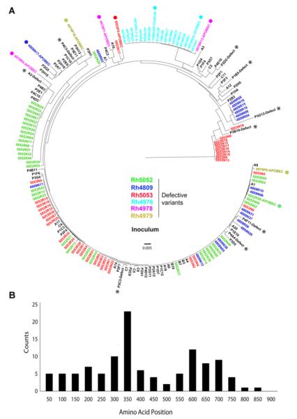
A better understanding of HIV-1 transmission is critical for developing preventative strategies. To that end, we analyzed 524 full-length env sequences of SIVmac251 at 6 and 10 days post intrarectal infection of rhesus macaques. There was no tissue compartmentalization of founder viruses across plasma, rectal and distal lymphatic tissues for most animals; however one animal has evidence of virus tissue compartmentalization. Despite identical viral inoculums, founder viruses were animal-specific, primarily derived from rare variants in the inoculum, and have a founder virus signature that can distinguish dominant founder variants from minor founder or untransmitted variants in the inoculum. Importantly, the sequences of post-transmission defective viruses were phylogenetically associated with competent viral variants in the inoculum and were mainly converted from competent viral variants by frameshift rather than APOBEC mediated mutations, suggesting the converting the transmitted viruses into defective viruses through frameshift mutation is an important component of rectal transmission bottleneck.

Significance: Anorectal receptive intercourse is a common route of HIV-1 transmission and a better understanding of the transmission mechanisms is critical for developing HIV-1 preventative strategies. Here, we report that there is no tissue compartmentalization of founder viruses during very early rectal transmission of SIV in the majority of rhesus macaques and founder viruses are preferentially derived from rare variant in the inoculum. We also found that founder viruses are animal-specific despite identical viral inoculums. After viruses cross the mucosal barriers, the host further reduces viral diversity by converting some of the transmitted functional viruses into defective viruses through frameshift rather than APOBEC derived mutations. To our knowledge, this is the first study of founder viruses at multiple tissue sites during very early rectal transmission.

Keywords: Defective variants; Founder virus signature; Genetic bottleneck; Rectal transmission; Tissue compartmentalization; Very early virological events.


Figures

Fig 1
Fig 1. The genetic make-up of SIVmac251 inoculum
Maximum likelihood tree (A) and highlighter plots (B) of full-length env nucleotide sequences after SGA and Sanger sequencing. Nucleotide polymorphisms in the highlighter plots are indicated by a colored mark. Thymine is represented in red, guanine in orange, adenine in green, cytosine in blue, pink filled circles denote APOBEC signatures, open diamonds represent G-to-A conversions, and gaps are shown by gray in the highlighter plots. (C) Maximum likelihood tree of full-length Env AA sequences. The red circle indicates the dominant virus variant group composed of 26 AA sequences with 99.5% identity; other variants were dispersed as rare variants. Bar length indicates 0.008 AA substitutions per site.
Fig 2
Fig 2. Maximum likelihood tree (A) and Highlighter plots (B) of SGA-derived env nucleotide sequences from 10 dpi macaque Rh4976
The sequences from rectum (green), axillary lymph node tissue (blue), plasma (red), or inoculum (black) are depicted by different colors. Nucleotide polymorphisms in the Highlighter plots are depicted by a colored tic mark (thymine in red, guanine in orange, adenine in green, cytosine in blue). Pink filled circles denote APOBEC signatures, open diamonds represent G-to-A conversions, and gaps are shown in gray).
Fig 3
Fig 3. The relationship between founder and inoculum variants as well as the founder virus signature (FVS) in 10 dpi macaque Rh4976
(A) Maximum likelihood tree of Env AA sequences of founder virus variants. Sequences derived from plasma are represented in red, rectum in green, lymph node in blue. Functional variants in inoculum are represented in black. The red circle indicates the dominant variant group of founder viruses with greater than 99.5% AA identity. Bar length indicates 0.009 AA substitutions per site. (B) The animal-specific founder virus signature (FVS) of the dominant founder variant group (top, highlighted in yellow). The variant nomenclature rule: the number represents the variant number of the cluster/group from which the consensus sequence was derived, followed by a space and the variant ID. The montage of MUSCLE alignment of Env AA sequences of minor founder variants and undetected functional variants in the inoculum are compared with the consensus sequence of the dominant founder variant group (red box).
Fig 4
Fig 4. Summary of the founder virus signature (FVS) from all animals
(A) The feature of FVS and their corresponding animals are listed, of which some are shared among animals in amino acid spatial position but are unique in sequence. A star indicates a particular sequence chosen from a group of sequence with similar position. A blue animal ID indicates 6 dpi and red indicates 10 dpi. The highlighted FVS are also depicted using the same colors on a 3D structure in panel B. (B) Localization of FVS on available 3D structures of SIV Env. The clipped V1V2 region is highlighted in green (truncated for easier crystallization), the V4 region is highlighted in red, and the FVS region on GP41 is highlighted in yellow.
Fig 5
Fig 5. Defective post-transmission variants were primarily caused by frameshift
(A) The phylogenetic relationship of defective post-transmission variants with all the variants in the inoculum. Defective post-transmission variants are color coded by animal ID and are phylogenetically associated with inoculum functional rather than defective variants. The majority of post-transmission defective variants are caused by frameshift. The APOBEC mediated mutations are labeled with solid circles. All variants in the inoculum are shown in black with defective variants labeled with stars. (B) The distribution of the first introduced stop codon in post-transmission defective variants. The peak of the first stop codon location is near the V3 region and very few are located within the cytoplasmic tail of GP41.

References

    1. Edwards C, Holmes E, Wilson D, Viscidi R, Abrams E, et al. Population genetic estimation of the loss of genetic diversity during horizontal transmission of HIV-1. BMC Evolutionary Biology. 2006;6:28. - PMC - PubMed
    1. Abrahams M-R, Anderson JA, Giorgi EE, Seoighe C, Mlisana K, et al. Quantitating the Multiplicity of Infection with Human Immunodeficiency Virus Type 1 Subtype C Reveals a Non-Poisson Distribution of Transmitted Variants. Journal of Virology. 2009;83:3556–3567. - PMC - PubMed
    1. Boeras DI, Hraber PT, Hurlston M, Evans-Strickfaden T, Bhattacharya T, et al. Role of donor genital tract HIV-1 diversity in the transmission bottleneck. Proceedings of the National Academy of Sciences. 2011;108:E1156–E1163. - PMC - PubMed
    1. Keele BF, Estes JD. Barriers to mucosal transmission of immunodeficiency viruses. Blood. 2011;118:839–846. - PMC - PubMed
    1. Stone M, Keele BF, Ma Z-M, Bailes E, Dutra J, et al. A Limited Number of Simian Immunodeficiency Virus (SIV) env Variants Are Transmitted to Rhesus Macaques Vaginally Inoculated with SIVmac251. Journal of Virology. 2010;84:7083–7095. - PMC - PubMed

Publication types

MeSH terms