. 2017 May;65(5):1543-1556.
doi: 10.1002/hep.29025. Epub 2017 Mar 30.

Targeting the mitochondrial pyruvate carrier attenuates fibrosis in a mouse model of nonalcoholic steatohepatitis

Affiliations

Targeting the mitochondrial pyruvate carrier attenuates fibrosis in a mouse model of nonalcoholic steatohepatitis

Kyle S McCommis et al. Hepatology. 2017 May.

Abstract

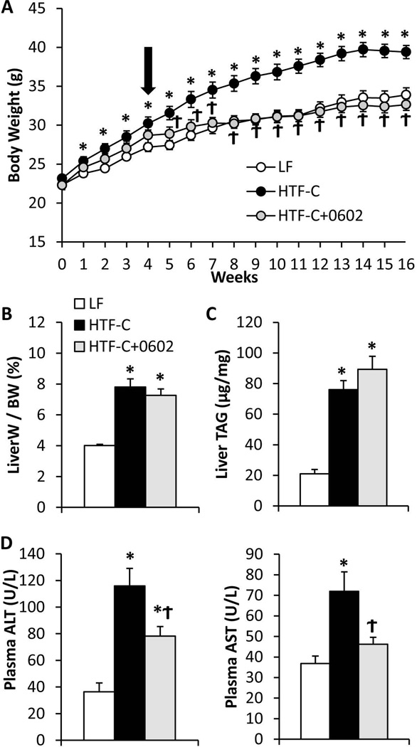
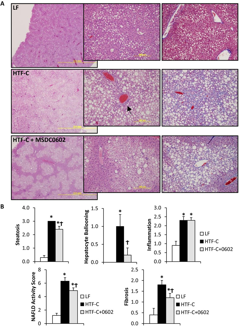
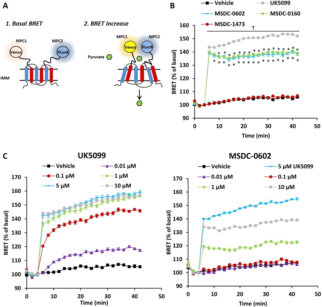
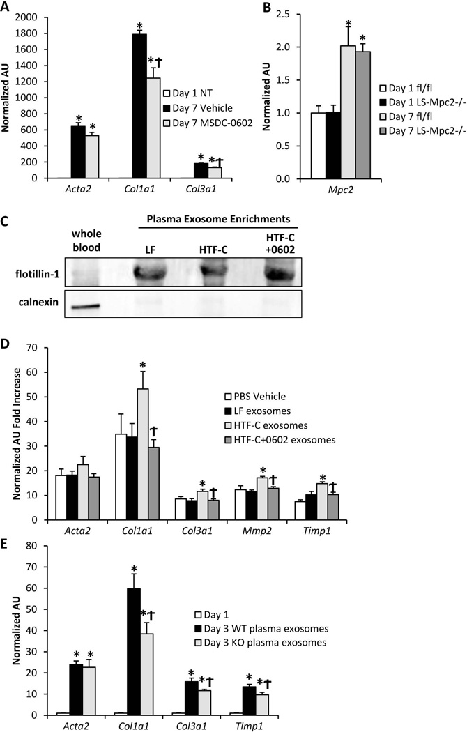
Diseases of the liver related to metabolic syndrome have emerged as the most common and undertreated hepatic ailments. The cause of nonalcoholic fatty liver disease is the aberrant accumulation of lipid in hepatocytes, though the mechanisms whereby this leads to hepatocyte dysfunction, death, and hepatic fibrosis are still unclear. Insulin-sensitizing thiazolidinediones have shown efficacy in treating nonalcoholic steatohepatitis (NASH), but their widespread use is constrained by dose-limiting side effects thought to be due to activation of the peroxisome proliferator-activated receptor γ. We sought to determine whether a next-generation thiazolidinedione with markedly diminished ability to activate peroxisome proliferator-activated receptor γ (MSDC-0602) would retain its efficacy for treating NASH in a rodent model. We also determined whether some or all of these beneficial effects would be mediated through an inhibitory interaction with the mitochondrial pyruvate carrier 2 (MPC2), which was recently identified as a mitochondrial binding site for thiazolidinediones, including MSDC-0602. We found that MSDC-0602 prevented and reversed liver fibrosis and suppressed expression of markers of stellate cell activation in livers of mice fed a diet rich in trans-fatty acids, fructose, and cholesterol. Moreover, mice with liver-specific deletion of MPC2 were protected from development of NASH on this diet. Finally, MSDC-0602 directly reduced hepatic stellate cell activation in vitro, and MSDC-0602 treatment or hepatocyte MPC2 deletion also limited stellate cell activation indirectly by affecting secretion of exosomes from hepatocytes.

Conclusion: Collectively, these data demonstrate the effectiveness of MSDC-0602 for attenuating NASH in a rodent model and suggest that targeting hepatic MPC2 may be an effective strategy for pharmacologic development. (Hepatology 2017;65:1543-1556).


Figures

Figure 1
Figure 1. MSDC-0602 prevents weight gain and increased circulating transaminases on HTF-C diet
[A] Body weight gain by C57BL6/J mice on diets that were low fat (LF) or high in trans fatty acids, fructose, and cholesterol (HTF-C). After 4 weeks on HTF-C diet, some mice were switched to HTF-C diet containing MSDC-0602. [B] Liver weight to body weight ratio for mice fed LF, HTF-C, or HTF-C containing MSDC-0602 chow. [C] Liver triglyceride content of mice fed LF, HTF-C, or HTF-C containing MSDC-0602 chow. [D] Plasma ALT and AST concentrations of mice fed LF, HTF-C, or HTF-C containing MSDC-0602 chow. Values are presented as the mean ± SEM. N=10, * indicates p<0.05 vs LF-fed group. ϯ indicates p<0.05 vs HTF-C fed group.
Figure 2
Figure 2. Histologic markers of NASH in HTF-C-fed mice are improved by MSDC-0602 treatment
[A] Representative H&E-stained liver sections from mice from each experimental group are shown. Magnification increases from left to right. [B] Liver NAFLD Activity Score (NAS) scoring (the sum of steatosis, ballooning, and inflammation scores) as well as fibrosis scoring was performed by a trained pathologist blinded to treatment group. Values are presented as the mean ± SEM. N=10, * indicates p<0.05 vs LF-fed group. ϯ indicates p<0.05 vs HTF-C fed group.
Figure 3
Figure 3. Reduced oxidative damage and expression of markers of stellate cell activation after treatment with MSDC-0602
[A] The graphs depict the liver content of toxic lipids and reactive nitrogen products in mice fed LF, HTF-C, or HTF-C containing MSDC-0602 chow. [B] The hepatic expression of indicated genes encoding proinflammatory cytokines, macrophage markers, and markers of stellate cell activation in mice fed LF, HTF-C, or HTF-C containing MSDC-0602 chow is shown. Values are presented as the mean ± SEM. N=10, * indicates p<0.05 vs LF-fed group. ϯ indicates p<0.05 vs HTF-C fed group.
Figure 4
Figure 4. MSDC-0602 can reverse liver injury after 16 weeks of HTF-C diet feeding
[A] Body weight gain by C57BL6/J mice on diets that were low fat (LF) or high in trans fatty acids, fructose, and cholesterol (HTF-C). After 4 weeks on HTF-C diet, some mice were switched to HTF-C diet containing MSDC-0602. Another sub-group was switched to HTF-C diet containing MSDC-0602 after 16 weeks. [B] Liver triglyceride content of mice fed LF, HTF-C, or HTF-C containing MSDC-0602 chow. [C] Plasma ALT and AST concentrations of mice fed LF, HTF-C, or HTF-C containing MSDC-0602 chow. [D] Liver NAS scoring and [E] fibrosis scoring was performed by a trained pathologist blinded to treatment group. [F] Hepatic expression of indicated fibrotic, stellate cell activation, and extracellular matrix regulating genes. Values are presented as the mean ± SEM. N=7–9, except for n=6 per group in panel F. * indicates p<0.05 vs LF-fed group. ϯ indicates p<0.05 vs HTF-C fed group. 1 indicates p<0.05 comparing the MSDC-0602 @4 and @16 groups.
Figure 5
Figure 5. MSDC-0602 stimulates the activity of a BRET-based biosensor of MPC activity
[A] Schematic of RESPYR BRET assay. [B] BRET kinetics of HEK-293 cells expressing RLuc8 fused to MPC2 and Venus yellow fluorescent protein fused to MPC1. Cells were stimulated after 5 minutes with vehicle (DMSO), 5 µM UK-5099, 10 µM MSDC-0602, 10 µM MSDC-0160, or 10 µM MSDC-1473. [C] Graphs represent BRET activity in dose response studies conducted with UK-5099 and MSDC-0602. Data were analyzed by repeated measures ANOVA in Prism. Post Hoc analysis was performed using Tukey’s multiple comparison tests. Values are presented as the mean ± SEM. N=10, 2 independent experiments with 4–5 technical replicates per experiment.
Figure 6
Figure 6. MPC2 deletion in hepatocytes protects mice from liver injury, fibrosis, and stellate cell activation
[A] Body weight gain by WT and LS-Mpc2−/− mice on HTF-C diet. After 4 weeks on HTF-C diet, some mice were switched to HTF-C diet containing MSDC-0602. [B] Plasma ALT and AST concentrations of WT and LS-Mpc2−/− mice on HTF-C diet or HTF-C diet containing MSDC-0602. [C] Liver fibrosis scoring was performed by a trained pathologist blinded to treatment group. [D] The hepatic expression of indicated genes encoding markers of stellate cell activation in WT and LS-Mpc2−/− mice on HTF-C diet or HTF-C diet containing MSDC-0602 is shown. [E] Liver triglyceride content of WT and LS-Mpc2−/− mice fed HTF-C or HTF-C containing MSDC-0602 chow. Values are presented as the mean ± SEM. N=7–10, * indicates p<0.05 vs fl/fl HTF-C-fed group.
Figure 7
Figure 7. MSDC-0602 or genetic deletion of MPC2 in hepatocytes controls stellate cell activation status via exosome release
[A] Expression of markers of stellate cell activation in day 1 quiescent (NT= not treated) and day 7 activated stellate cells treated with vehicle or 15 µM MSDC-0602. Values are normalized (=1.0) to day 1 quiescent expression levels. [B] Hepatic stellate cells were isolated from WT or LS-Mpc2−/− mice and activated in vitro. The expression of Mpc2 was quantified in day 1 quiescent and day 7 activated cells. [C] Western blot of either whole blood or plasma exosome-enriched protein lysates probed for the membrane-bound vesicle marker flotillin-1 and the cell marker calnexin [D] Hepatic stellate cells were isolated from WT mice and treated with exosomes isolated from plasma of mice fed LF diet, HTF-C diet, or HTF-C diet containing MSDC-0602 as well as vehicle control (PBS). The expression of markers of stellate cell activation was quantified after 24 h exposure to exosomes between day 2 and 3 in culture. Values are normalized (=1.0) to day 1 quiescent expression levels. [E] Hepatic stellate cells were isolated from WT mice and treated with exosomes isolated from plasma of WT or LS-Mpc2−/− mice for 24 h between days 2 and 3 in culture. Values are normalized (=1.0) to day 1 quiescent expression levels. Values are presented as the mean ± SEM. N=6, with 2 independent stellate cell isolations and 3 technical replicates per experiment.

Comment in

References

    1. Younossi ZM, Koenig AB, Abdelatif D, Fazel Y, Henry L, Wymer M. Global epidemiology of nonalcoholic fatty liver disease-Meta-analytic assessment of prevalence, incidence, and outcomes. Hepatology. 2016;64:73–84. - PubMed
    1. Chalasani N, Younossi Z, Lavine JE, Diehl AM, Brunt EM, Cusi K, Charlton M, et al. The diagnosis and management of non-alcoholic fatty liver disease: practice Guideline by the American Association for the Study of Liver Diseases, American College of Gastroenterology, and the American Gastroenterological Association. Hepatology. 2012;55:2005–2023. - PubMed
    1. Baffy G, Brunt EM, Caldwell SH. Hepatocellular carcinoma in non-alcoholic fatty liver disease: an emerging menace. J Hepatol. 2012;56:1384–1391. - PubMed
    1. Charlton MR, Burns JM, Pedersen RA, Watt KD, Heimbach JK, Dierkhising RA. Frequency and outcomes of liver transplantation for nonalcoholic steatohepatitis in the United States. Gastroenterology. 2011;141:1249–1253. - PubMed
    1. Wong RJ, Aguilar M, Cheung R, Perumpail RB, Harrison SA, Younossi ZM, Ahmed A. Nonalcoholic steatohepatitis is the second leading etiology of liver disease among adults awaiting liver transplantation in the United States. Gastroenterology. 2015;148:547–555. - PubMed

Publication types

MeSH terms