. 2017 Feb 23;129(8):970-980.
doi: 10.1182/blood-2016-08-732800. Epub 2016 Dec 27.

Incomplete inhibition by eculizumab: mechanistic evidence for residual C5 activity during strong complement activation

Affiliations

Incomplete inhibition by eculizumab: mechanistic evidence for residual C5 activity during strong complement activation

Markus J Harder et al. Blood. .

Abstract

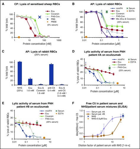
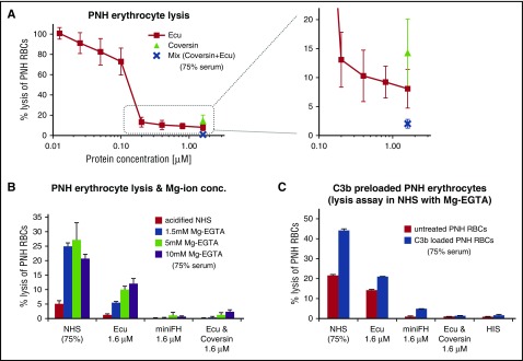
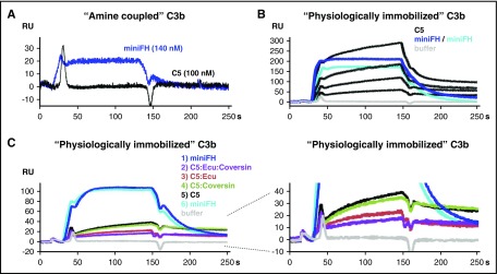
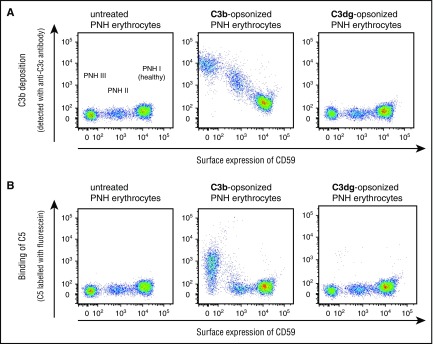
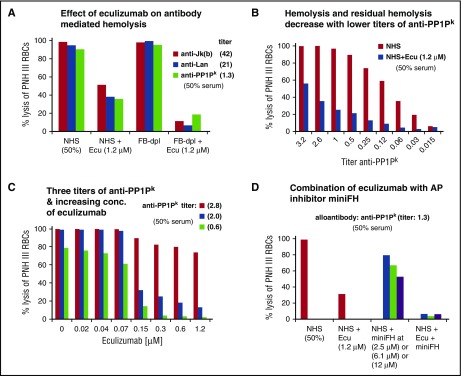
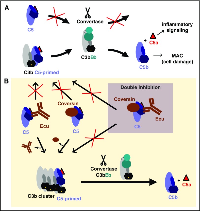
Eculizumab inhibits the terminal, lytic pathway of complement by blocking the activation of the complement protein C5 and shows remarkable clinical benefits in certain complement-mediated diseases. However, several reports suggest that activation of C5 is not always completely suppressed in patients even under excess of eculizumab over C5, indicating that residual C5 activity may derogate the drug's therapeutic benefit under certain conditions. By using eculizumab and the tick-derived C5 inhibitor coversin, we determined conditions ex vivo in which C5 inhibition is incomplete. The degree of such residual lytic activity depended on the strength of the complement activator and the resulting surface density of the complement activation product C3b, which autoamplifies via the alternative pathway (AP) amplification loop. We show that at high C3b densities required for binding and activation of C5, both inhibitors reduce but do not abolish this interaction. The decrease of C5 binding to C3b clusters in the presence of C5 inhibitors correlated with the levels of residual hemolysis. However, by employing different C5 inhibitors simultaneously, residual hemolytic activity could be abolished. The importance of AP-produced C3b clusters for C5 activation in the presence of eculizumab was corroborated by the finding that residual hemolysis after forceful activation of the classical pathway could be reduced by blocking the AP. By providing insights into C5 activation and inhibition, our study delivers the rationale for the clinically observed phenomenon of residual terminal pathway activity under eculizumab treatment with important implications for anti-C5 therapy in general.


Figures

Figure 1.
Figure 1.
Different C5 inhibitors reveal residual TP activity on foreign activating cells. (A) CP-mediated lysis of shRBCs. Hemolysin-treated shRBCs were mixed with 5% serum in the presence of specific inhibitors. Released hemoglobin was measured as a marker of hemolysis (the average of 3 independent assays with standard deviation [SD] is shown). (B) AP-mediated lysis of rabbit erythrocytes. Rabbit erythrocytes were incubated in 25% human serum in presence of inhibitors or controls (average of 3 independent assays with SD is shown). (C) Different combinations of C5 inhibitors protect rRBCs. As in panel B, but eculizumab and another commercial anti-C5 antibody (clone: 046-10.2.2) were used at concentrations that substantially exceed the typical C5 concentration of the serum dilution in this assay (average of 3 independent assays with SD is shown). (D-E) TP activity of serum from PNH patients on eculizumab. rRBCs were incubated in 25% human serum derived from 2 PNH patients (#A and #B) on eculizumab in absence or presence of additional complement inhibitors as indicated (average of 2 independent assays with SD is shown). (F) Determination of free C5 levels through sandwich enzyme-linked immunosorbent assay using eculizumab as capture antibody and a polyclonal anti-C5 as a detection antibody. Eculizumab deposited on the microtiter plate captures free C5 from NHS, but not when NHS has been premixed with 0.5 µM eculizumab. No free C5 was captured from patient serum or patient serum that had been diluted 1:1 (1-in-2) with NHS (average of 3 independent assays with SD is shown; the NHS/eculizumab mixture [negative control] was only assayed twice). Ecu, eculizumab; PAS-Cov., PASylated Coversin; PBS, phosphate-buffered saline; PBSE, PBS supplemented with EDTA (to 5 mM).
Figure 2.
Figure 2.
Forceful AP activation reveals TP activity in C5-inhibited serum on PNH cells. (A) Protection of PNH cells from complement alternative pathway mediated lysis. PNH-RBCs were incubated for 24h in acidified human serum mixed with complement inhibitors (75% final serum concentration; average of 3 independent assays with SD). (B) As in panel A, but with addition of Mg-EGTA (whereby EGTA chelates Ca ions to block CP activity) to the serum, with an incubation time at 37°C for 2 hours. Hemolysis was measured by determining the release of hemoglobin at 405 nm (1 typical assay out of 3 independent assays with average values of triplicate data points with SD is shown; for the other 2 assays, see supplemental Figure 5 A-B). (C) Residual hemolysis of PNH-RBCs preloaded with C3b in C5-inhibited serum. PNH-RBCs were preloaded with C3b in human factor I–depleted serum under combined C5 inhibition (with eculizumab and coversin) to allow for efficient C3b opsonization without lysis. C3b preloaded cells were washed and then exposed to 75% human serum in absence or presence of the inhibitors. The C3b-preloaded PNH cells were incubated for 2 hours in acidified human serum supplemented with 5 mM Mg-EGTA (1 typical assay out of 3 independent assays with average values of duplicate data points with SD is shown; for the 2 other assays, see supplemental Figure 5C-D). Ecu, eculizumab; HIS, heat-inactivated serum.
Figure 3.
Figure 3.
SPR analysis of C5 binding to C3b. (A) Binding of C5 and mini-FH to C3b immobilized on a carboxymethyldextran-coated sensor chip by amine coupling (800 RUs). Mini-FH at a concentration of 140 nM or C5 at 100 nM was applied to the C3b surface (reference-subtracted responses are shown throughout). (B) C5 binding to “physiologically,” convertase-immobilized C3b. C3 convertases were assembled onto the amine coupled C3b sensor chip followed by flow of C3, which becomes activated in situ and binds covalently. According to this procedure, another 900 RUs of C3b were immobilized in a physiological manner via the convertase onto the 800 RUs of amine-coupled C3b. Mini-FH was assayed at 1.5 µM in 2 consecutive injections (2 overlaid dark blue curves), followed by C5 at 1.1, 0.6, 0.3, and 0.1 µM (black curves) and then another injection of mini-FH at 1.5 µM (cyan). The lower amount of RUs achieved for the third injection of mini-FH indicates that the C3b surface was not completely regenerated after being exposed to higher C5 concentrations. (C) As in panel B, but analytes were injected in the following sequence: mini-FH at 140 nM, C5 at 110 nM mixed with both eculizumab at 1.7 µM and coversin at 1.7 µM (C5/eculizumab /coversin), C5/eculizumab (110 nM:1.7 µM), C5/coversin (110 nM:1.7 µM), and C5 alone at 110 nM. The C5 inhibitors themselves did not bind to the sensor chip (not depicted). Ecu, eculizumab.
Figure 4.
Figure 4.
FACS analysis of C5 binding to PNH-RBCs loaded with C3b or C3dg. PNH-PBCs were loaded with C3b in human factor I–depleted serum under combined C5 inhibition (with eculizumab and coversin) to allow for efficient C3b opsonization without lysis. A portion of the C3b loaded cells were exposed to factor I to allow for processing of the surface-bound C3b to C3dg. (A) Correlation of C3b deposition levels with PNH subtype. Detection of expression levels of CD59 (x-axis) allows classification of PNH-RBCs in type I (normal CD59 levels), type II (reduced level), and type III erythrocytes (absence of CD59). C3b deposition (y-axis) is more pronounced for type III PNH-RBCs than for type II or type I. (B) Correlation of C5 binding with C3b surface levels. C5 binding on PNH-RBCs coincides with high C3b density (1 representative assay from 2 independent experiments is shown).
Figure 5.
Figure 5.
Residual hemolysis of antibody-sensitized PNH erythrocytes in presence of eculizumab. (A) Residual TP activity under eculizumab after CP activation detected through hemolysis of PNH-RBCs. Three different alloantibodies were each incubated with erythrocytes from PNH patients (all of ABO blood group 0, positive for the antigens Jkb, Lan, and PP1Pk), washed and exposed to standardized NHS, standardized FB-dpl serum, or serum mixed with eculizumab. The final serum content was 50%, and the final eculizumab concentration was 1.2 µM. Erythrocytes from PNH patient D were used. (Results from 1 out of 3 independent assays are shown; for the 2 other assays with erythrocytes from PNH patients E and F, see supplemental Figure 6A-B). (B) Levels of hemolysis and residual hemolysis under eculizumab correlate with the concentration of anti-PP1Pk antibody used for sensitization. PNH-RBCs were incubated with anti-PP1Pk alloantibody at different titers, washed, and then exposed to NHS or NHS supplemented with eculizumab. The final serum content was 50%. Erythrocytes from PNH patient F were used. (Results from 1 out of 3 independent assays are shown; for the 2 other assays with erythrocytes from PNH patients G and F with alloantibody anti-Jka and anti-Lan, see supplemental Figure 7). (C) Effect of increasing eculizumab concentrations on hemolysis of PNH-RBCs sensitized with different titers of alloantibody. Reactions were performed as in panel B. Anti-PP1Pk alloantibody and erythrocytes from PNH patient F were used. (Results from 1 out of 3 independent assays are shown; for the 2 other assays with erythrocytes from PNH patients C and F with alloantibody anti-Jra, see supplemental Figure 8). (D) Effect of combined complement inhibition by eculizumab and the alternative pathway inhibitor mini-FH (which specifically accelerates the natural decay of alternative pathway convertases but is a cofactor for factor I–mediated cleavage of all, classical/lectin and alternative pathway produced C3b molecules). Assay was performed as in panel B, with PNH-RBCs of patient F and anti-PP1Pk alloantibody. (Results from 1 out of 3 independent assays are shown; for other assays with erythrocytes from PNH patients E and F and anti-Jkb alloantibody, see supplemental Figure 9). Ecu, eculizumab.
Figure 6.
Figure 6.
Hypothetical model of C5 activation and inhibition. (A) Adaptation of a model of C5 activation proposed by Jore et al. The scissile bond in C5 (located near the anaphylatoxin domain, C5a, which is depicted as a red triangle) is not readily accessible in free C5 for proteolytic activation by the convertase C3bBb. When C5 binds to C3b molecules, it undergoes a conformational change (a “priming event”) that renders the scissile bond accessible for proteolytic activation by a nearby convertase. (B) C5 inhibitors stabilize the unproductive C5 conformation, thus hindering the conformational priming event and, thus, the proteolytic activation of C5. However, surfaces bearing densely packed C3b molecules can compete (to some extent) with C5 inhibitors for C5 priming (ie, binding and conformational reorientation), resulting in a residual C5 activation level. Simultaneous inhibition with 2 orthogonal C5 inhibitors more effectively stabilizes the unproductive C5 conformation and prevents activation by the convertase even in presence of high C3b densities. Ecu, eculizumab; MAC, membrane attack complex.

Comment in

  • Eculizumab: another breakthrough.
    Brodsky RA. Brodsky RA. Blood. 2017 Feb 23;129(8):922-923. doi: 10.1182/blood-2017-01-760496. Blood. 2017. PMID: 28232621 No abstract available.

References

    1. Hillmen P, Young NS, Schubert J, et al. . The complement inhibitor eculizumab in paroxysmal nocturnal hemoglobinuria. N Engl J Med. 2006;355(12):1233-1243. - PubMed
    1. Brodsky RA, Young NS, Antonioli E, et al. . Multicenter phase 3 study of the complement inhibitor eculizumab for the treatment of patients with paroxysmal nocturnal hemoglobinuria. Blood. 2008;111(4):1840-1847. - PubMed
    1. Legendre CM, Licht C, Muus P, et al. . Terminal complement inhibitor eculizumab in atypical hemolytic-uremic syndrome. N Engl J Med. 2013;368(23):2169-2181. - PubMed
    1. Brodsky RA. Paroxysmal nocturnal hemoglobinuria. Blood. 2014;124(18):2804-2811. - PMC - PubMed
    1. Noris M, Remuzzi G. Glomerular diseases dependent on complement activation, including atypical hemolytic uremic syndrome, membranoproliferative glomerulonephritis, and C3 glomerulopathy: core curriculum 2015. Am J Kidney Dis. 2015;66(2):359-375. - PMC - PubMed

Publication types

MeSH terms