Review
. 2017 Feb 9;129(6):680-692.
doi: 10.1182/blood-2016-10-695957. Epub 2016 Dec 27.

Diagnosis, risk stratification, and response evaluation in classical myeloproliferative neoplasms

Affiliations
Review

Diagnosis, risk stratification, and response evaluation in classical myeloproliferative neoplasms

Elisa Rumi et al. Blood. .

Abstract

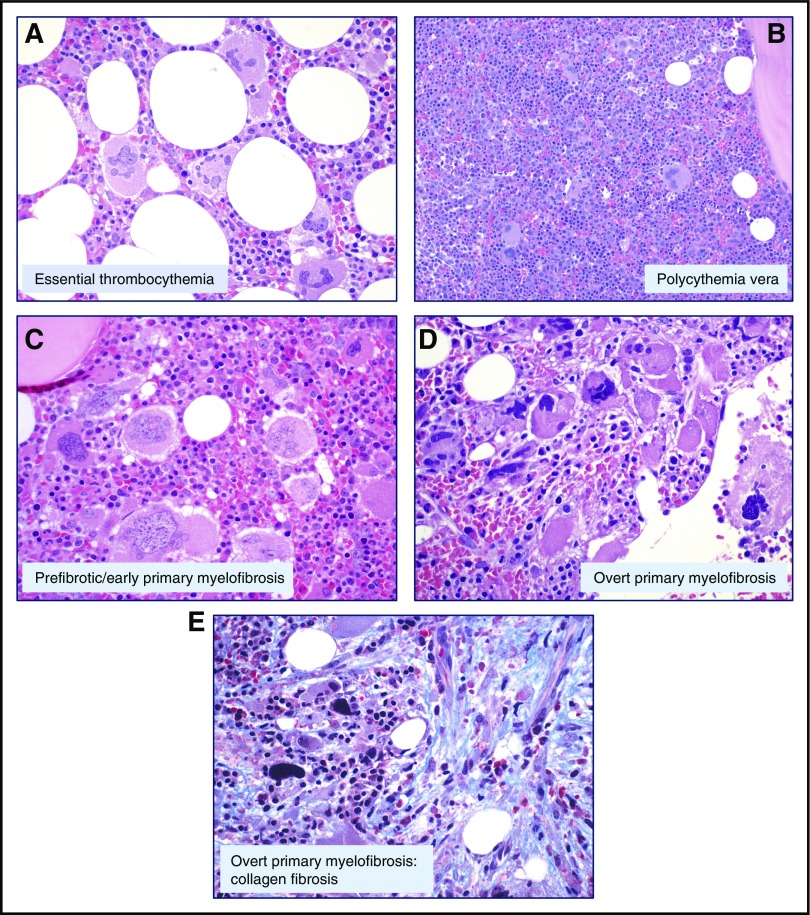
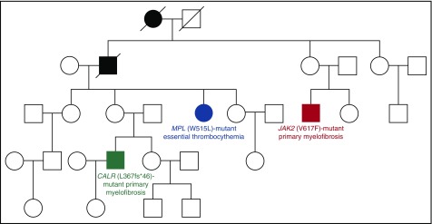
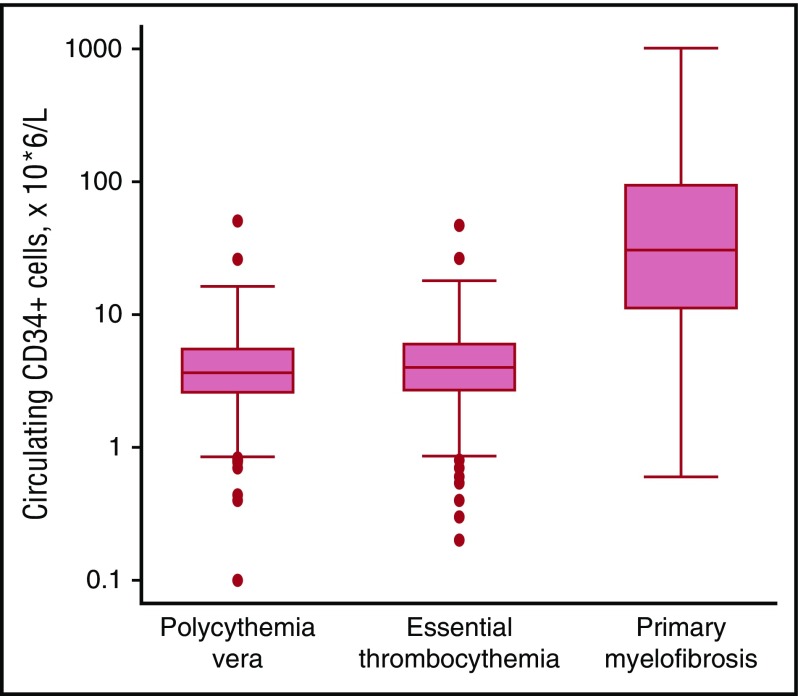
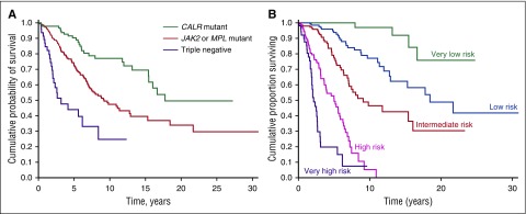
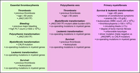
Philadelphia-negative classical myeloproliferative neoplasms (MPNs) include polycythemia vera (PV), essential thrombocythemia (ET), and primary myelofibrosis (PMF). The 2016 revision of the WHO Classification of Tumours of Haematopoietic and Lymphoid Tissues includes new criteria for the diagnosis of these disorders. Somatic mutations in the 3 driver genes, that is, JAK2, CALR, and MPL, represent major diagnostic criteria in combination with hematologic and morphological abnormalities. PV is characterized by erythrocytosis with suppressed endogenous erythropoietin production, bone marrow panmyelosis, and JAK2 mutation. Thrombocytosis, bone marrow megakaryocytic proliferation, and presence of JAK2, CALR, or MPL mutation are the main diagnostic criteria for ET. PMF is characterized by bone marrow megakaryocytic proliferation, reticulin and/or collagen fibrosis, and presence of JAK2, CALR, or MPL mutation. Prefibrotic myelofibrosis represents an early phase of myelofibrosis, and is characterized by granulocytic/megakaryocytic proliferation and lack of reticulin fibrosis in the bone marrow. The genomic landscape of MPNs is more complex than initially thought and involves several mutant genes beyond the 3 drivers. Comutated, myeloid tumor-suppressor genes contribute to phenotypic variability, phenotypic shifts, and progression to more aggressive disorders. Patients with myeloid neoplasms are at variable risk of vascular complications, including arterial or venous thrombosis and bleeding. Current prognostic models are mainly based on clinical and hematologic parameters, but innovative models that include genetic data are being developed for both clinical and trial settings. In perspective, molecular profiling of MPNs might also allow for accurate evaluation and monitoring of response to innovative drugs that target the mutant clone.


Figures

Figure 1.
Figure 1.
Representative bone marrow biopsies from patients with MPNs. (A) ET: Normocellular marrow, proliferation of giant megakaryocytes with hyperlobulated nuclei, scattered or in loose clusters (hematoxylin and eosin [H&E], original magnification ×40). (B) PV: Hypercellular marrow with erythroid proliferation and scattered pleomorphic megakaryocytes (H&E, original magnification ×20). (C) PMF: Hypercellular marrow with granulocytic proliferation and large megakaryocytes with atypical bulbous nuclei (H&E, original magnification ×40). (D) Overt PMF: Hypercellular marrow, proliferation of atypical megakaryocytes forming dense clusters, and dilated vessels with intraluminal hematopoiesis (H&E, original magnification ×40). (E) Overt PMF (collagen fibrosis): Bands of collagen fibrosis within hematopoietic lacunae (Masson trichrome staining, original magnification ×40). Courtesy of Emanuela Boveri, Fondazione Istituto di Ricovero e Cura a Carattere Scientifico Policlinico San Matteo, Pavia, Italy.
Figure 2.
Figure 2.
Four-generation Australian pedigree with different types of familial MPNs associated with different driver mutations. Full symbols indicate affected individuals. In this pedigree, a germ line mutation (R1569H) in the RBBP6 gene segregated with an MPN phenotype. The RBBP6 protein is a RING finger E3 ubiquitin ligase that contributes to ubiquitinate and degrade p53 in association with MDM2: mutant RBBP6 may cause an elevation in somatic mutagenesis rates through inhibition of p53 function and deregulation of cell cycle. The fact that individuals with the germ line mutation acquired somatic mutations in different genes supports the notion of genetic predisposition. Modified from Harutyunyan et al with permission.
Figure 3.
Figure 3.
Circulating CD34+ cells in patients with MPNs. Flow cytometry enumeration of circulating CD34+ cells in patients with PV (n = 239), ET (n = 391), or PMF (n = 106). Data are shown in a box plot depicting the upper and lower adjacent values (highest and lowest horizontal line, respectively), upper and lower quartile with median value (box), and outside values (dots). Values found in PMF patients are significantly higher than those found in PV or ET patients (P < .0001): the existence of overlaps is consistent with the notion that abnormal stem cell trafficking may be found also in some patients with PV or ET, especially those with advance disease. Overall, the available evidence indicates that flow cytometry enumeration of circulating CD34+ cells represents a simple, useful tool for estimating abnormal stem cell trafficking in patients with MPNs.
Figure 4.
Figure 4.
Kaplan-Meier analysis of survival of PMF patients stratified according to their driver mutation or a clinical-molecular prognostic model that includes IPSS variables and driver mutation. (A) Patients stratified according to their driver mutation. This analysis illustrates the prognostic significance of the driver mutation: although all patients have a similar PMF phenotype (that is, bone marrow megakaryocytic proliferation with atypia, fibrosis grades 2/3, and abnormal stem cell trafficking), their outcome is largely determined by the driver mutant gene. (B) Patients stratified according to a clinical-molecular prognostic model. As a proof of concept, this analysis illustrates the potential of integrating clinical and molecular data for improving the prognostication precision in clinical practice and in designing clinical trials. The clinical-molecular prognostic model depicted here includes JAK2, CALR, and MPL mutation status in addition to the IPSS variables. Modified from Rumi et al with permission.
Figure 5.
Figure 5.
Conventional and molecular risk factors for patients with MPNs. Information is from studies discussed in the “Risk stratification” section.

References

    1. Dameshek W. Some speculations on the myeloproliferative syndromes. Blood. 1951;6(4):372-375. - PubMed
    1. Swerdlow SH, Campo E, Harris NL, et al. . WHO Classification of Tumours of Haematopoietic and Lymphoid Tissues. Lyon, France: IARC; 2008.
    1. Arber DA, Orazi A, Hasserjian R, et al. . The 2016 revision to the World Health Organization classification of myeloid neoplasms and acute leukemia. Blood. 2016;127(20):2391-2405. - PubMed
    1. James C, Ugo V, Le Couédic JP, et al. . A unique clonal JAK2 mutation leading to constitutive signalling causes polycythaemia vera. Nature. 2005;434(7037):1144-1148. - PubMed
    1. Kralovics R, Passamonti F, Buser AS, et al. . A gain-of-function mutation of JAK2 in myeloproliferative disorders. N Engl J Med. 2005;352(17):1779-1790. - PubMed

MeSH terms