. 2017 Jun;28(6):1792-1801.
doi: 10.1681/ASN.2016080849. Epub 2016 Dec 27.

Diversification of Cell Lineages in Ureter Development

Affiliations

Diversification of Cell Lineages in Ureter Development

Tobias Bohnenpoll et al. J Am Soc Nephrol. 2017 Jun.

Abstract

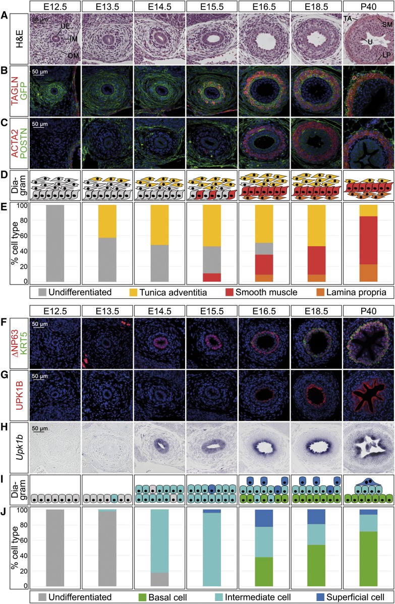
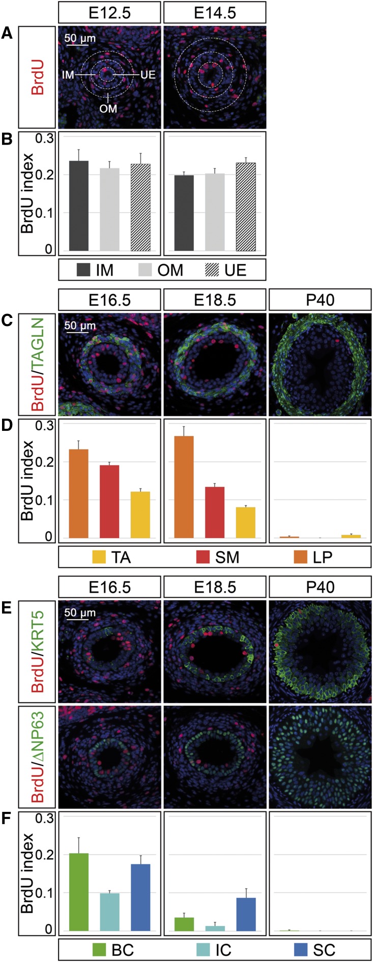
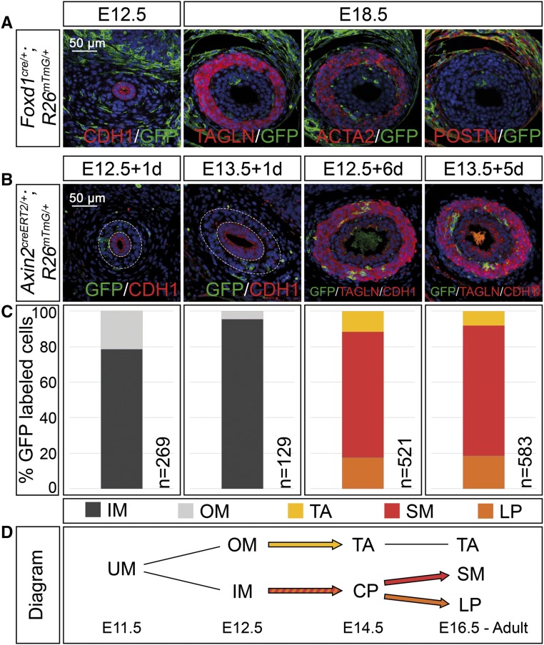
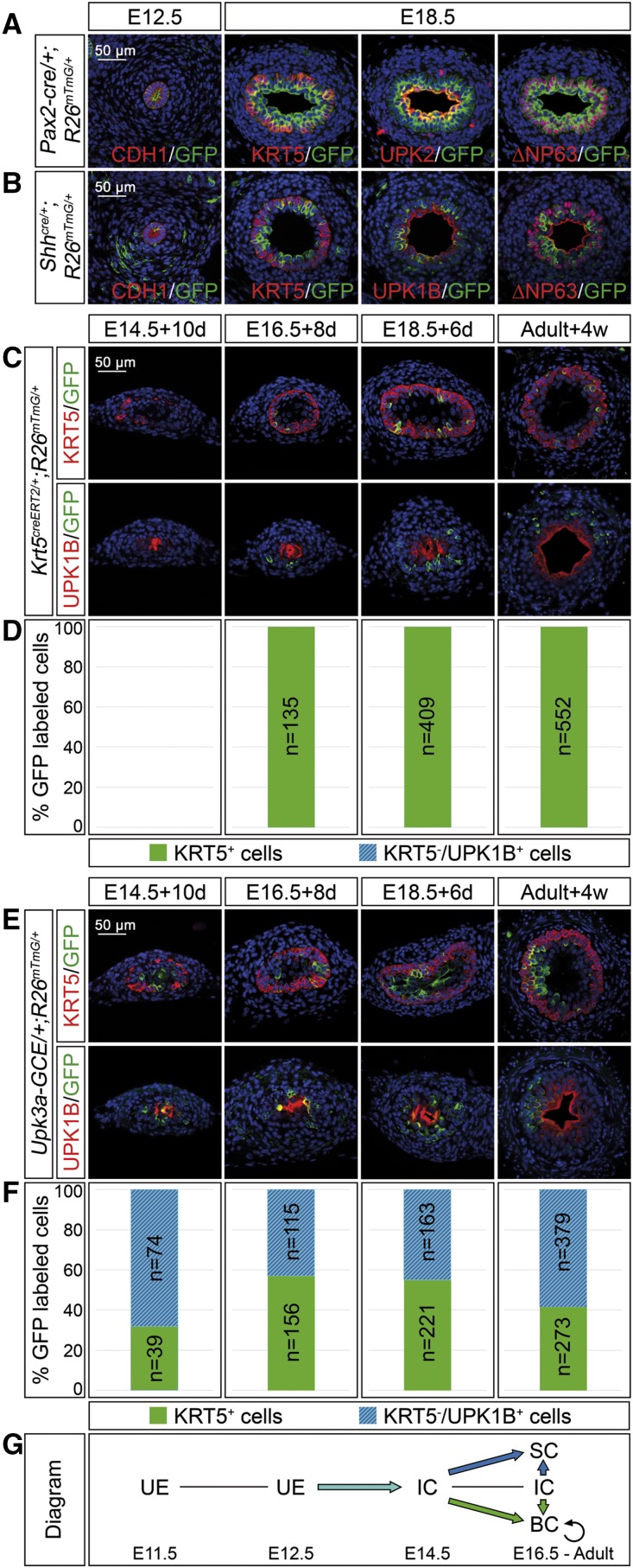
The mammalian ureter consists of a mesenchymal wall composed of smooth muscle cells and surrounding fibrocytes of the tunica adventitia and the lamina propria and an inner epithelial lining composed of layers of basal, intermediate, and superficial cells. How these cell types arise from multipotent progenitors is poorly understood. Here, we performed marker analysis, cell proliferation assays, and genetic lineage tracing to define the lineage relations and restrictions of the mesenchymal and epithelial cell types in the developing and mature mouse ureter. At embryonic day (E) 12.5, the mesenchymal precursor pool began to subdivide into an inner and outer compartment that began to express markers of smooth muscle precursors and adventitial fibrocytes, respectively, by E13.5. Smooth muscle precursors further diversified into lamina propria cells directly adjacent to the ureteric epithelium and differentiated smooth muscle cells from E16.5 onwards. Uncommitted epithelial progenitors of the ureter differentiated into intermediate cells at E14.5. After stratification into two layers at E15.5 and three cell layers at E18.5, intermediate cells differentiated into basal cells and superficial cells. In homeostasis, proliferation of all epithelial and mesenchymal cell types remained low but intermediate cells still gave rise to basal cells, whereas basal cells divided only into basal cells. These studies provide a framework to further determine the molecular mechanisms of cell differentiation in the tissues of the developing ureter.

Keywords: genetics and development; kidney development; molecular genetics; renal cell biology; renal development; ureteric bud.


Figures

Figure 1.
Figure 1.
Cell differentiation in ureter development occurs in a temporally and spatially controlled manner. (A–E) Time course of cell differentiation in the ureteric mesenchyme. (A) Hematoxylin and eosin (H&E) staining on transverse sections of the proximal ureter. At E12.5, the inner and outer mesenchymal region (IM and OM) and the ureteric epithelium (UE) are indicated. At P40, the three subregions of the mesenchyme, the lamina propria (LP), the smooth muscle (SM), and the tunica adventitia (TA) are marked adjacent to the urothelium (U). (B and C) Coimmunofluorescence analysis of expression of the SMC marker TAGLN and the lineage marker GFP (B) and of the SMC marker ACTA2 and the adventitial marker POSTN (C) on transverse sections of the proximal ureter in Tbx18cre/+;R26mTmG/+ mice. Nuclei are counterstained with DAPI (blue). (D) Schematic representation of cell differentiation in the ureteric mesenchyme. Fibrocytes of the tunica adventitia (yellow) are defined as POSTN+GFP+, SMCs (red) as TAGLN+ACTA2+GFP+, and lamina propria cells (orange) as TAGLNGFP+. (E) Quantification of differentiated cell types in the ureteric mesenchyme on the basis of marker expression as explained in (D). For numbers see Supplemental Table 1A. (F–J) Time course of epithelial differentiation in the ureter. (F and G) Coimmunofluorescence analysis of expression of the B cell marker KRT5, the B and I cell marker NP63 (F), and the I and S cell marker UPK1B (G) with DAPI-stained nuclei (blue), and (H) in situ hybridization for Upk1b expression on transverse sections of the proximal ureter in Tbx18cre/+;R26mTmG/+ mice. (I) Schematic representation of cell expansion and differentiation in the ureteric epithelium. B cells (green) are defined as KRT5+NP63+UPK1BUpk1b, I cells (turquois) as KRT5NP63+UPK1B+(low)Upk1b+, and S cells (blue) as KRT5NP63UPK1B+(high)Upk1b+. (J) Quantification of differentiated cell types in the ureteric epithelium on the basis of marker expression as explained in (I). For numbers see Supplemental Table 1B.
Figure 2.
Figure 2.
Proliferation rates explain differential cell-type expansion in the developing ureter. (A) Coimmunofluorescence analysis on proximal ureter sections at E12.5 and E14.5 for BrdU and the nuclear counterstain DAPI (blue). White dotted lines demarcate the ureteric epithelium (UE), and the inner and outer mesenchymal cell populations (IM and OM). (B) Quantification of cell proliferation by the BrdU index in the areas indicated in (A). (C) Coimmunofluorescence analysis on proximal ureter sections at E16.5, E18.5, and P40 for BrdU, the nuclear counterstain DAPI (blue), and the SM marker TAGLN. (D) Quantification of cell proliferation by the BrdU index in TAGLN outer mesenchymal cells of the tunica adventitia (TA), in TAGLN+ smooth muscle cells (SM), and in TAGLN inner mesenchymal cells of the lamina propria (LP). (E) Coimmunofluorescence on proximal ureter sections at E16.5, E18.5, and P40 for BrdU, the nuclear counterstain DAPI (blue), and the B cell marker KRT5 (upper row) and BrdU, the nuclear counterstain DAPI (blue), and the B and I cell marker NP63 (lower row). (F) Quantification of cell proliferation by the BrdU index in KRT5+ B cells (BC), in KRT5+NP63+ I cells (IC), and KRT5NP63 S cells (SC). For numbers see Supplemental Table 2, A–C.
Figure 3.
Figure 3.
Adventitial cells are separated early in mesenchymal development whereas lamina propria cells derive from SMC precursors in the ureter. (A) Coimmunofluorescence analysis on transverse sections of the proximal ureter for the lineage marker GFP and the epithelial marker CDH1 at E12.5, with the SMC markers TAGLN and ACTA2, and the adventitial cell marker POSTN at E18.5 shows recombination in outer fibrocytes only in Foxd1cre/+;R26mTmG/+ embryos. (B) Coimmunofluorescence of the lineage marker GFP with the epithelial marker CDH1 and the SMC marker TAGLN on proximal sections of ureters from Axin2creERT2/+;R26mTmG/+ embryos that were tamoxifen-induced at E12.5 or E13.5 and harvested after 1 and 6 or 5 days to detect localization of recombined cells. (C) Relative distribution of GFP+ cells localized to the inner and outer ureteric mesenchyme (IM and OM), and to the tunica adventitia (TA), the smooth muscle (SM), and the lamina propria (LP) in ureters shown in (B). The number of counted GFP+ cells (n) is given. For additional numbers see Supplemental Table 3. (D) Schematic representation of the lineage relations in the ureteric mesenchyme. Abbreviations are as explained in (C). Inner mesenchymal cells contribute to the SM layer and the lamina propria but not to the tunica adventitia. UM, undifferentiated mesenchyme; CP, common precursor.
Figure 4.
Figure 4.
I cells are progenitors of B and S cells in ureter development and homeostasis. (A and B) Coimmunofluorescence analysis on transverse sections of the proximal ureter of the lineage marker GFP with the epithelial marker CDH1 at E12.5, and the B cell marker KRT5, the B and I cell marker NP63, and the S cell markers UPK1B/UPK2 in Pax2-cre/+;R26mTmG/+ embryos (A) and in Shhcre/+;R26mTmG/+ embryos (B) shows that Pax2+ and Shh+ cells of the early ureteric bud contribute to all urothelial cell types in the ureter but not the surrounding mesenchyme. (C and E) Coimmunofluorescence analysis of the lineage marker GFP with the B cell marker KRT5 (upper panel) and the S cell marker UPK1B (lower panel) on transverse sections of ureters isolated at E14.5, E16.5, and E18.5, induced with 500 nM 4-hydroxytamoxifen for the first 24 hours and cultured for 10, 8, and 6 days; and in adult ureters 4 weeks after tamoxifen administration in Krt5creERT2/+;R26mTmG/+ mice (C) and Upk3a-GCE/+;R26mTmG/+ mice (E). (D and F) The bar diagrams display the percentage of lineage-labeled (GFP+) cells contributing to KRT5+UPK1B B cells or to KRT5UPK1B+ I and S cells in the ureter of Krt5creERT2/+;R26mTmG/+ mice (D) and of Upk3a-GCE/+;R26mTmG/+ mice (F) at the indicated stages and conditions. The number of counted GFP+ cells (n) is given. For additional numbers see Supplemental Table 4, A and B. (G) Schematic representation of the lineage relations of urothelial cell types. KRT5+ cells only give rise to B cells whereas UPK3A+ cells give rise to B, I, and S cells. BC, basal cell; IC, intermediate cell; SC, superficial cell; UE, undifferentiated epithelium.

References

    1. Velardo JT: Histology of the ureter. In: The ureter, 2nd Ed., edited by Bergman H, New York, Springer-Verlag, 1981
    1. Yu J, Manabe M, Wu XR, Xu C, Surya B, Sun TT: Uroplakin I: A 27-kD protein associated with the asymmetric unit membrane of mammalian urothelium. J Cell Biol 111: 1207–1216, 1990 - PMC - PubMed
    1. Weiss RM, Guo S, Shan A, Shi H, Romano RA, Sinha S, Cantley LG, Guo JK: Brg1 determines urothelial cell fate during ureter development. J Am Soc Nephrol 24: 618–626, 2013 - PMC - PubMed
    1. Bohnenpoll T, Kispert A: Ureter growth and differentiation. Semin Cell Dev Biol 36: 21–30, 2014 - PubMed
    1. Airik R, Trowe MO, Foik A, Farin HF, Petry M, Schuster-Gossler K, Schweizer M, Scherer G, Kist R, Kispert A: Hydroureternephrosis due to loss of Sox9-regulated smooth muscle cell differentiation of the ureteric mesenchyme. Hum Mol Genet 19: 4918–4929, 2010 - PubMed

LinkOut - more resources