. 2017 Feb 8;37(6):1439-1452.
doi: 10.1523/JNEUROSCI.2984-16.2016. Epub 2016 Dec 27.

Adenosine Shifts Plasticity Regimes between Associative and Homeostatic by Modulating Heterosynaptic Changes

Affiliations

Adenosine Shifts Plasticity Regimes between Associative and Homeostatic by Modulating Heterosynaptic Changes

Nicholas M Bannon et al. J Neurosci. .

Abstract

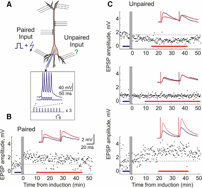
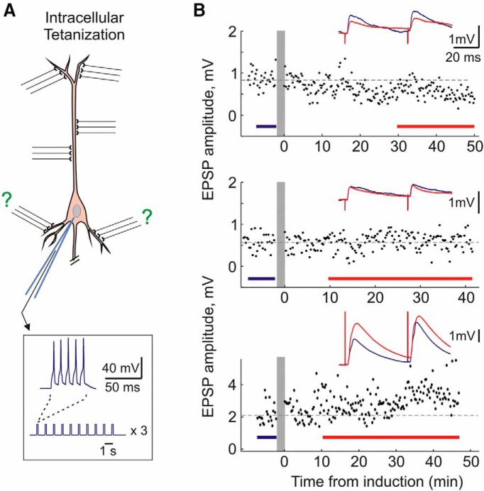
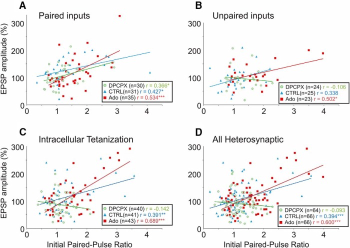
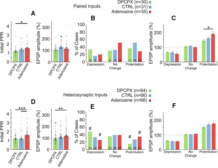
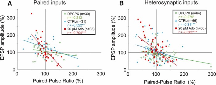
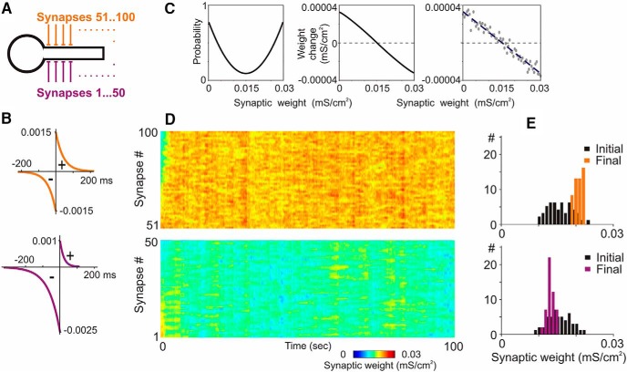
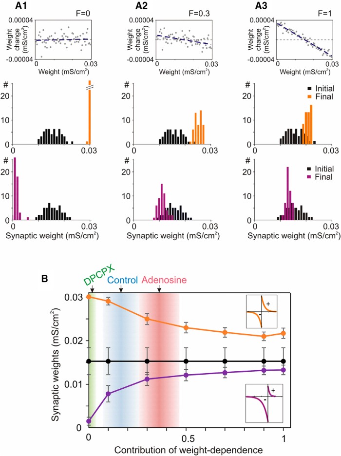
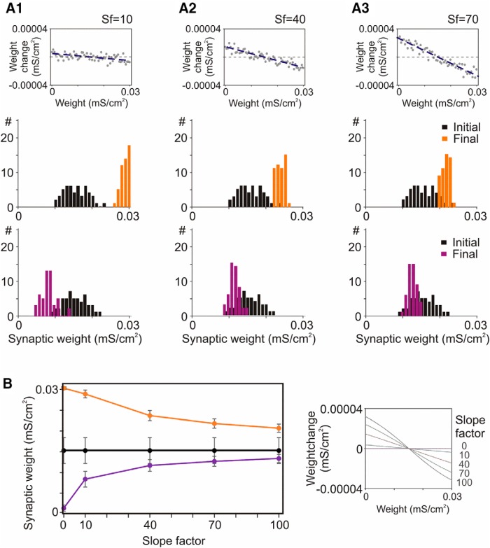
Endogenous extracellular adenosine level fluctuates in an activity-dependent manner and with sleep-wake cycle, modulating synaptic transmission and short-term plasticity. Hebbian-type long-term plasticity introduces intrinsic positive feedback on synaptic weight changes, making them prone to runaway dynamics. We previously demonstrated that co-occurring, weight-dependent heterosynaptic plasticity can robustly prevent runaway dynamics. Here we show that at neocortical synapses in slices from rat visual cortex, adenosine modulates the weight dependence of heterosynaptic plasticity: blockade of adenosine A1 receptors abolished weight dependence, while increased adenosine level strengthened it. Using model simulations, we found that the strength of weight dependence determines the ability of heterosynaptic plasticity to prevent runaway dynamics of synaptic weights imposed by Hebbian-type learning. Changing the weight dependence of heterosynaptic plasticity within an experimentally observed range gradually shifted the operating point of neurons between an unbalancing regime dominated by associative plasticity and a homeostatic regime of tightly constrained synaptic changes. Because adenosine tone is a natural correlate of activity level (activity increases adenosine tone) and brain state (elevated adenosine tone increases sleep pressure), modulation of heterosynaptic plasticity by adenosine represents an endogenous mechanism that translates changes of the brain state into a shift of the regime of synaptic plasticity and learning. We speculate that adenosine modulation may provide a mechanism for fine-tuning of plasticity and learning according to brain state and activity.SIGNIFICANCE STATEMENT Associative learning depends on brain state and is impaired when the subject is sleepy or tired. However, the link between changes of brain state and modulation of synaptic plasticity and learning remains elusive. Here we show that adenosine regulates weight dependence of heterosynaptic plasticity: adenosine strengthened weight dependence of heterosynaptic plasticity; blockade of adenosine A1 receptors abolished it. In model neurons, such changes of the weight dependence of heterosynaptic plasticity shifted their operating point between regimes dominated by associative plasticity or by synaptic homeostasis. Because adenosine tone is a natural correlate of activity level and brain state, modulation of plasticity by adenosine represents an endogenous mechanism for translation of brain state changes into a shift of the regime of synaptic plasticity and learning.

Keywords: adenosine; heterosynaptic plasticity; learning rules; neuron models; synaptic plasticity; visual cortex.


Figures

Figure 1.
Figure 1.
Induction of long-term plasticity by pairing procedure. A, Scheme of pairing protocol. Two independent inputs to a layer 2/3 pyramidal neuron are stimulated by bipolar electrodes. Pairing consisted of three trains (1/min) of 10 bursts (1 Hz) of five pulses (5 ms, 100 Hz, 0.4–1.5 nA) through the recording electrode, with an EPSP evoked at one input 10 ms before each burst of spikes (paired input). Unpaired inputs were not stimulated during the induction. B, C, Example time course of EPSP amplitudes evoked by the first pulse in a paired-pulse paradigm. Vertical gray bars show timing of plasticity induction. Color-coded insets show averaged responses to paired-pulse stimuli before and after plasticity induction. Examples of potentiation at paired inputs, and depression, no change, and potentiation at unpaired inputs in control solution. These examples are also representative of plasticity induced under DPCPX or adenosine.
Figure 2.
Figure 2.
Induction of long-term plasticity by intracellular tetanization. A, Scheme of intracellular tetanization protocol. Intracellular tetanization consisted of the same postsynaptic firing as pairing: three trains (1/min) of 10 bursts (1 Hz) of five pulses (5 ms, 100 Hz, 0.4–1.5 nA) through the recording electrode. However, no inputs were stimulated during the intracellular tetanization. B, Examples of potentiation, no changes, and depression induced by intracellular tetanization. Same conventions as in Figure 1.
Figure 3.
Figure 3.
Homosynaptic and heterosynaptic plasticity are weight dependent. A–D, Correlation between initial PPR and changes of EPSP amplitude following induction protocol at paired inputs (A), at unpaired inputs (B), following intracellular tetanization (C), and at all heterosynaptic sites (D, pooled data from B and C). Recordings were conducted in either control ACSF (CTRL, blue triangles), or on the background of 30 nm selective A1R antagonist DPCPX (green circles) or 20 μm adenosine (red squares). A, At paired inputs (homosynaptic sites), changes in EPSP amplitudes were positively correlated with the initial PPR in all experimental series, regardless of background adenosine receptor ligands. B, C, At unpaired inputs and inputs undergoing intracellular tetanization (both of which may be considered heterosynaptic), changes of EPSP amplitudes were positively correlated with the initial PPR in control solution and on the background of 20 μm adenosine. On the background of A1R blockade with DPCPX, there were still cases of potentiation and depression, but the relationship of EPSP change to initial synaptic strength (initial PPR) was abolished. D, Pooled data (from B and C) representing all heterosynaptic sites. *p < 0.05; **p < 0.01; ***p < 0.001.
Figure 4.
Figure 4.
Effects of adenosine on PPR and net plastic outcome at homosynaptic and heterosynaptic inputs. A, Initial (preinduction) PPR (left) and mean EPSP amplitude change (right) induced at paired inputs in three drug conditions. Error bars show SEM; gray dash marks denote individual cases. B, Percentage of inputs that expressed significant potentiation or depression, or did not change (t test comparing baseline period to after induction period for each input). Data for paired inputs, for three drug groups. Inset in each bar shows number of cases. A χ2 test revealed no significant relationship between adenosine receptor manipulation and the frequency of potentiation, depression, or no change. Note that potentiation is generally a more common outcome than depression. C, Mean changes of EPSP amplitude in inputs that underwent potentiation, that underwent depression, or that did not change. Paired inputs in three drug conditions. The mean magnitude of potentiation was larger under adenosine than in the DPCPX group. D, Initial PPR (left) and mean EPSP amplitude change (right) induced at heterosynaptic inputs in three drug conditions. Conventions as in A. E, Percentage of heterosynaptic inputs that expressed significant potentiation or depression, or did not change, in three drug groups. Blocking tonic A1R activation (DPCPX group) results in more cases of depression and fewer cases of potentiation than expected (χ2 test). The adenosine group exhibited fewer cases of depression and more cases of potentiation than expected. Standardized residuals of ≥1.4 are denoted with #, indicating that these counts deviated the most from expected observations. F, Mean changes of EPSP amplitude in heterosynaptic inputs that underwent potentiation, that underwent depression, or that did not change. No significant effects of adenosine receptor manipulation on magnitude of EPSP change.
Figure 5.
Figure 5.
Homosynaptic and heterosynaptic plasticity are expressed partially via presynaptic mechanisms. A, B, Correlation of changes in the PPR with changes in EPSP amplitude at paired inputs (A) and heterosynaptic inputs (B; unpaired inputs and after intracellular tetanization). Data for three drug groups: control ACSF (blue triangles); 30 nm A1R antagonist DPCPX (green circles); 20 μm adenosine (red squares). Changes in the PPR were negatively correlated with EPSP amplitude changes. This relationship was generally strengthened as a function of adenosine receptor activation. Compared with control, r values were higher in adenosine, but lower in DPCPX groups.
Figure 6.
Figure 6.
A neuron model with homosynaptic and heterosynaptic plasticity. A–C, A model neuron with 100 input synapses expressing homosynaptic and heterosynaptic plasticity. STDP rules (B) were symmetrical at 50 synapses (orange), and depression-biased at the other 50 synapses (violet). Heterosynaptic plasticity rules were the same at all synapses, and included weight dependence of the probability and magnitude of change (C, left and middle; Eqs. 10 and 11). Rightmost plot in C shows example relation between initial weights and weight changes with a random component (Eq. 11, σ) calculated for initial synaptic weights from 0 to 0.03 mS/cm2 (0.0005 mS/cm2 increment), and regression line through these points. D, E, Dynamics of synaptic weights (D) and distributions of synaptic weights in the beginning and at the end of simulation (E) for two groups of synapses, with symmetrical (top) and depression-biased STDP (bottom) and heterosynaptic plasticity. Each synapse was driven by individual spike trains with Poisson-distributed interspike intervals (averaged frequency, 3 Hz; averaged correlation between spike trains, 0.5).
Figure 7.
Figure 7.
Prevention of runaway dynamics in the model neuron depends on the weight-dependent component to heterosynaptic plasticity. A1–A3, Example relations between initial weights and heterosynaptic weight changes with contribution of the weight-dependent component (Eq. 12, F) of 0, 0.3, and 1; and initial and final distributions of weights of two groups of synapses in models implementing these relations. Two groups of synapses had symmetrical or depression-biased STDP rules as shown in the insets in B. B, Dependence of the final weights (mean ± SD) of two groups of synapses on the contribution of the weight-dependent component of heterosynaptic plasticity. Black are mean ± SD of initial synaptic weights. Arrows (top) show R2 of correlations between initial PPR and amplitude changes at heterosynaptic inputs measured in electrophysiological experiments with DPCPX, control, and adenosine. Gradient areas show mean ± SD of R2 for each group obtained with bootstrapping (1000 resamplings; 0.0172 ± 0.019; 0.165 ± 0.091; 0.362 ± 0.104; for the three groups).
Figure 8.
Figure 8.
Prevention of runaway dynamics in the model depends on the slope of weight dependence of heterosynaptic plasticity. A1–A3, Example relations between heterosynaptic weight changes and initial weights for slope factors (Sf) 10, 40, and 70, as indicated, and initial and final distributions of weights of two groups of synapses in models implementing these relations. B, Dependence of the final weights (mean ± SD) of two groups of synapses on the slope of weight dependence of heterosynaptic plasticity, as shown on the right (Eq. 11, SlopeFactor). Black are mean ± SD of initial synaptic weights. Two groups of synapses had STDP rules as in Figure 6 model.

References

    1. Bannon NM, Zhang P, Ilin V, Chistiakova M, Volgushev M (2014) Modulation of synaptic transmission by adenosine in layer 2/3 of the rat visual cortex in vitro. Neuroscience 260:171–184. 10.1016/j.neuroscience.2013.12.018 - DOI - PMC - PubMed
    1. Bazhenov M, Timofeev I, Steriade M, Sejnowski TJ (2002) Model of thalamocortical slow-wave sleep oscillations and transitions to activated states. J Neurosci 22:8691–8704. - PMC - PubMed
    1. Benington JH, Kodali SK, Heller HC (1995) Stimulation of A1 adenosine receptors mimics the electroencephalographic effects of sleep deprivation. Brain Res 692:79–85. 10.1016/0006-8993(95)00590-M - DOI - PubMed
    1. Bjorness TE, Greene RW (2009) Adenosine and sleep. Curr Neuropharmacol 7:238–245. 10.2174/157015909789152182 - DOI - PMC - PubMed
    1. Bjorness TE, Kelly CL, Gao T, Poffenberger V, Greene RW (2009) Control and function of the homeostatic sleep response by adenosine A1 receptors. J Neurosci 29:1267–1276. 10.1523/JNEUROSCI.2942-08.2009 - DOI - PMC - PubMed

Publication types

LinkOut - more resources