. 2017 Jan 10;114(2):310-315.
doi: 10.1073/pnas.1612322114. Epub 2016 Dec 27.

Bax transmembrane domain interacts with prosurvival Bcl-2 proteins in biological membranes

Affiliations

Bax transmembrane domain interacts with prosurvival Bcl-2 proteins in biological membranes

Vicente Andreu-Fernández et al. Proc Natl Acad Sci U S A. .

Erratum in

Abstract

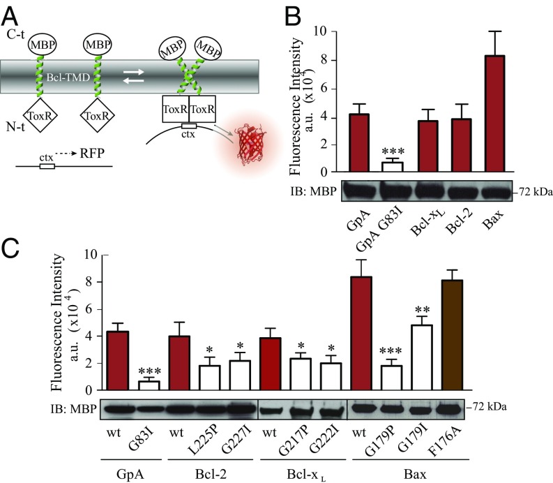
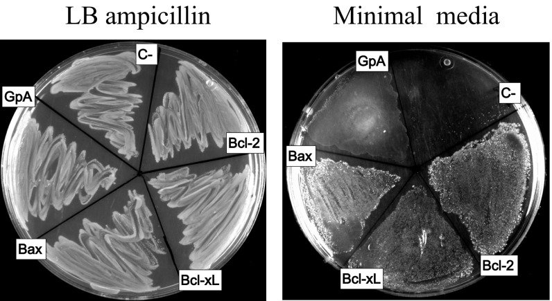
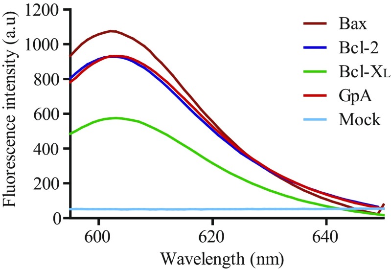
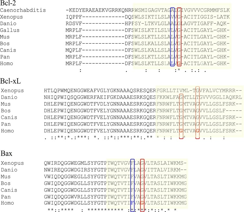
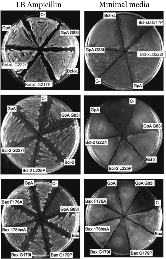
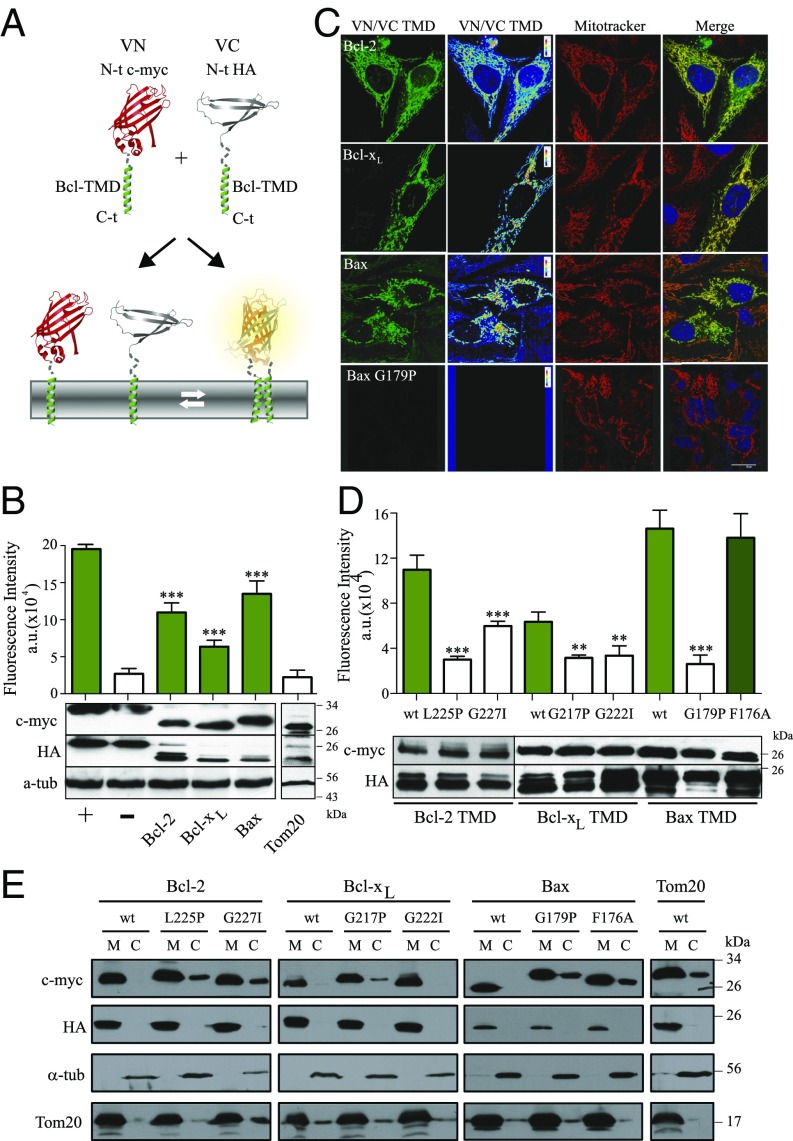
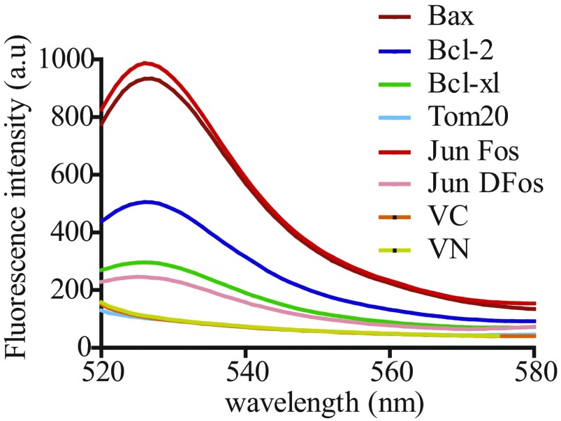
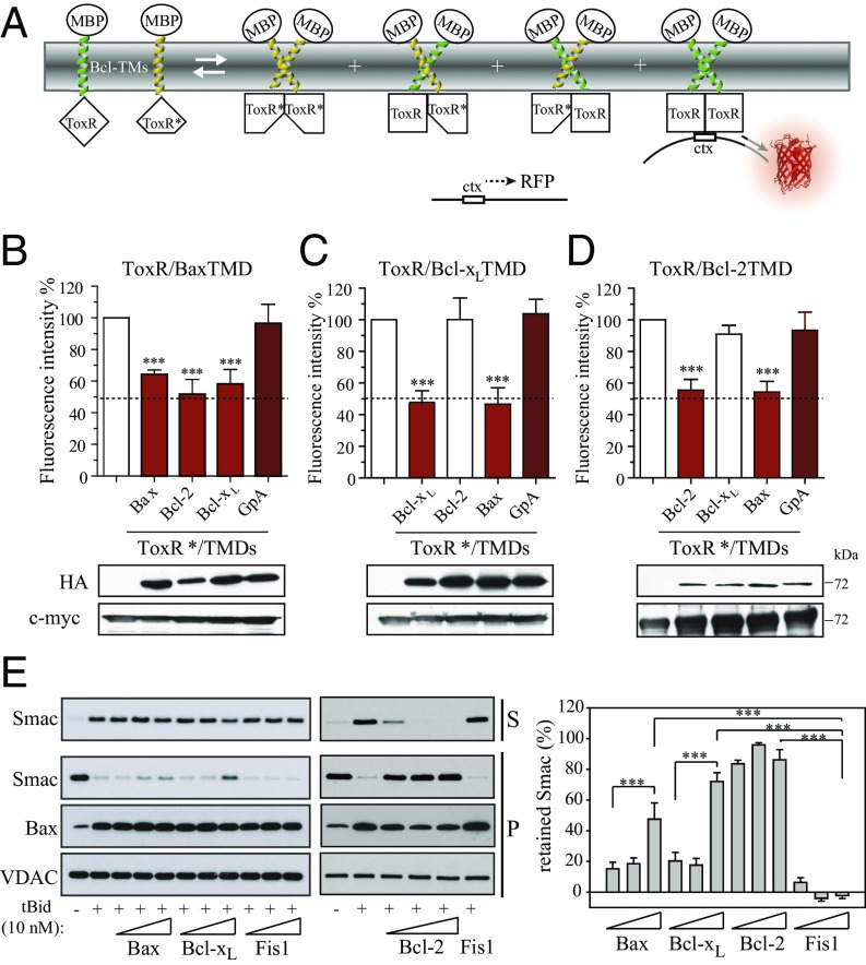
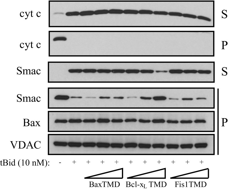
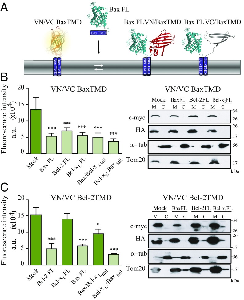
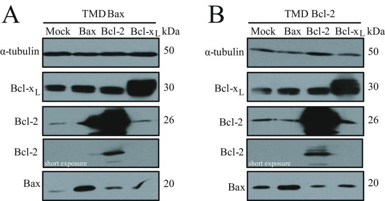
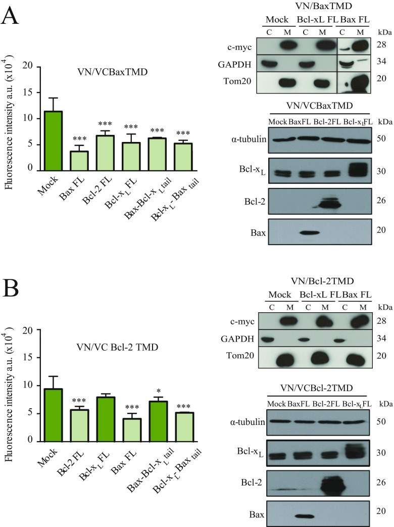
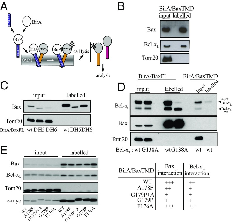
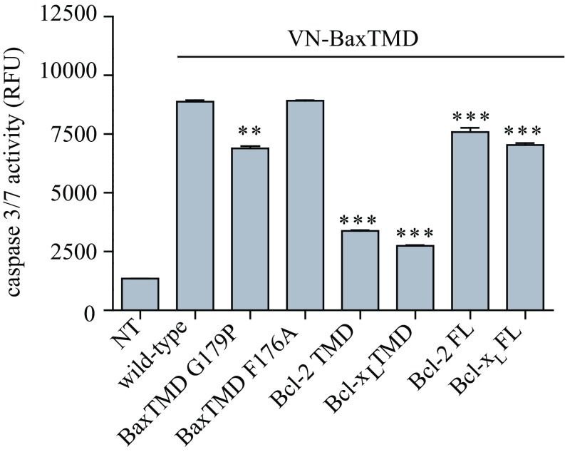
The Bcl-2 (B-cell lymphoma 2) protein Bax (Bcl-2 associated X, apoptosis regulator) can commit cells to apoptosis via outer mitochondrial membrane permeabilization. Bax activity is controlled in healthy cells by prosurvival Bcl-2 proteins. C-terminal Bax transmembrane domain interactions were implicated recently in Bax pore formation. Here, we show that the isolated transmembrane domains of Bax, Bcl-xL (B-cell lymphoma-extra large), and Bcl-2 can mediate interactions between Bax and prosurvival proteins inside the membrane in the absence of apoptotic stimuli. Bcl-2 protein transmembrane domains specifically homooligomerize and heterooligomerize in bacterial and mitochondrial membranes. Their interactions participate in the regulation of Bcl-2 proteins, thus modulating apoptotic activity. Our results suggest that interactions between the transmembrane domains of Bax and antiapoptotic Bcl-2 proteins represent a previously unappreciated level of apoptosis regulation.

Keywords: Bcl-2; apoptosis; mitochondria; oligomerization; transmembrane.


Conflict of interest statement

The authors declare no conflict of interest.

Figures

Fig. 1.
Fig. 1.
Bax, Bcl-xL, and Bcl-2 TMDs homooligomerize in membranes. (A) TMD constructs fused to ToxR were expressed in the inner membrane of E. coli. The TMD interaction reconstituted active ToxR and activated ctx promoter-regulated RFP expression. The C-terminal MBP domain was exposed to the periplasm. (B) ToxR-TMD-MBP constructs (Table 1) were transformed into MM39 cells. The fluorescence signal (ʎexc 570, ʎem 620 nm) obtained with the ToxRed system for Bax, Bcl-xL, and Bcl-2 TMDs is shown. Red bars highlight interactions. The GpA TMD and nondimerizing TMD GpA G83I variant served as positive and negative controls, respectively. Western blotting of MBP revealed equivalent expression levels. (C) Homooligomerization assay of wild-type and variants of the Bax, Bcl-xL, and Bcl-2 TMDs. Fusion protein functionality, orientation, and membrane insertion of all variants were controlled by growth on minimum medium (Fig. S4). Reduction in fluorescence indicates the disruption of homooligomerization (white bars). Results obtained with mutations that apparently do not interfere with homooligomerization are depicted in brown. Error bars represent the mean ± SD; n ≥ 3. Western blotting of MBP revealed equivalent expression levels for all chimeras. P values according to Dunnett's test are displayed. *P < 0.05; **P < 0.01; ***P < 0.001.
Fig. S1.
Fig. S1.
Maltose complementation assay to test ToxR-TMD-MBP chimera topology. MalE-deficient E. coli MM39 cells were transformed with appropriate ToxR-GpA-MBP and ToxR-Bcl-MBP vectors. The MalE complementation assay indicates that chimeras containing GpA TMD and Bcl-2–related TMDs are expressed and integrated into the inner membrane of E. coli. The constructs were cultured on either complete medium LB (Left) or M9 agar containing 0.4% maltose (Right). Because the MM39 cell line is deficient in MBP, only cells with properly integrated ToxR chimera can survive on maltose minimal medium.
Fig. S2.
Fig. S2.
Emission spectra for the ToxRed assay. For fluorescence measurements of RFP, samples from bacterial lysates were excited at 584 nm, and emission spectra were recorded from 595 to 650 nm. The mock line is for whole-cell lysates containing a control plasmid without a ctx::mCherry reporter.
Fig. S3.
Fig. S3.
Multisequence alignment of Bcl-2 TMDs from different species. Evolutionarily conserved glycine residues are boxed in red, and mutated leucine and phenylalanine residues from Bcl-2 and Bax sequences, respectively, are boxed in blue.
Fig. S4.
Fig. S4.
Maltose complementation assay to test ToxR-TMD-MBP chimera topology in Bcl-2 mutants. MalE-deficient E. coli MM39 transformed with the different ToxR-TMD-MBP mutant constructs were cultured on either on complete medium LB (Left) or M9 agar containing 0.4% maltose (Right) as in Fig. S1.
Fig. 2.
Fig. 2.
Bax, Bcl-2, and Bcl-xL homooligomerize in the OMM. (A) BiFC assay analyzing TMD homooligomerization by reconstitution of venus fluorescence from separate N and C termini fragments fused to TMD segments. (B) Bax, Bcl-2, and Bcl-xL TMD homooligomerization measured by BiFC in HCT116 cells. Fusions of b-Fos and b-Jun protein domains were used as positive controls (+), and the Δb-Fos/bJun pair served as a negative control (−); n = 3. Significant increases compared with negative control were analyzed by using Dunnett's multiple comparison test (95% confidence interval). Chimeric protein expression of VN (c-myc) and VC (HA) constructs is compared in B, Lower; α-tubulin was used as loading control. (C) Confocal images of HCT116 cells transfected with VC and VN constructs of the Bcl-2 and Bcl-xL, Bax, and Bax G197P TMDs. Formation of homooligomers (green and rainbow scale, first and second column, respectively) and mitochondria (red, third column), colocalized (yellow, fourth column). (Scale bar, 20 μm.) (D) Self-association assays of wild-type and single amino acid substitution variants of the Bax, Bcl-2, and Bcl-xL TMDs measured by BiFC in HCT116 cells. Error bars represent the mean ± SD, n ≥ 3. P values according to Dunnett's test are displayed. *P < 0.05; **P < 0.01; ***P < 0.001. (E) Subcellular fractionation of HCT116 cells transfected with TMD constructs was controlled by using Tom20 (mitochondrial fraction; M) and α-tubulin (cytosol; C).
Fig. S5.
Fig. S5.
Emission spectra for the BiFC assay. For fluorescence measurements, samples from cellular lysates were excited at 515 nm, and emission spectra were recorded from 520 to 580 nm. Nonoligomerizing Tom20 TMD, VN, and VC constructs were included as controls.
Fig. 3.
Fig. 3.
The Bax TMD interacts with antiapoptotic Bcl-xL and Bcl-2 TMDs. (A) Dominant-negative ToxR assay to analyze Bcl-2 protein TMD heterooligomerization. Coexpression of TMD constructs fused to wild-type ToxR (green) and TMD constructs fused to an inactive ToxR* mutant (yellow) in E. coli can result in heterooligomerization, leading to reduced RFP synthesis. (B) Effect of the Bax, Bcl-2, and Bcl-xL TMDs on ToxR/Bax TMD homooligomerization. The GpA TMD served as a control. (C) Effect of the Bax, Bcl-2, and Bcl-xL TMDs on Bcl-xL TMD homooligomerization as in B. (D) Effect of the Bax, Bcl-2, and Bcl-xL TMDs on Bcl-2 TMD homooligomerization as in B. The GpA TMD served as a control. (E) Smac release by endogenous Bax with and without tBid in the absence and presence of the Bax, Bcl-xL, Bcl-2, or Fis1 TMD peptides from purified HCT116 Bak KO mitochondria. Smac was monitored in the supernatant (S) and pellet (P) by Western blot. Bax and VDAC served as controls. Error bars represent the mean ± SD, n ≥ 3. P values according to Dunnett's test are displayed. *P < 0.05; **P < 0.01; ***P < 0.001.
Fig. S6.
Fig. S6.
Influence of Bcl-2 protein TMDs on Smac and cyt c release. Smac and cyt c release by Bax with and without tBid in the absence or presence of Bax TMD, Bcl-xL TMD, or Fis1 TMD from purified HCT116 Bak KO mitochondria. Smac and cyt c were monitored in the supernatant and pellet by Western blot. VDAC served as a loading control.
Fig. 4.
Fig. 4.
Bcl-2 protein TMDs interact with full-length Bcl-2 proteins. (A) Heterooligomerization studies with the BiFC system using venus fluorescent protein (VFP) reconstituted from VN/VC BaxTMD chimeras and full-length Bax (BaxFL). In this assay, a decrease in fluorescence indicates the formation of heterooligomers between venus-derived chimeras and full-length Bcl-2 proteins. (B) Oligomerization analysis of the Bax TMD in the presence of full-length Bax, Bcl-2, Bcl-xL, Bax-Bcl-xL tail, and Bcl-xL-Baxtail. VFP reconstitution by VN/VC BaxTMD chimeras was challenged with the indicated full-length proteins in HCT116 cells (B, Left). Subcellular fractionation showed mitochondrial (M) localization of VN/Bax and VC/Bax in the absence or presence of full-length proteins. C indicates the cytosolic fraction. (C) Analysis of Bcl-2 TMD oligomerization in the presence of full-length Bax, Bcl-2, Bcl-xL, Bax–Bcl-xL tail, and Bcl-xL–Baxtail proteins in HCT116 cells. Subcellular fractionation showed Bcl-2 TMD-derived constructs localization in the absence or the presence of full-length proteins. Tom20 and α-tubulin served as controls. *P < 0.05; ***P < 0.001.
Fig. S7.
Fig. S7.
Expression of FL proteins in VN/VC Bax TMD (A) and VN/VC Bcl-2 TMD (B) competition experiments using full-length Bcl-2–derived proteins from Fig. 4 in the HCT116 wild-type cell line.
Fig. S8.
Fig. S8.
Bax, Bcl-2, and Bcl-xL homooligomerize in the OMM of HCT116 Bax/Bak DKO cells. Bax, Bcl-2, and Bcl-xL TMD homooligomerization measured by BiFC in HCT-116 Bax/Bak DKO cells is shown. Fusion b-Fos and b-Jun proteins were used as positive controls (+), and the Δb-Fos/b-Jun pair served as negative controls (−); n = 3. Significant increases compared with the negative control were analyzed by using Dunnett's multiple comparison test (95% confidence interval). Chimeric protein expression is compared in Lower by Western blotting with the appropriate antibodies. Error bars represent the mean ± SD; n ≥ 3. ***P < 0.001.
Fig. S9.
Fig. S9.
Bcl-2 TMDs interact with full-length proteins in the OMM of HCT116 Bax/Bak DKO cells. (A) Oligomerization analysis of the Bax TMD in the presence of full-length Bax, Bcl-2, Bcl-xL, Bax–Bcl-xLtail, and Bcl-xL–Baxtail. VFP reconstitution by VN/VC BaxTMD chimeras was challenged with the indicated full-length proteins in HCT116 cells (A, Left). Subcellular fractionation showed mitochondrial (M) localization of VN/Bax and VC/Bax in the absence or presence of full-length Bax and Bcl-xL proteins (A, Right Upper). Expression of full-length proteins was analyzed by Western blot (A, Right Lower). (B) Oligomerization analysis of the Bcl-2 TMD in the presence of full-length Bax, Bcl-2, Bcl-xL, Bax–Bcl-xLtail, and Bcl-xL–Baxtail. Subcellular fractionation showed mitochondrial (M) localization of VN/Bcl-2 and VC/Bcl-2 in the absence or presence of full-length Bax and Bcl-xL proteins (B, Right Upper). Expression of full-length proteins was analyzed by Western blot (A, Right Lower). *P < 0.05; ***P < 0.001.
Fig. 5.
Fig. 5.
Bax TMD interacts with endogenous Bax and Bcl-xL. (A) The BirA in situ proximal biotinylation assay was used to identify interactions between the Bax TMD and endogenous Bax and Bcl-xL proteins in human cells. After the expression of BirA/BaxTMD, proteins interacting with Bax TMD (blue) were labeled with biotin (star) by BirA. Crude cell extract was subsequently applied to a biotin affinity matrix (solid line). Bound (labeled) proteins were analyzed by SDS/PAGE and Western blot. (B) Input and labeled proteins of the biotin affinity matrix from HCT116 cells expressing BirA/BaxTMD fusion were analyzed by Western blot. Tom20 served as a control. (C) BirA fusion assay analyzing BirA/BaxFL-interactions with wild-type Bax compared with Bax ΔH5 and Bax ΔH6 present in the labeled fraction (L) compared with the input (I) by Western blot. n = 3. (D) BirA fusion assay comparing BirA/BaxFL and BirA/ BaxTMD interactions with either endogenous Bcl-xL (Bcl-xL WT) or ectopically expressed wild-type myc-Bcl-xL or the myc-Bcl-xL G138A variant by Western blot. Tom20 serves as control. n = 3. (E) The BirA fusion assay was used to identify interactions between Bax TMD mutants and endogenous Bax and Bcl-xL proteins in human cells.
Fig. 6.
Fig. 6.
TMD interactions modulate apoptotic response. Effect of VN/BaxTMD variants, VN/Bcl-2TMD and VN/Bcl-xLTMD chimeric proteins, or Bcl-2 and Bcl-xL full-length proteins on caspase-3/7 activity induced by VN-BaxTMD in HCT116 cells is shown. Caspase 3/7 activity was analyzed in cytosolic extracts 24 h after transfection. Error bars represent the mean ± SD, n = 3. P values according to Dunnett's test are displayed. **P < 0.01; ***P < 0.001.

Similar articles

Cited by

References

    1. Chipuk JE, Green DR. How do BCL-2 proteins induce mitochondrial outer membrane permeabilization? Trends Cell Biol. 2008;18(4):157–164. - PMC - PubMed
    1. Youle RJ, Strasser A. The BCL-2 protein family: Opposing activities that mediate cell death. Nat Rev Mol Cell Biol. 2008;9(1):47–59. - PubMed
    1. Billen LP, Kokoski CL, Lovell JF, Leber B, Andrews DW. Bcl-XL inhibits membrane permeabilization by competing with Bax. PLoS Biol. 2008;6(6):e147. - PMC - PubMed
    1. Kim H, et al. Hierarchical regulation of mitochondrion-dependent apoptosis by BCL-2 subfamilies. Nat Cell Biol. 2006;8(12):1348–1358. - PubMed
    1. Kuwana T, et al. BH3 domains of BH3-only proteins differentially regulate Bax-mediated mitochondrial membrane permeabilization both directly and indirectly. Mol Cell. 2005;17(4):525–535. - PubMed

Publication types

MeSH terms