. 2016 Dec 23;2(12):e1601756.
doi: 10.1126/sciadv.1601756. eCollection 2016 Dec.

Syrosingopine sensitizes cancer cells to killing by metformin

Affiliations

Syrosingopine sensitizes cancer cells to killing by metformin

Don Benjamin et al. Sci Adv. .

Abstract

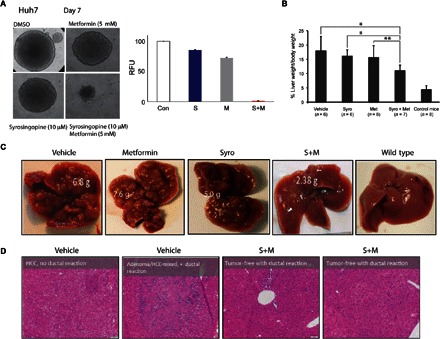
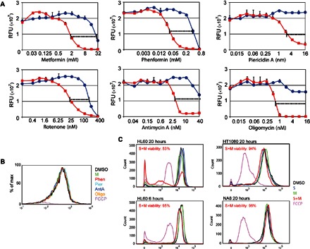
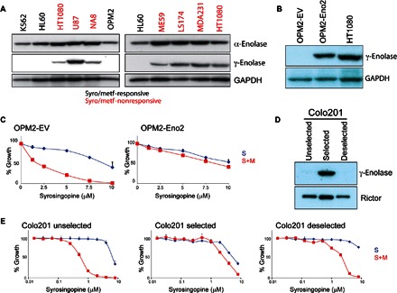
We report that the anticancer activity of the widely used diabetic drug metformin is strongly potentiated by syrosingopine. Synthetic lethality elicited by combining the two drugs is synergistic and specific to transformed cells. This effect is unrelated to syrosingopine's known role as an inhibitor of the vesicular monoamine transporters. Syrosingopine binds to the glycolytic enzyme α-enolase in vitro, and the expression of the γ-enolase isoform correlates with nonresponsiveness to the drug combination. Syrosingopine sensitized cancer cells to metformin and its more potent derivative phenformin far below the individual toxic threshold of each compound. Thus, combining syrosingopine and codrugs is a promising therapeutic strategy for clinical application for the treatment of cancer.

Keywords: Cancer; Mitochondria; biguanide; metabolism; metformin; synthetic lethality; syrosingopine.


Figures

Fig. 1
Fig. 1. Synthetic lethality between syrosingopine and metformin.
(A) Proliferation assay of murine 6.5 cells titrated with increasing amounts of syrosingopine (S) alone and in the presence of 4 mM metformin (S+M). (B and C) Similar titration in various human cancer cell lines: HL60 (promyelocytic leukemia), OPM2 (multiple myeloma), and HT1080 (fibrosarcoma). (D) Titration of ex vivo human leukemic blasts (AML7991) and similar titration with peripheral blood cells (PBC1) from a healthy donor. (E) Annexin V staining for apoptotic cells was performed on HL60 cells after 20 hours of treatment with syrosingopine (Syro; 5 μM), metformin (Met; 5 mM), or in combination. Viable cells are in the lower left quadrant, and early apoptotic and late apoptotic cells are in the lower right and upper right quadrants, respectively. Bottom: Phase-contrast microscopy of the same cells. Ctrl, control. (F) Fluorescence-activated cell sorting (FACS) profile of annexin V–stained CML7359 leukemic blast cells treated for 60 hours with syrosingopine (5 μM), metformin (5 mM), and in combination (S+M). The cells were also treated with staurosporine (Stauro; 1 μM) as a positive control (Con) for apoptosis.
Fig. 2
Fig. 2. Efficacy of drug combination in mouse and solid tumor models.
(A) Hepatospheres of Huh7 hepatocellular carcinoma cells treated with syrosingopine and metformin as indicated. Right: Quantitation of surviving cells by resazurin staining. DMSO, dimethyl sulfoxide; RFU, relative fluorescence units. (B) Reduction in the ratio of liver weight/body weight in the mouse liver cancer model treated with syrosingopine-metformin. *P = 0.9 and **P = 0.95. (C) External liver appearance after 2 weeks of drug treatment. (D) Histological sections from vehicle- and drug combination–treated livers and accompanying pathological report. HCC, hepatocellular carcinoma.
Fig. 3
Fig. 3. Syrosingopine is synthetic lethal with mitochondrial ETC inhibition.
(A) Proliferation assay of 6.5 cells treated with syrosingopine alone (blue lines) or in combination (red lines) with various mitochondrial inhibitors: piericidin A (1 nM), sodium malonate (30 mM), antimycin A (5 nM), sodium azide (1 mM), oligomycin (1 nM), and FCCP (10 μM). Data shown are non-normalized, and the y axis intercept shows the effect on cell growth of each mitochondrial inhibitor by itself in the absence of syrosingopine. (B) Immunoblot of 6.5, HL60, and OPM2 parental and ρ0-derived lines for mitochondrial genome–encoded MT-Co1 (mitochondrially encoded cytochrome c oxidase I). GAPDH, glyceraldehyde-3-phosphate dehydrogenase. (C) Proliferation assay of 6.5, HL60, and OPM2 parental cells (blue lines) and ρ0 derivatives (red lines), titrated with increasing concentrations of syrosingopine. Growth was measured after 3 days of treatment.
Fig. 4
Fig. 4. Syrosingopine strongly potentiates the effect of mitochondrial inhibitors.
(A) HL60 cells treated with increasing amounts of various mitochondrial inhibitors in the absence (blue lines) or presence of 5 μM syrosingopine (red lines). (B) Determination of mitochondrial membrane potential with TMRM staining. HL60 cells were treated for 20 hours with the following compounds: 5 mM metformin, 100 μM phenformin (Phen), 8 nM piericidin A (Pier), 5 nM antimycin A (AntA), 1 nM oligomycin (Oligo), 10 μM FCCP (positive control), and 5 μM syrosingopine. (C) Mitochondrial membrane potential of cells treated with the indicated drugs for the indicated length of time measured by TMRM staining. Viability of syrosingopine-metformin–treated cells was measured by trypan blue staining, followed by automated cell counting.
Fig. 5
Fig. 5. Syrosingopine-elicited synthetic lethality is unrelated to VMAT inhibition.
(A) 6.5 and HL60 cells treated with syrosingopine (S), syrosingopine in the presence of 4 mM metformin (S+M), reserpine (R), and reserpine in the presence of 4 mM metformin (R+M). (B) 6.5ρ0 and HL60ρ0 cells treated with syrosingopine (S) and reserpine (R). (C) 6.5 and HL60 cells treated with tetrabenazine (T), tetrabenazine in the presence of 4 mM metformin (T+M), and ρ0 cells with tetrabenazine alone (gray lines). Growth was measured after 3 days of treatment.
Fig. 6
Fig. 6. Syrosingopine binds the glycolytic enzyme α-enolase.
(A) Coomassie-stained SDS–polyacrylamide gel electrophoresis (SDS-PAGE) gels of HL60 cell lysate after a DARTS assay (±50 μM syrosingopine). Band marked with asterisk was excised, and proteins were eluted for mass spectrometry. (B) α-Enolase peptides from the excised band were identified by mass spectrometry. (C) Thermophoretic profile of binding interaction between syrosingopine and recombinant α-enolase (black curve) and γ-enolase (gray curve). (D) Enolase activity assay performed at room temperature for HL60 lysates treated as indicated. (E) Measurement in 6.5ρ0 cells of ATP levels and extracellular lactate after treatment with selected drugs for 7 hours. **P = 0.99. ns, not significant. RLU, relative luminescence units. (F) Proliferation assay of HL60 cells titrated with NaF in the presence or absence of 4 mM metformin for 3 days.
Fig. 7
Fig. 7. γ-Enolase correlates with syrosingopine-metformin insensitivity.
(A) Cell panel of syrosingopine-metformin–responsive and syrosingopine-metformin–nonresponsive cell lines immunoblotted for α-enolase (Eno1) and γ-enolase (Eno2). (B) Immunoblot for γ-enolase expression in OPM2 cells transfected with an Eno2 ORF (open reading frame)–bearing virus. (C) Proliferation assay of OPM2–EV (empty vector) and OPM2-Eno2 cells treated with syrosingopine-metformin for 3 days. (D) Immunoblot for γ-enolase in Colo201 cells undergoing selection with syrosingopine-metformin and with selection removed. (E) Proliferation assay of Colo201 cells titrated with syrosingopine in the presence or absence of 4 mM metformin for 4 days.

References

    1. Evans J. M. M., Donnelly L. A., Emslie-Smith A. M., Alessi D. R., Morris A. D., Metformin and reduced risk of cancer in diabetic patients. BMJ 330, 1304–1305 (2005). - PMC - PubMed
    1. Libby G., Donnelly L. A., Donnan P. T., Alessi D. R., Morris A. D., Evans J. M. M., New users of metformin are at low risk of incident cancer: A cohort study among people with type 2 diabetes. Diabetes Care 32, 1620–1625 (2009). - PMC - PubMed
    1. Jiralerspong S., Palla S. L., Giordano S. H., Meric-Bernstam F., Liedtke C., Barnett C. M., Hsu L., Hung M.-C., Hortobagyi G. N., Gonzalez-Angulo A. M., Metformin and pathologic complete responses to neoadjuvant chemotherapy in diabetic patients with breast cancer. J. Clin. Oncol. 27, 3297–3302 (2009). - PMC - PubMed
    1. Decensi A., Puntoni M., Goodwin P., Cazzaniga M., Gennari A., Bonanni B., Gandini S., Metformin and cancer risk in diabetic patients: A systematic review and meta-analysis. Cancer Prev. Res. 3, 1451–1461 (2010). - PubMed
    1. Noto H., Goto A., Tsujimoto T., Noda M., Cancer risk in diabetic patients treated with metformin: A systematic review and meta-analysis. PLOS ONE 7, e33411 (2012). - PMC - PubMed

LinkOut - more resources