Review
. 2017 Jul;37(4):907-935.
doi: 10.1002/med.21426. Epub 2016 Dec 28.

Trends in Regenerative Medicine: Repigmentation in Vitiligo Through Melanocyte Stem Cell Mobilization

Affiliations
Review

Trends in Regenerative Medicine: Repigmentation in Vitiligo Through Melanocyte Stem Cell Mobilization

Stanca A Birlea et al. Med Res Rev. 2017 Jul.

Abstract

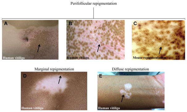
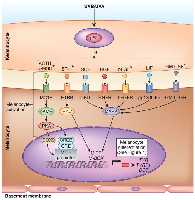
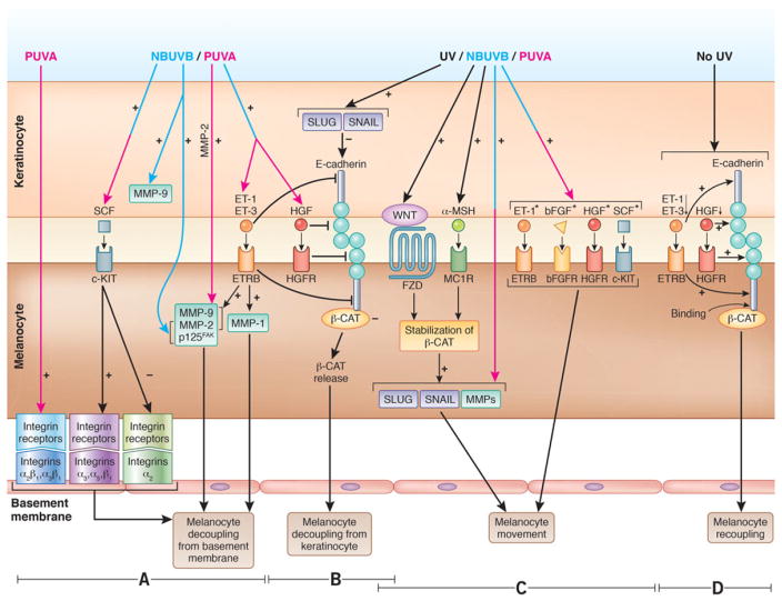
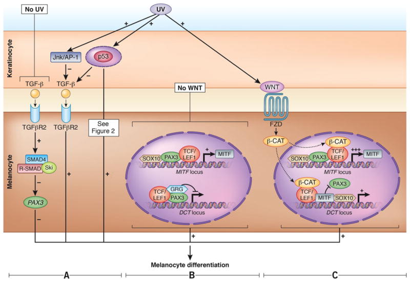
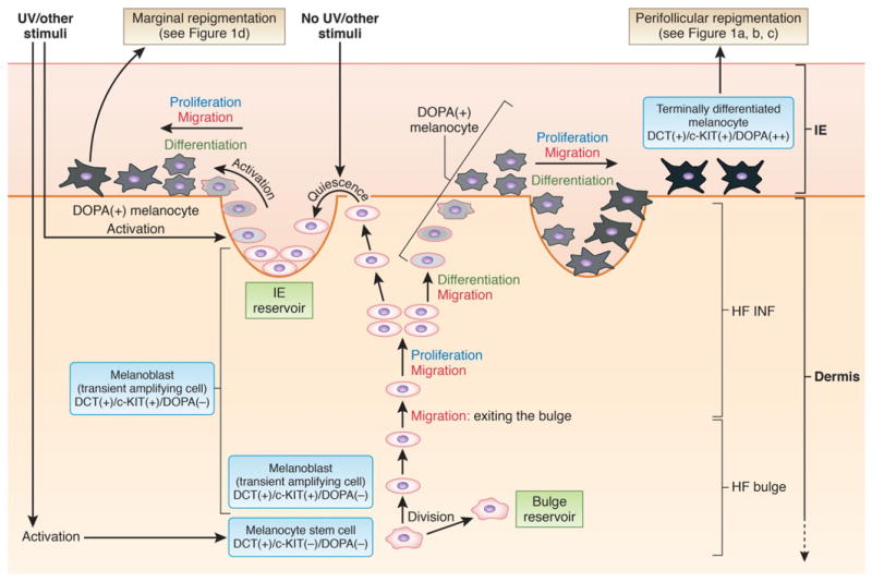
Vitiligo is the most frequent human pigmentary disorder, characterized by progressive autoimmune destruction of mature epidermal melanocytes. Of the current treatments offering partial and temporary relief, ultraviolet (UV) light is the most effective, coordinating an intricate network of keratinocyte and melanocyte factors that control numerous cellular and molecular signaling pathways. This UV-activated process is a classic example of regenerative medicine, inducing functional melanocyte stem cell populations in the hair follicle to divide, migrate, and differentiate into mature melanocytes that regenerate the epidermis through a complex process involving melanocytes and other cell lineages in the skin. Using an in-depth correlative analysis of multiple experimental and clinical data sets, we generated a modern molecular research platform that can be used as a working model for further research of vitiligo repigmentation. Our analysis emphasizes the active participation of defined molecular pathways that regulate the balance between stemness and differentiation states of melanocytes and keratinocytes: p53 and its downstream effectors controlling melanogenesis; Wnt/β-catenin with proliferative, migratory, and differentiation roles in different pigmentation systems; integrins, cadherins, tetraspanins, and metalloproteinases, with promigratory effects on melanocytes; TGF-β and its effector PAX3, which control differentiation. Our long-term goal is to design pharmacological compounds that can specifically activate melanocyte precursors in the hair follicle in order to obtain faster, better, and durable repigmentation.

Keywords: melanoblast; melanocyte stem cell; regeneration; repigmentation; vitiligo.


Figures

Figure 1
Figure 1
Repigmentation patterns in human vitiligo and mouse model. (A, B, C) Perifollicular repigmentation pattern: (A, B) in human vitiligo after narrow band ultraviolet B (NBUVB) therapy; (C) in K14-Steel factor (SLF)/Dct-lacZ double transgenic mouse treated with c-Kit-blocking antibodies. (D) Marginal repigmentation pattern in human vitiligo. (E) Diffuse repigmentation pattern in human vitiligo. Panels A–C: Reproduced from [28]. Panel D: Reproduced from [3]. Panel E: Authors’ original. Arrows point toward the specific repigmentation pattern described in each panel.
Figure 2
Figure 2
Melanocyte activation integrated in the schematic view of the UV-induced pigmentation pathway in the normal skin. Once stimulated by UV light, p53 initiates the release by keratinocyte of melanogenic paracrine growth factors and cytokines: adrenocorticotropic hormone (ACTH), α-melanocyte-stimulating hormone (α-MSH),, endothelin-1 (ET-1), stem cell factor (SCF), hepatocyte growth factor (HGF), basic fibroblast growth factor (bFGF),, leukemia inhibitory factor (LIF), and granulocyte macrophage colony-stimulating factor (GM-CSF)., The keratinocyte factors further interact with their corresponding receptors on melanocytes, and induce melanocyte activation, with subsequent stimulation of microphtalmia-associated transcription factor (MITF), and its downstream targets, the melanogenic enzymes Tyrosinase (TYR), Tyrosinase-related protein 1 (TYRP1), and Dopachrome tautomerase (DCT); this cascade leads to synthesis of melanin and melanocyte differentiation (see Fig. 4). MITF, the master regulator of melanogenic pathway, is activated through three signaling pathways regulated by cAMP,, protein kinase C (PKC),,, and mitogen-activated protein kinase (MAPK), to subsequently induce proliferation and differentiation of human melanocytes. Figure based and modified from [62, 63]. UVB was shown to induce all keratinocyte factors included in the figure. *Factors activated by both UVB and UVA. Figure 2 was done by the medical illustrator Debbie Maizels.
Figure 3
Figure 3
Component processes of melanocyte migration (A–C). (A) Melanocyte decoupling from basement membrane. Left-hand side section: PUVA induces the expression of α2 βl and α3 βl receptors in human melanocyte, which mediates melanocyte detachment from basement membrane. Middle section: SCF downregulates the expression of α2 receptor, and upregulates the expression of α3, α5, and β1 integrin receptors in human neonatal melanocytes, which enhance melanocyte decoupling. Right-hand side section: NBUVB increases the expression of endothelin receptor B (ETRB)-induced metalloproteinases (MMPs) and p125FAK in melanocyte, melanoma cells, and keratinocyte,, which enhances melanocytic cells decoupling from the basement membrane. PUVA also induces significant upregulation of MMP-2. (B) Decoupling of melanocyte from keratinocyte. Melanocyte decoupling from keratinocytes is orchestrated by ET-1 and hepatocyte growth factor (HGF), both inhibitors of E-cadherin; decreased expression of E-cadherin in UVB-irradiated melanocytic cells and keratinocytes produces loss of cell–cell adhesions. E-cadherin is also repressed in human keratinocytes by transcription factors SLUG and SNAIL, whose expression is elevated by UV, possibly through WNT/β-catenin activation. (C) Melanocyte movement. UV-induced loss of E-cadherin releases β-catenin, which forms a complex with a Frizzled (FZD) receptor, and further binds a WNT ligand expressed by keratinocyte; these events lead to β-catenin translocation from cytoplasm to the nucleus where β-catenin induces the expression of WNT target genes (SNAIL/SLUG, MMP-2, MMP-9) which under UV, stimulate melanocyte migration., ,– Melanocyte migration is also stimulated by α-MSH-MC1R, and SCF/c-KIT signaling pathways, ET-1, bFGF, and HGF., (D) Melanocyte recoupling. Melanocyte recoupling to keratinocyte to reform the epidermal melanin unit can be supposedly initiated by increased expression of E-cadherin in absence of UV-induced stimulation of ET-1, ET-3, and HGF. Subsequently, E-cadherin can bind β-catenin, sequestering it in the cytoplasm, thus reestablishing the melanocyte–keratinocyte attachment. Experimental evidence used for this figure was generated in human, and mouse models, or using data from in vitro experiments performed on melanocyte/melanoma/epidermoid carcinoma cells. Pink, blue, and pink–blue arrows denote the action of either UVA or UVB, or of both, respectively. *Factors that stimulatemelanocyte proliferation. Figure 3 was done by the medical illustrator Debbie Maizels.
Figure 4
Figure 4
Melanocyte differentiation. Transforming growth factor β (TGF-β) signaling pathway. (A) In the absence of UV radiation (left-hand side section), keratinocytes express TGF-β that blocks melanocyte differentiation via SMAD signaling, by repressing PAX3. In the presence of UV, the JNK/AP-1 pathway downregulates TGF-β (middle section). TGF-β is itself repressed in epidermal keratinocytes by p53 that promotes melanocyte differentiation., (B, C) Canonical WNT signaling pathway. (B) In the absence of WNT: Upper side in the melanocyte nucleus-the complex of PAX3-SOX10 can activate the expression of MITF. Lower side in the melanocytes nucleus-the transcriptional repressor complex PAX3-LEF1-groucho-related proteins (GRGs) represses DCT transcription. (C) In the presence of WNT: Upper side in the melanocyte nucleus-β-catenin is activated by WNT and, together with LEF1, upregulates expression of MITF. Lower side in the melanocyte nucleus-activated β-catenin also forms an activator complex on the DCT promoter with MITF and LEF1 and displaces the repressor complex containing PAX3. SOX10 can also synergistically activate DCT with MITF. Experimental evidence used for this figure was generated in human and mouse. Parts B and C of Figure 4 are based on and modified from [97]. Figure 4 was done by the medical illustrator Debbie Maizels.
Figure 5
Figure 5
Working model of the repigmentation pathway. This scheme was reproduced and modified from [48] and is based on data produced in a K14-Steel factor (SLF); Dct-lacZ double transgenic mouse, in a non-vitiligo Dct-LacZ+ mouse after wounding or UVB exposure, or in C57 black male mice after PUVA, and in human vitiligo after NBUVB or PUVA treatment, or after application of topical Chinese herbal medication. DCT, dopachrome tautomerase; DOPA, 3,4-dihydroxyphenylalanine; HF, hair follicle; IE, interfollicular epidermis; INF, infundibulum. Figure 5 was done by the medical illustrator Debbie Maizels.

Similar articles

Cited by

References

    1. Birlea SA, Spritz RA, Norris DA. Vitiligo. In: Wolff K, Goldsmith LA, Katz SI, Gilchrest BA, Paller AS, Leffell DJ, editors. Fitzpatrick’s Dermatology in General Medicine. 8. New York, NY: McGraw-Hill; 2012. pp. 792–803.
    1. Le Poole IC, van den Wijngaard RM, Westerhof W, Dutrieux RP, Das PK. Presence or absence of melanocytes in vitiligo lesions: An immunohistochemical investigation. J Invest Dermatol. 1993;100:816–822. - PubMed
    1. Falabella R. Vitiligo and the melanocyte reservoir. Indian J Dermatol. 2009;54:313–318. - PMC - PubMed
    1. Goldstein NB, Koster MI, Hoaglin LG, Spoelstra NS, Kechris KJ, Robinson SE, Robinson WA, Roop DR, Norris DA, Birlea SA. Narrow band ultraviolet B treatment for human vitiligo is associated with proliferation, migration, and differentiation of melanocyte precursors. J Invest Dermatol. 2015;135:2068–2076. - PMC - PubMed
    1. Ito T, Ito N, Saatoff M, Hashizume H, Fukamizu H, Nickoloff BJ, Takigawa M, Paus R. Maintenance of hair follicle immune privilege is linked to prevention of NK cell attack. J Invest Dermatol. 2008;128:1196–1206. - PubMed

Publication types