. 2018 Sep;38(9):1618-1630.
doi: 10.1177/0271678X16682509. Epub 2016 Dec 28.

Hemodynamic mechanisms underlying elevated oxygen extraction fraction (OEF) in moyamoya and sickle cell anemia patients

Affiliations

Hemodynamic mechanisms underlying elevated oxygen extraction fraction (OEF) in moyamoya and sickle cell anemia patients

Jennifer M Watchmaker et al. J Cereb Blood Flow Metab. 2018 Sep.

Abstract

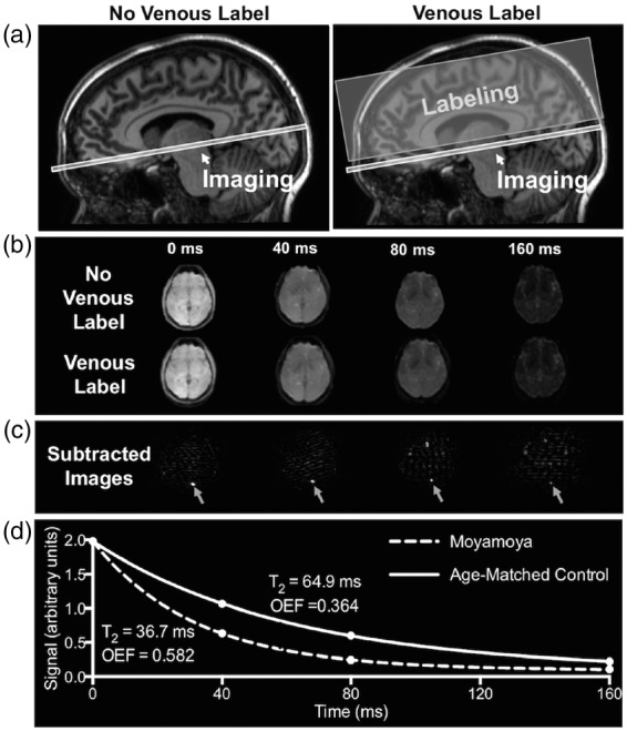
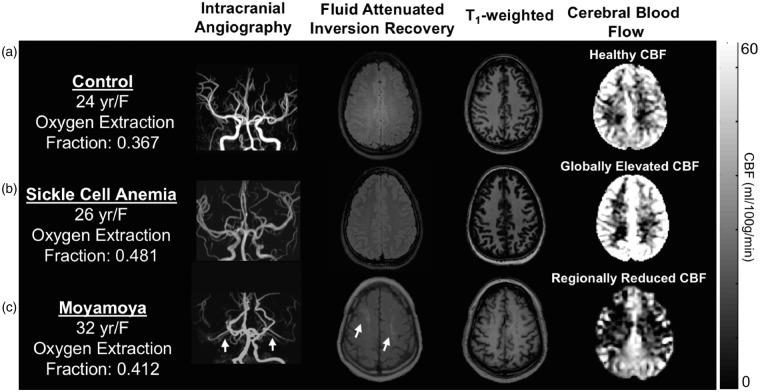
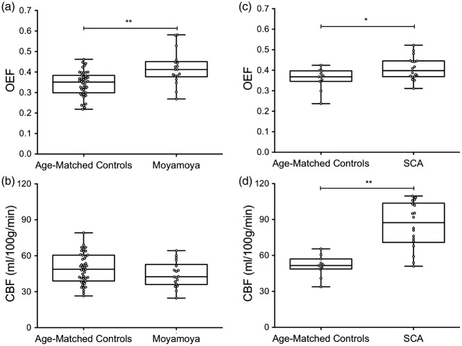
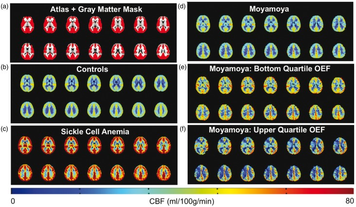
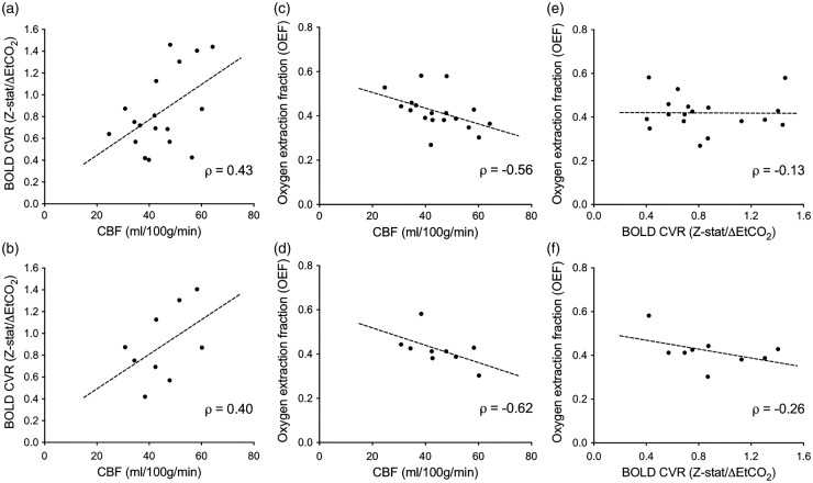
Moyamoya is a bilateral, complex cerebrovascular condition characterized by progressive non-atherosclerotic intracranial stenosis and collateral vessel formation. Moyamoya treatment focuses on restoring cerebral blood flow (CBF) through surgical revascularization, however stratifying patients for revascularization requires abilities to quantify how well parenchyma is compensating for arterial steno-occlusion. Globally elevated oxygen extraction fraction (OEF) secondary to CBF reduction may serve as a biomarker for tissue health in moyamoya patients, as suggested in patients with sickle cell anemia (SCA) and reduced oxygen carrying capacity. Here, OEF was measured (TRUST-MRI) to test the hypothesis that OEF is globally elevated in patients with moyamoya (n = 18) and SCA (n = 18) relative to age-matched controls (n = 43). Mechanisms underlying the hypothesized OEF increases were evaluated by performing sequential CBF-weighted, cerebrovascular reactivity (CVR)-weighted, and structural MRI. Patients were stratified by treatment and non-parametric tests applied to compare study variables (significance: two-sided P < 0.05). OEF was significantly elevated in moyamoya participants (interquartile range = 0.38-0.45) compared to controls (interquartile range = 0.29-0.38), similar to participants with SCA (interquartile range = 0.37-0.45). CBF was inversely correlated with OEF in moyamoya participants. Elevated OEF was only weakly related to reductions in CVR, consistent with basal CBF level, rather than vascular reserve capacity, being most closely associated with OEF.

Keywords: MRI; Oxygen extraction fraction; moyamoya; neurophysiology; stroke.


Figures

Figure 1.
Figure 1.
T2-relaxation-under-spin-tagging (TRUST) MRI for non-invasive quantification of whole-brain oxygen extraction fraction (OEF). TRUST-MRI is used to determine the Carr–Purcell–Meiboom–Gill (CPMG) T2 in the superior sagittal sinus, which has a known relationship with venous oxygenation (Yv). Arterial oxygenation (Ya) is measured by pulse oximetry. By taking the fractional difference in oxygenation (Ya-Yv)/Ya, OEF is calculated. A schematic of the venous blood imaging is shown in panel (a). Volume-selective venous labeling pulses are applied to invert inflowing venous blood water magnetization; these labeling acquisitions are interleaved with an image with no labeling (control image). Panel (b) shows representative control and label images at four different effective echo times (eTEs). Panel (c) shows the subtraction of the label image from control image, which allows for isolation of signal in the superior sagittal sinus. Signal intensities from the subtraction image (control-label) at the four eTEs are fitted to a known model (d) and venous blood water T2 is determined. Panel (d) shows T2 decay for a control with a normal OEF (T2 = 64.9 ms; Yv = 0.62; OEF = 0.364) and a participant with moyamoya (T2 = 36.7 ms; Yv = 0.41; OEF = 0.582).
Figure 2.
Figure 2.
Individual cases of structural, cerebral blood flow (CBF), and oxygen extraction fraction (OEF) imaging. (a) A healthy control participant with a normal OEF, and normal findings on structural and CBF imaging. (b) A participant with sickle cell anemia (SCA) and (c) moyamoya. The participant with SCA demonstrates elevated CBF in the setting of decreased oxygen carrying capacity of hemoglobin and increased OEF (OEF = 0.481). The participant with bilateral moyamoya with lenticulostriate collaterals (white arrows on intracranial angiography) demonstrates right and left anterior and middle cerebral artery territory infarcts seen on FLAIR, bilateral anterior-territory hypoperfusion, and globally elevated OEF (OEF = 0.412) relative to control data.
Figure 3.
Figure 3.
Oxygen extraction fraction (OEF) and cerebral blood flow (CBF) in moyamoya, sickle cell anemia (SCA), and age-matched control participants. A two-sided Wilcoxon rank-sum test was performed to test the primary hypothesis that OEF was elevated in moyamoya and SCA participants compared to age-matched controls, and that CBF was decreased in moyamoya participants compared to controls. (a) OEF is elevated in moyamoya participants compared to age-matched controls (P < 0.001). (b) Reduced mean gray matter CBF in moyamoya participants relative to controls, but this reduction was not significant, likely due to preserved posterior territory CBF in many patients and regions of high ASL intravascular signal. (c) OEF is elevated in SCA participants compared to age-matched controls (P = 0.045). (d) CBF is elevated in SCA participants compared to age-matched controls (P < 0.001). *P < 0.05, **P < 0.01. In the moyamoya arm of the study, a sub-analysis was performed that evaluated only females in both the control (n = 15) and moyamoya (n = 17) groups. Trends were identical, and the OEF was elevated in the moyamoya participants compared to the controls (P = 0.06), and CBF was decreased in the moyamoya participants compared to controls (P = 0.02).
Figure 4.
Figure 4.
Non-invasive cerebral blood flow (CBF) imaging using pseudo-continuous arterial spin labeling. Central slices of a T1-weighted atlas with gray matter mask (red) overlaid are shown in (a). Mean CBF maps in controls (n = 43) and participants with sickle cell anemia (n = 18) are shown in (b,c). (d) shows mean CBF maps from moyamoya participants (n = 18). Mean CBF maps from moyamoya participants with bottom quartile OEF values (OEF < 0.38), and moyamoya participants with upper quartile OEF values (OEF > 0.45) are shown in (e) and (f), respectively.
Figure 5.
Figure 5.
Relationship between cerebral blood flow (CBF), cerebrovascular reactivity (CVR), and oxygen extraction fraction (OEF) in participants with moyamoya. A Spearman’s rho (ρ) was calculated to test the relationships between CBF, CVR, and OEF. A sub-analysis was performed in only participants who had not undergone surgical revascularization (n = 9; lower). CBF trended positively with blood oxygenation level-dependent (BOLD) hypercapnia-induced cerebrovascular reactivity measured as mean gray-matter z-statistic divided by mean change in end-tidal CO2 (ΔEtCO2) in mmHg (Z-stat/ΔEtCO2) when considering all moyamoya participants (a; ρ = 0.43, P = 0.073) and moyamoya participants who had not undergone surgical revascularization (b; ρ = 0.40, P = 0.29). The larger range in CBF was attributable to known, regional hyperintensity artifacts in ASL due to residual intravascular signal at the time of imaging. CBF was inversely correlated with OEF when considering all moyamoya participants (c; ρ = −0.56, P = 0.016) and moyamoya participants who had not undergone surgical revascularization (d; ρ = −0.62, P = 0.082). There was a non-significant inverse relationship between BOLD reactivity and OEF when considering all moyamoya patients (e; ρ = −0.13, P = 0.61) and moyamoya participants who had not undergone surgical revascularization (f; ρ = −0.26, P = 0.50). The mean change in EtCO2 during the hyercapnic CVR study was 4.82 ± 2.15 mmHg. Additionally, the BOLD signal change during hypercapnia normalized by baseline signal strongly correlated with z-statistic values (ρ = 0.80, P ≤ 0.0001).

References

    1. Scott RM, Smith ER. Moyamoya disease and moyamoya syndrome. New Engl J Med 2009; 360: 1226–1237. - PubMed
    1. Hallemeier CL, Rich KM, Grubb RL, Jr., et al. Clinical features and outcome in North American adults with moyamoya phenomenon. Stroke 2006; 37: 1490–1496. - PubMed
    1. Arias EJ, Derdeyn CP, Dacey RG, Jr., et al. Advances and surgical considerations in the treatment of moyamoya disease. Neurosurgery 2014; 74(Suppl 1): S116–S125. - PubMed
    1. Liebeskind DS, Cotsonis GA, Saver JL, et al. Collaterals dramatically alter stroke risk in intracranial atherosclerosis. Ann Neurol 2011; 69: 963–974. - PMC - PubMed
    1. Strother MK, Anderson MD, Singer RJ, et al. Cerebrovascular collaterals correlate with disease severity in adult North American patients with Moyamoya disease. AJNR Am J Neuroradiol 2014; 35: 1318–1324. - PMC - PubMed

Publication types