. 2017 Jan 17;8(3):5233-5246.
doi: 10.18632/oncotarget.14125.

The pseudogene DUXAP10 promotes an aggressive phenotype through binding with LSD1 and repressing LATS2 and RRAD in non small cell lung cancer

Affiliations

The pseudogene DUXAP10 promotes an aggressive phenotype through binding with LSD1 and repressing LATS2 and RRAD in non small cell lung cancer

Chen-Chen Wei et al. Oncotarget. .

Abstract

Pseudogenes have been considered as non-functional transcriptional relics of human genomic for long time. However, recent studies revealed that they play a plethora of roles in diverse physiological and pathological processes, especially in cancer, and many pseudogenes are transcribed into long noncoding RNAs and emerging as a novel class of lncRNAs. However, the biological roles and underlying mechanism of pseudogenes in the pathogenesis of non small cell lung cancer are still incompletely elucidated. This study identifies a putative oncogenic pseudogene DUXAP10 in NSCLC, which is located in 14q11.2 and 2398 nt in length. Firstly, we found that DUXAP10 was significantly up-regulated in 93 human NSCLC tissues and cell lines, and increased DUXAP10 was associated with patients poorer prognosis and short survival time. Furthermore, the loss and gain of functional studies including growth curves, migration, invasion assays and in vivo studies verify the oncogenic roles of DUXAP10 in NSCLC. Finally, the mechanistic experiments indicate that DUXAP10 could interact with Histone demethylase Lysine specific demethylase1 (LSD1) and repress tumor suppressors Large tumor suppressor 2 (LATS2) and Ras-related associated with diabetes (RRAD) transcription in NSCLC cells. Taken together, these findings demonstrate DUXAP10 exerts the oncogenic roles through binding with LSD1 and epigenetic silencing LATS2 and RRAD expression. Our investigation reveals the novel roles of pseudogene in NSCLC, which may serve as new target for NSCLC diagnosis and therapy.

Keywords: DUXAP10; LSD1; migration and invasion; proliferation; pseudogene.


Conflict of interest statement

CONFLICTS OF INTEREST

The authors declare no conflicts of interest.

Figures

Figure 1
Figure 1. Relative DUXAP10 expression levels in NSCLC tissues and its clinical significance
A, B. Relative expression of DUXAP10 in NSCLC tissues compared with normal tissue was analyzed by using GEO datasets GSE31210 and GSE19188. C. Relative expression of DUXAP10 in 93 pairs NSCLC tissues compared with corresponding non-tumor tissues was examined by qPCR, and normalized to GAPDH expression. D. The patients were divided into two groups according to DUXAP10 expression. E. Kaplan–Meier overall survival and progression-free survival curves according to DUXAP10 expression levels.
Figure 2
Figure 2. Effects of DUXAP10 on NSCLC cell proliferation in vitro
A. DUXAP10 expression levels of NSCLC cell lines (A549, H1975, SPC-A1, H1299, H226 and PC-9), compared with that in normal human bronchial epithelial cells (16HBE). B. A549 and H1975 cells were transfected with si-DUXAP10, PC-9 cells were transfected with pCDNA-DUXAP10. C. MTT assays were performed to determine the cell viability for si-DUXAP10-transfected A549 and H1975, and PC-9 cells transfected with pCDNA-DUXAP10. D, E. Colony-forming assays and EDU staining assays were used to determine the proliferation of si-DUXAP10-transfected A549 and H1975 cells. *P<0.05, **P<0.01.
Figure 3
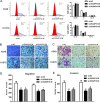
Figure 3. Knockdown of DUXAP10 inhibited cell cycle and cell migration and invasion in vitro
A. Flow cytometry assays were performed to analysis the cell cycle progression when NSCLC cells transfected with si-DUXAP10. The bar chart represented the percentage of cells in G0/G1, S, or G2/M phase, as indicated. All experiments were performed in biological triplicates with three technical replicates. B to E. Effect of knockdown of DUXAP10 on cell migration and invasion. Data are presented as mean ± SD. *P<0.05, **P<0.01.
Figure 4
Figure 4. The stable DUXAP10 knockdown A549 cells were used for the in vivo study
A and B. The nude mice carrying tumors from respective groups were shown and tumor growth curves were measured after the injection of A549 cells. Tumor volume was calculated every 4 days. C. Tumor weights are represented as means of tumor weights ±S.D. D. qPCR assay was performed to determine the average expression of DUXAP10 in xenograft tumors. E. Tumors developed from sh-DUXAP10 transfected A549 cells showed lower Ki67 protein levels than tumors developed by control cells. Upper: H & E staining; Lower: immunostaining.
Figure 5
Figure 5. DUXAP10 could inhibit LATS2 and RRAD expression
A. DUXAP10 expression levels in cell cytoplasm or nucleus of NSCLC cell lines A549 and H1975 were detected by qPCR. GAPDH was used as a cytosol marker and U1 was used as a nucleus marker. B. RIP with rabbit monoclonal anti-LSD1, rabbit monoclonal anti-EZH2, rabbit monoclonal anti-SUZ12, preimmune IgG, or 10% input from A549 and H1975 cell extracts. RNA levels in immunoprecipitates were detected by qPCR. Expression levels of DUXAP10 RNA are presented as fold enrichment in LSD1 relative to IgG immunoprecipitates. C. RNA pulldown and western blotting assays were performed and the results revealed that DUXAP10 could bind to LSD1. D and E. The qPCR and western blot assay were conducted to detect the levels of LATS2 and RRAD mRNA in A549 and H1975 cells transfected with si-DUXAP10 and results are expressed relative to the corresponding values for control cells. F and G. QPCR and Western blot assays were used to detect the LATS2 and RRAD expression both in mRNA and protein levels in A549 and H1975 cells transfected with si-LSD1. H and I. ChIP–qPCR of LSD1 occupancy and H3K4-2me binding in the LATS2 and RRAD promoter in A549 and H1975 cells, and IgG as a negative control. At 48h after transfection, ChIP–qPCR of LSD1 occupancy and H3K4-2me binding in the LATS2 and RRAD promoter in A549 and H1975 cells treated with si-DUXAP10 or scrambled siRNA. *P<0.05, **P<0.01.
Figure 6
Figure 6. Effect of LATS2 and RRAD of overexpression on A549 cell in vitro
A, B. The mRNA levels and protein levels of LATS2 and RRAD in A549 cells transfected with pCDNA–LATS2 or pCDNA-RRAD was detected by qPCR analysis. C, D. MTT assays and Edu staining assays were used to determine the cell viability. Values represent the mean ± s.d. from three independent experiments. E. Cell cycle was analyzed by flow cytometry. The bar chart represents the percentage of cells in G1–G0, S, or G2–M phase, as indicated. *P < 0.05 and **P < 0.01.
Figure 7
Figure 7. DUXAP10 negatively regulates expression of LATS2 and RRAD by rescue assays
A, B, C, D. MTT, edu and colony formation assays were used to determine the cell proliferation ability for A549 cells transfected with pCDNA-DUXAP10 and pCDNA-LATS2 and pCDNA-RRAD and co-transfected with pCDNA-DUXAP10 and pCDNA-LATS2 or pCDNA-DUXAP10 and pCDNA-RRAD. E. QPCR analyzed the LATS2 and RRAD mRNA levels in 20 pairs NSCLC tissues and found that there was a significantly negative correlation between DUXAP10 and RRAD or LATS2. Values represent the mean ± s.d. from three independent experiments.

References

    1. Siegel R, Naishadham D, Jemal A. Cancer statistics, 2013. CA Cancer J Clin. 2013;63:11–30. - PubMed
    1. Verdecchia A, Francisci S, Brenner H, Gatta G, Micheli A, Mangone L, Kunkler I. Recent cancer survival in Europe: a 2000-02 period analysis of EUROCARE-4 data. Lancet Oncol. 2007;8:784–796. - PubMed
    1. Wistuba II. Genetics of preneoplasia: lessons from lung cancer. Curr Mol Med. 2007;7:3–14. - PubMed
    1. Klebe S, Henderson DW. Facts and fiction: premalignant lesions of lung tissues. Pathology. 2013;45:305–315. - PubMed
    1. Poliseno L, Salmena L, Zhang J, Carver B, Haveman WJ, Pandolfi PP. A coding-independent function of gene and pseudogene mRNAs regulates tumour biology. Nature. 2010;465:1033–1038. - PMC - PubMed

MeSH terms