. 2017 Jan;140(1):146-157.
doi: 10.1093/brain/aww260. Epub 2016 Nov 8.

Molecular magnetic resonance imaging discloses endothelial activation after transient ischaemic attack

Affiliations

Molecular magnetic resonance imaging discloses endothelial activation after transient ischaemic attack

Aurélien Quenault et al. Brain. 2017 Jan.

Abstract

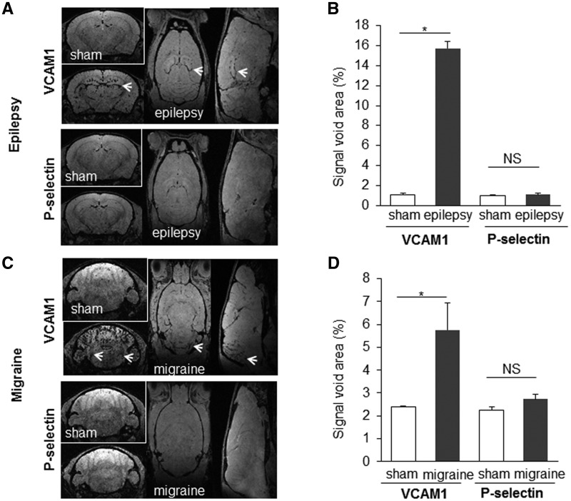
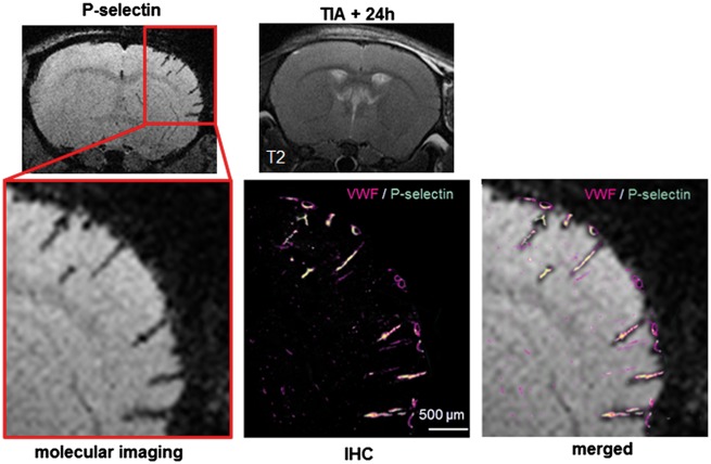
SEE SUN ET AL DOI101093/AWW306 FOR A SCIENTIFIC COMMENTARY ON THIS ARTICLE: About 20% of patients with ischaemic stroke have a preceding transient ischaemic attack, which is clinically defined as focal neurological symptoms of ischaemic origin resolving spontaneously. Failure to diagnose transient ischaemic attack is a wasted opportunity to prevent recurrent disabling stroke. Unfortunately, diagnosis can be difficult, due to numerous mimics, and to the absence of a specific test. New diagnostic tools are thus needed, in particular for radiologically silent cases, which correspond to the recommended tissue-based definition of transient ischaemic attack. As endothelial activation is a hallmark of cerebrovascular events, we postulated that this may also be true for transient ischaemic attack, and that it would be clinically relevant to develop non-invasive in vivo imaging to detect this endothelial activation. Using transcriptional and immunohistological analyses for adhesion molecules in a mouse model, we identified brain endothelial P-selectin as a potential biomarker for transient ischaemic attack. We thus developed ultra-sensitive molecular magnetic resonance imaging using antibody-based microparticles of iron oxide targeting P-selectin. This highly sensitive imaging strategy unmasked activated endothelial cells after experimental transient ischaemic attack and allowed discriminating transient ischaemic attack from epilepsy and migraine, two important transient ischaemic attack mimics. We provide preclinical evidence that combining conventional magnetic resonance imaging with molecular magnetic resonance imaging targeting P-selectin might aid in the diagnosis of transient ischaemic attack.

Keywords: P-selectin; cerebrovascular inflammation; mimics; molecular imaging; transient ischaemic attack.


Figures

Figure 1
Figure 1
Vascular, radiological and neurological characterization of a preclinical mouse model corresponding to the tissue-based definition of TIA in humans. Compression-induced MCA occlusion leads to transient focal brain ischaemia. (A) Schematic description of the mouse model of TIA: after craniotomy, the parietal branch bifurcation of the MCA was exposed, without altering the integrity of the dura. An electrophysiology pipette was affixed and pushed to compress the MCA. (B) Compression-induced MCAO leads to transient focal brain ischaemia, as shown by regional cerebral blood velocity (rCBV) Doppler recordings (mean profiles of 10 mice per group). (C and D) A threshold of duration of MCAO distinguishes TIA from ischaemic stroke. (C) Representative T2 images 24 h after MCAO of different durations (0/sham, 15 or 60 min, and permanent occlusion). (D) Lesion volumes measured by MRI (hypersignals on T2) 24 h after MCAO of different durations. MCAO lasting up to 15 min, induced no brain lesions, while longer occlusions led to infarction that increased with the duration of ischaemia. Individual values are plotted as diamond shapes, and bars represent the mean volume lesion ± standard error of the mean (SEM) (n = 10 per group, *P < 0.05, **P < 0.01 and ***P < 0.001 versus sham). (E) Evidence of reversible functional deficits: P1 and N1 cortical waves were consistently recorded with a mean latency measured before occlusion of 11.2 ± 0.7 ms and 17.6 ± 2.0 ms, respectively, and a mean peak-to-peak amplitude of 77.2 ± 55.3 mV. The figure shows a horizontally stacked representative co-monitoring of regional cerebral blood velocity (continuous) and SEPs (every minute) for one representative mouse subjected to MCAO for 15 min. On the first curve, the P1 and N1 waves are highlighted. On this animal, P1 wave disappears 6 min after initiating MCAO and reappears (asterisk) 1 min after the end of occlusion, first inconstantly and then definitively 5 min after. The mean delays of disappearance and reappearance of SEPs relative to MCAO and MCA de-occlusion, respectively are reported on the top (mean ± SEM; n = 6).
Figure 2
Figure 2
Vascular adhesion molecules as putative biomarkers to discriminate ischaemic stroke from TIA. (A) RT-PCR analyses of mRNA cortical levels for Vcam1 and Selp (P-selectin), two key markers of endothelial activation 24 h after MCAO of different durations. Bars are mean values for expression in ipsilateral and contralateral cortices normalized to expression levels of Gapdh mRNA (n = 3 per group; *P < 0.05 versus sham). (B) Representative immunohistological images of slices stained with antibodies against VWF, P-selectin, VCAM1 in the cortex of sham, TIA and stroke mice and corresponding quantifications (C). A total of 5095, 5452 and 5537 VWF-positive vessels were counted on slices obtained from sham, TIA and ischaemic mice, respectively. The proportion of vessels co-stained with VWF and either P-selectin or VCAM1 was then calculated (*P < 0.05 versus sham, n = 3).
Figure 3
Figure 3
Development and optimization of MPIO-αP-selectin for ultrasensitive molecular MRI of cerebrovascular inflammation. (A) Schematic description of targeted MPIOs. (B and C) Stereotaxic injection of 1 µg of lipopolysaccharide (LPS) in striatum was performed in anaesthetized mice. Molecular MRI acquired 20 min after intravenous (I.V.) administration of targeted-MPIOs (24 h after lipopolysaccharide, n = 3 per group) showed signal voids corresponding to MPIOs binding. MPIO conjugate to IgG did not induce signal void. AF737-coupled MPIO revealed better contrast enhancement than RB40.34-coupled MPIO (*P < 0.05 versus sham). (D) Bright field and confocal imaging of a cerebral vessel immunostained with antibodies against VWF and P-selectin showed in vivo ability of targeted MPIOs to bind P-selectin at the endothelial surface. (E and F) Kinetics of AF737-coupled MPIO clearance from ischaemic stroke mice (n = 3) and corresponding quantification. Signal voids decreased gradually with time after MPIO-αP-selectin injection.
Figure 4
Figure 4
High resolution molecular MRI of P-selectin positively diagnoses TIA. (A) Representative MRA, T2 and T2* images and molecular imaging [serial T2* gradient echo imaging with flow compensation (GEFC) images] of P-selectin and VCAM1, 24 h post-surgery in sham, TIA and ischaemic stroke mice and corresponding quantifications (B) of signal void areas (ipsi/contralateral MCA territory; n = 5 per group; *P < 0.05 versus sham).
Figure 5
Figure 5
High resolution molecular MRI of P-selectin discriminates TIA from epilepsy and migraine, two TIA mimics. Representative images and corresponding quantifications of molecular MRI of P-selectin and VCAM1 in sham animals (saline) and 24 h after inducing epilepsy (kainate 30 mg/kg; intraperitoneally) (A and B) or migraine (nitroglycerin 10 mg/kg; intraperitoneally) (C and D) (n = 5 per group; *P < 0.05 versus sham). Areas expected to be active in each condition are designated by an arrow: the hippocampus for epilepsy and the trigeminal nucleus for migraine.
Figure 6
Figure 6
Illustration of the current state of TIA imaging in the clinical setting (i.e. no brain infarct on T2 or DWI images) and its potential future improvement evidencing a cerebrovascular inflammation revealed by molecular MRI of P-selectin. A superimposition of molecular MRI and regular immunostaining showed that MPIOs targeted for P-selectin are specific signals.

Comment in

References

    1. Arsava EM, Gurer G, Gursoy-Ozdemir Y, Karatas H, Dalkara T. A new model of transient focal cerebral ischemia for inducing selective neuronal necrosis. Brain Res Bull 2009; 78: 226–31. - PubMed
    1. Battistini L, Piccio L, Rossi B, Bach S, Galgani S, Gasperini C, et al.CD8+ T cells from patients with acute multiple sclerosis display selective increase of adhesiveness in brain venules: a critical role for P-selectin glycoprotein ligand-1. Blood 2003; 101: 4775–82. - PubMed
    1. Berti R, Williams AJ, Moffett JR, Hale SL, Velarde LC, Elliott PJ, et al.Quantitative real-time RT-PCR analysis of inflammatory gene expression associated with ischemia-reperfusion brain injury. J Cereb Blood Flow Metab 2002; 22: 1068–79. - PubMed
    1. Beziere N, von Schacky C, Kosanke Y, Kimm M, Nunes A, Licha K, et al.Optoacoustic imaging and staging of inflammation in a murine model of arthritis. Arthritis Rheumatol 2014; 66: 2071–8. - PubMed
    1. Easton JD, Saver JL, Albers GW, Alberts MJ, Chaturvedi S, Feldmann E, et al.Definition and evaluation of transient ischemic attack: a scientific statement for healthcare professionals from the American Heart Association/American Stroke Association Stroke Council; Council on Cardiovascular Surgery and Anesthesia; Council on Cardiovascular Radiology and Intervention; Council on Cardiovascular Nursing; and the Interdisciplinary Council on Peripheral Vascular Disease. The American Academy of Neurology affirms the value of this statement as an educational tool for neurologists. Stroke 2009; 40: 2276–93. - PubMed