Review
. 2017 Jan 2;14(1):1-15.
doi: 10.21873/cgp.20015.

The Multiple Roles of Exosomes in Metastasis

Affiliations
Review

The Multiple Roles of Exosomes in Metastasis

Ulrich H Weidle et al. Cancer Genomics Proteomics. .

Abstract

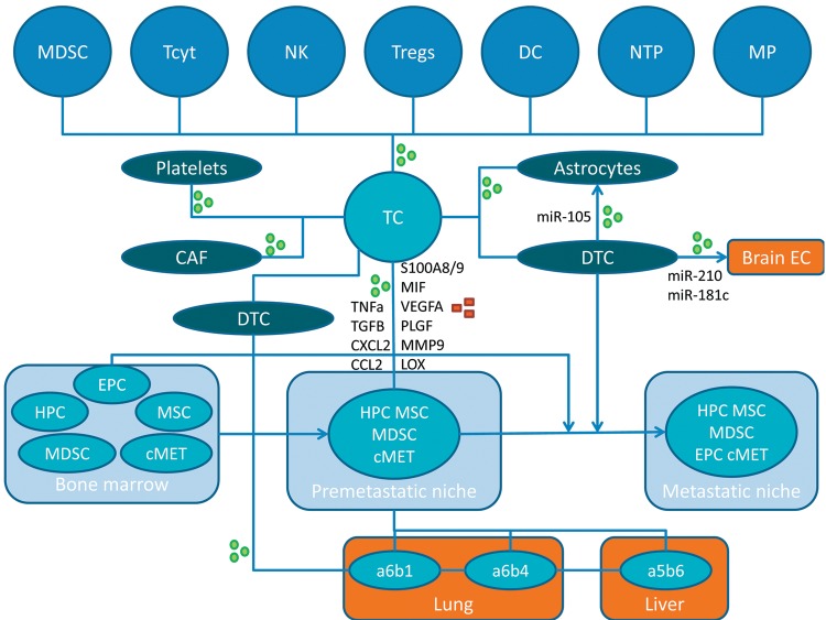
Exosomes are important contributors to cell-cell communication and their role as diagnostic markers for cancer and the pathogenesis for cancer is under intensive investigation. Here, we focus on their role in metastasis-related processes. We discuss their impact regarding promotion of invasion and migration of tumor cells, conditioning of lymph nodes, generation of premetastatic niches and organotropism of metastasis. Furthermore, we highlight interactions of exosomes with bone marrow and stromal components such as fibroblasts, endothelial cells, myeloid- and other immune-related cells in the context of metastases. For all processes as described above, we outline molecular and cellular components for therapeutic intervention with metastatic processes.

Keywords: Exosome interaction with stromal cells; organ tropism of metastasis; pre- and metastatic niche; review.


Figures

Figure 1
Figure 1. Interplay between tumor cells, other cell types, secreted factors and exosomes in the creation of pre- and metastatic niches. Exosomes and secreted factors are indicated by green circles or red squares, respectively. α5β6, α6β1 and α6β4,integrins; CAF, cancer-associated fibroblast; CCL2, CC chemokine ligand 2; cMET, tyrosine kinase receptor c-MET; CXCL2, chemokine (C-X-C motif) ligand 2; DC, dendritic cell; DTC, disseminated tumor cell; EC, endothelial cell; EPC, endothelial progenitor cell; HPC, hematopoietic progenitor cell; LOX, lysyl oxidase; MDSC, myeloid-derived suppressor cell; MIF, macrophage migration inhibitory factor; MMP9, matrix metalloprotease 9; MP, macrophage; MSC, mesenchymal stem cell; NK, natural killer cell; NTP, neutrophile; PLGF, pacental growth factor; S100A8/9, S100 calcium binding protein A8 or A9; TC, tumor cell; Tcyt, cytotoxic T-cell; TGFβ, transforming growth factor β; TNFα, tumor necrosis factor α; Tregs, regulatory T-cells; VEGFA, vascular endothelial growth factor, isoform A.
Figure 2
Figure 2. Expression of selected microRNAs in breast cancer in comparison to matched normal tissues. Steady-state RNA levels corresponding to miR-105, miR-181c and miR-210 based on 1,084 invasive breast cancer samples and 104 matched normal samples derived from cohorts of The Cancer Genome Atlas (TCGA) are shown. Expression was quantified by RNA sequencing and is shown as log2 of normalized read counts. The red lines indicate low versus higher expression. Expression data are shown as box plots. The line in the middle of the box represents the data median, the rectangles show the upper and lower 25% quartile, therefore 50% of all data points are included in the rectangle. All other data points, except for outliers are located within the upper and lower whiskers.
Figure 3
Figure 3. Expression of niche-related factors in breast cancer, melanoma and pancreatic tumors. Steady-state levels form RNA encoding the corresponding factors are shown. Three cohorts derived from The Cancer Genome Atlas (TCGA) are displayed: breast invasive carcinoma (n=1,100), melanoma (n=472) and pancreatic adenocarcinoma (n=179). Expression was quantitated by RNA sequencing and is shown as log2 normalized read counts. The red lines indicate low versus higher expression. Expression data are shown as box plots. The line in the middle of the box represents the data median, the rectangles show the upper and lower 25% quartile, therefore 50% of all data points are included in the rectangle. All other data points, except for outliers are located within the upper and lower whiskers. CCL2, CC chemokine ligand 2; CXCL2, chemokine (C-X-C motif) ligand 2; G-CSF, granulocyte stimulating factor; LOX, lysine oxidase; MIF, macrophage migration inhibitory factor; MMP9, macrophage migration inhibitory factor; TGFβ, transforming growth factor β1; TNFα, tumor necrosis factor α.

References

    1. Langley RR, Fidler IJ. Tumor cell-organ microenvironment interactions in the pathogenesis of cancer metastases. Endocr Reviews. 2007;28:297–321. - PubMed
    1. Obenauf AC, Massague J. Surviving at a distance. Trends Cancer. 2015;1:76–91. - PMC - PubMed
    1. Massague J, Obenauf AC. Metastatic colonization by circulating tumor cells. Nature. 2016;529:298–306. - PMC - PubMed
    1. Nagrath S, Sequist LV, Maheswaran S, Bell DW, Irima D, Ulkus L, Smith MR, Kwak EL, Digumarthy S, Muzikansky A, Pyan P, Balis UJ, Tomkins RG, Haber DA, Toner M. Isolation of rare circulating tumor cells in cancer patients by microchip technology. Nature. 2007;450:1235–1239. - PMC - PubMed
    1. Hüsemann Y, Geigl JB, Schubert F, Meyer M, Burghart E, Forni G, Eils R, Fehm T, Rietmüller G, Klein CA. Systemic spread is an early step in breast cancer. Cancer Cell. 2008;13:58–68. - PubMed

MeSH terms