Review
. 2016 Dec 28;81(1):e00040-16.
doi: 10.1128/MMBR.00040-16. Print 2017 Mar.

Phosphoribosyl Diphosphate (PRPP): Biosynthesis, Enzymology, Utilization, and Metabolic Significance

Affiliations
Review

Phosphoribosyl Diphosphate (PRPP): Biosynthesis, Enzymology, Utilization, and Metabolic Significance

Bjarne Hove-Jensen et al. Microbiol Mol Biol Rev. .

Abstract

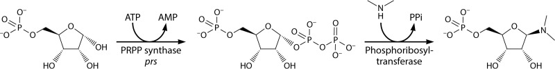
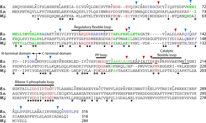
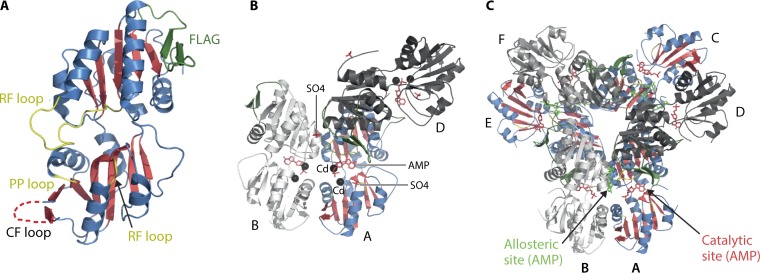
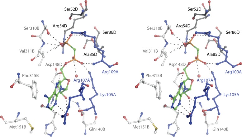
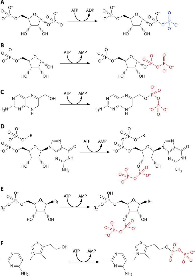
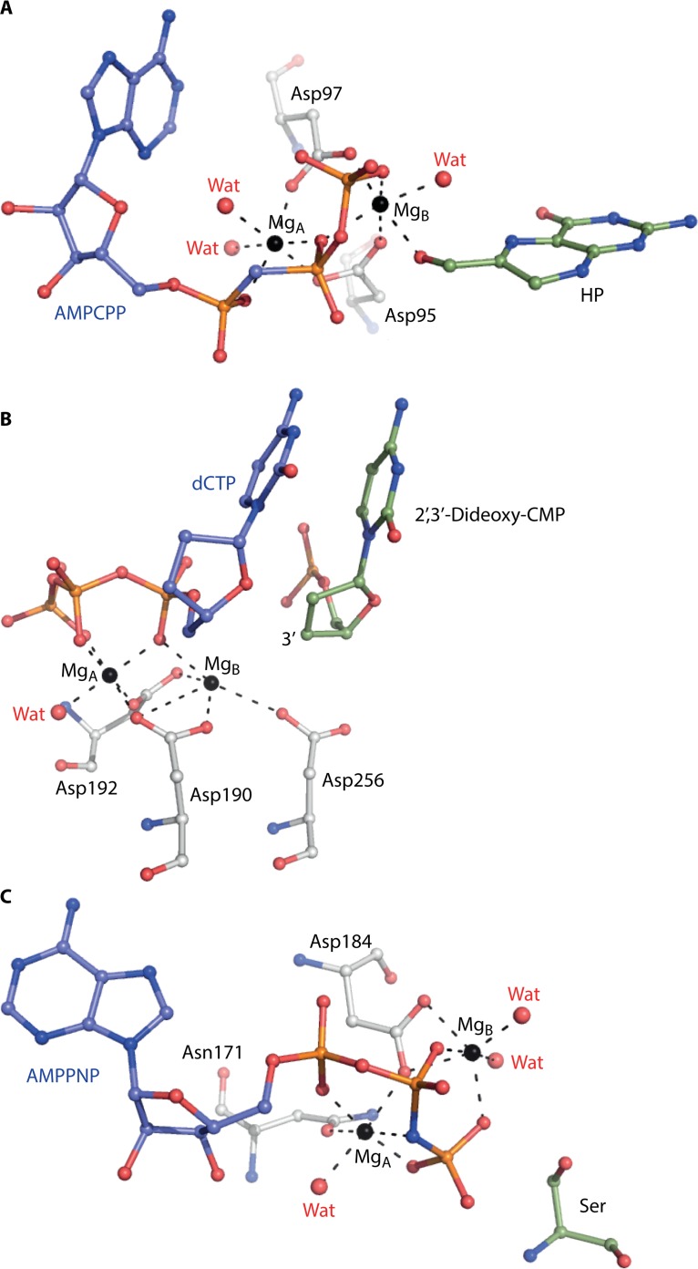
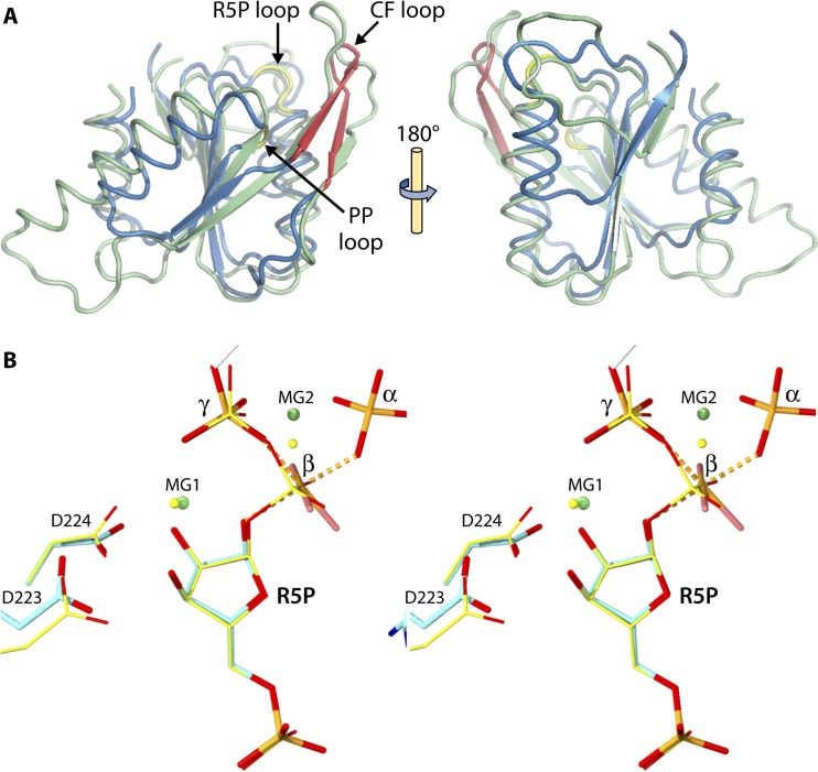
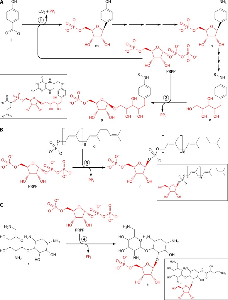
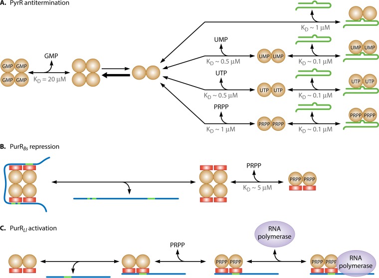
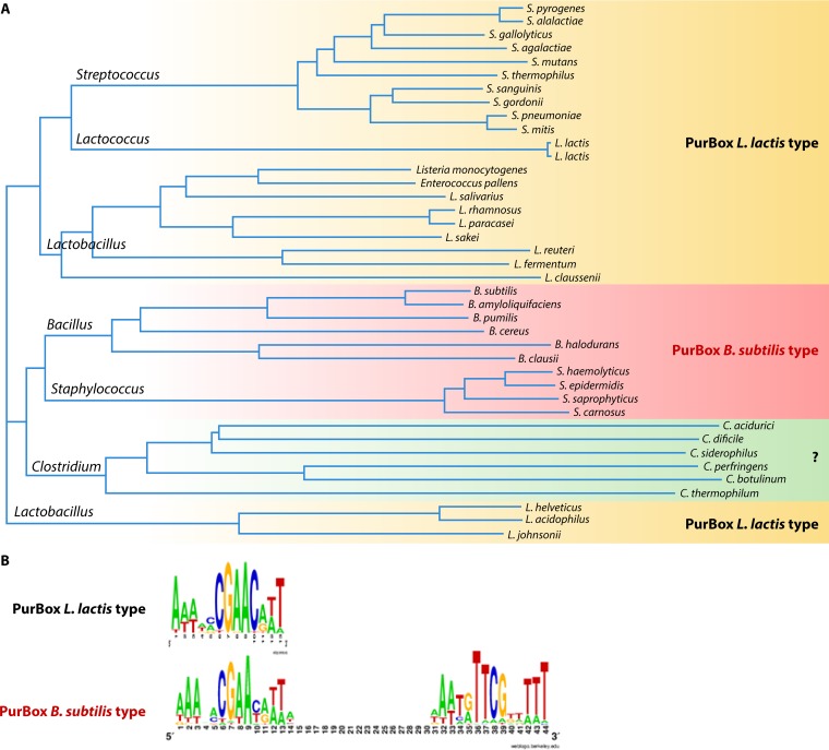
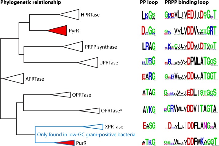
Phosphoribosyl diphosphate (PRPP) is an important intermediate in cellular metabolism. PRPP is synthesized by PRPP synthase, as follows: ribose 5-phosphate + ATP → PRPP + AMP. PRPP is ubiquitously found in living organisms and is used in substitution reactions with the formation of glycosidic bonds. PRPP is utilized in the biosynthesis of purine and pyrimidine nucleotides, the amino acids histidine and tryptophan, the cofactors NAD and tetrahydromethanopterin, arabinosyl monophosphodecaprenol, and certain aminoglycoside antibiotics. The participation of PRPP in each of these metabolic pathways is reviewed. Central to the metabolism of PRPP is PRPP synthase, which has been studied from all kingdoms of life by classical mechanistic procedures. The results of these analyses are unified with recent progress in molecular enzymology and the elucidation of the three-dimensional structures of PRPP synthases from eubacteria, archaea, and humans. The structures and mechanisms of catalysis of the five diphosphoryltransferases are compared, as are those of selected enzymes of diphosphoryl transfer, phosphoryl transfer, and nucleotidyl transfer reactions. PRPP is used as a substrate by a large number phosphoribosyltransferases. The protein structures and reaction mechanisms of these phosphoribosyltransferases vary and demonstrate the versatility of PRPP as an intermediate in cellular physiology. PRPP synthases appear to have originated from a phosphoribosyltransferase during evolution, as demonstrated by phylogenetic analysis. PRPP, furthermore, is an effector molecule of purine and pyrimidine nucleotide biosynthesis, either by binding to PurR or PyrR regulatory proteins or as an allosteric activator of carbamoylphosphate synthetase. Genetic analyses have disclosed a number of mutants altered in the PRPP synthase-specifying genes in humans as well as bacterial species.

Keywords: amino acid metabolism; diphosphoryl transfer; nucleotide metabolism; phosphoribosyl pyrophosphate; protein structure-function.


Figures

FIG 1
FIG 1
Biochemical function of PRPP. The β,γ-diphosphoryl group of ATP is transferred to ribose 5-phosphate with the generation of PRPP in a reaction catalyzed by PRPP synthase, which is encoded by a prs gene. PRPP is a substrate in a number of substitution reactions, most of which involve a nitrogen-containing compound. The reaction occurs at C-1 of the ribosyl moiety and proceeds with inversion of the configuration of this carbon and with PPi as a leaving group.
FIG 2
FIG 2
Alignment of amino acid sequences of PRPP synthases from B. subtilis (B.s.), S. oleracea isozyme 4 (S.o.), and M. jannaschii (M.j.). The B. subtilis PRPP synthase amino acid sequence is numbered from the N-terminal serine, as the original N-terminal methionine is removed in the mature protein. Functional elements of the B. subtilis PRPP synthase are indicated by bars above the B. subtilis amino acid sequence. Amino acid residues identical in all three sequences are indicated by asterisks below the M. jannaschii PRPP synthase amino acid sequence, whereas conserved amino acid residues are indicated by dots. Conserved amino acids are valine, leucine, and isoleucine; phenylalanine and tyrosine; aspartate and glutamate; arginine and lysine; serine and threonine; and glycine and alanine. The division of the N- and C-terminal domains (150-Leu-Met-151) of B. subtilis PRPP synthase is indicated by a vertical line. B. subtilis PRPP synthase amino acid residues, which are located at the active site, are shown in red and those which are located at the allosteric, regulatory site are shown in blue, whereas those involved in subunit-subunit interactions are shown in green. Amino acid residues involved in formation of the bent dimer are shown in bold, whereas those involved in formation of the parallel dimer are shown in italics. Whenever amino acid residues of the PRPP synthase of S. oleracea or M. jannaschii are identical to those of the B. subtilis enzyme, the color code of the latter enzyme is applied to the S. oleracea and M. jannaschii residues as well. The underlined amino acid residues Val178-Asp196, Arg198, and Asn209-Val211 are involved in the formation of a tightly packed interface necessary for allosteric inhibition. Amino acid residues were selected on the basis of the three-dimensional structures previously published (49, 50, 54). Vertical arrowheads point to B. subtilis PRPP synthase amino acids, which are homologous to amino acids altered in the human PRPP synthase isozyme 1 due to point mutations in the PRSP1 gene. Red arrowheads point to amino acid alterations resulting in increased PRPP synthase activity, whereas blue arrowheads point to amino acid alterations resulting in decreased PRPP synthase activity. The amino acid alterations and properties of the human PRPP synthase variants are described further in the text and are summarized in Table 4.
FIG 3
FIG 3
Three-dimensional structure of B. subtilis PRPP synthase. (A) Monomer drawn on the basis of the SO42− PRPP synthase structure (PDB code 1dkr) (49). The N-terminal domain is at the top. Shown are the five-stranded parallel β-sheets (red), helices (blue), flag region (green), regulatory flexible (RF) loop, the ribose 5-phosphate (R5P) loop, and the PP loop (yellow). The unresolved catalytic flexible (CF) loop is shown as a dotted line. (B) Bent and parallel dimers drawn on the basis of the Cd2+ PRPP synthase structure (PDB code 1ibs) (50). Subunit A is colored similar to the monomer in panel A. Shown are the Cd2+ (black), AMP of the active site (red), and sulfate bound at the position of the phosphate moiety of ribose 5-phosphate and at the position of the α-phosphate of ADP of the allosteric site (red). (C) Hexameric propeller structure drawn on the basis of the mADP PRPP synthase structure (PDB code 1dku) (49). Subunit A (as well as subunits C and E) are colored as described for the monomer in panel A. Shown are the positions of the methylene ADP moieties (red) and methylene ADP molecules (green), both modeled to only AMP, of the ATP binding sites and the allosteric sites, respectively.
FIG 4
FIG 4
Allosteric site of B. subtilis PRPP synthase. Stereo view based on the mADP PRPP synthase structure (PDB code 1dku) (49). The site is occupied by methylene ADP. Amino acid residues contributing to methylene ADP binding are provide by three subunits, labeled A, B, and D, as in Fig. 3. Amino acid residues of subunit A are shown in blue, amino acid residues of subunit B are in light gray, and amino acid residues of subunit D are shown in dark gray.
FIG 5
FIG 5
Positions of nonhomologous regions. Each line of bars represents polypeptides of a PRPP synthase, with the amino terminus at the left end. S.c.1, S. cerevisiae Prs1; S.c.3, S. cerevisiae Prs3; S.c.5, S. cerevisiae Prs5; S.p.1, S. pombe Prs1; S.p.2, S. pombe Prs2; S.p.3, S. pombe Prs3; PAP39, human PRPP synthase-associated protein 39; PAP41, human PRPP synthase-associated protein 41. S. cerevisiae and S. pombe Prs3 are shown at the top as a bar consisting of three segments (I, II, and III) represented in different shades of blue. This bar could represent most class I PRPP synthases. The left and right ends of segment II are located within the regulatory and catalytic flexible loops, respectively. An NHR is shown in red. The number of amino acid residues of each NHR is shown below the bars. The three NHRs of S. cerevisiae Prs1 and -5 are designated NHR1, NHR5-1, and NHR5-2. According to this nomenclature, S. cerevisiae Prs1 has the structure segment 1–segment 2–NHR1–segment 3; S. cerevisiae Prs5 has the structure segment 1–NHR5-1–segment 2–NHR1–segment 3; S. pombe PRPP synthase 1 has the structure segment I–segment II–NHR–segment III; S. pombe PRPP synthase 2 has the structure segment I–NHR–segment II–segment III; human PAP39 and -41 have the structure segment I–segment II–NHR–segment III.
FIG 6
FIG 6
Catalytic mechanism of PRPP synthase. (A) Closure of the catalytic flexible loop of T. volcanium PRPP synthase by superimposition of the open and closed structures (PDB codes 3lrt and 3mbi, respectively). Structural elements are colored as described for Fig. 3A. A 17-Å movement of the catalytic flexible loop, consisting of the β10 and β11 strands, results in the closed conformation necessary for catalysis. (B) Close-up view of the binding of substrates at the active site of T. volcanium PRPP synthase, with open and closed catalytic flexible loops. In the open conformation, the triphosphate chain of ATP, modeled here to only ADP, forms a more or less linear arrangement. In the closed conformation, the triphosphate chain, again modeled to only ADP, bends with the β-phosphate, resulting in a position ideal for attack of O-1 of ribose 5-phosphate on the β-phosphorus. An Mg2+ of the closed conformation is shown as a black sphere (138). (C) Stereo view of the binding of ribose 5-phosphate, Mg2+, and the transition state analog AlF3 to the active site of B. subtilis PRPP synthase, AlF3 PRPP synthase. (Reproduced from reference with permission.) α indicates the α-phosphate of ATP provided by an AMP molecule; β indicates the β-phosphate of ATP provided by Al3+ (bound to three F ions); γ indicates the γ-phosphate of ATP provided by the phosphate of a second AMP molecule. The two Mg2+ are indicated by MG1 and MG2. Relevant amino acid residues His135, Asp174, Lys197, and Arg199 are included as well. (D) Stereo view of the binding of ribose 5-phosphate, α,β-methylene ATP, Mg2+, and Ca2+ to the active site of B. subtilis PRPP synthase in the GDP PRPP synthase complex. (Reproduced from reference with permission.) Ca2+ (designated CA1) coordinates to the hydroxyls at C-1, C-2, and C-3 of ribose 5-phosphate, oxygen of the β- and γ-phosphates of α,β-methylene ATP, Asp174, as well as a water molecule. The Mg2+ (designated MG2) coordinates to the oxygen of C-2′ of the ribosyl moiety as well as oxygen of the α- and γ-phosphates of α,β-methylene ATP, as well as to three water molecules. Thus, there is no coordination to oxygen of the β-phosphate of α,β-methylene ATP.
FIG 7
FIG 7
Reactions catalyzed by diphosphoryltransferases and alternative biosynthesis of PRPP. In some cases, ATP may be replaced by dATP. The diphosphoryl and phosphoryl moieties of the products are shown in red and blue, respectively. (A) Reaction catalyzed by phosphoribosyl bisphosphate phosphokinase. The substrate is ribosyl 1,5-bisphosphate, the product is PRPP. (B) Reaction catalyzed by PRPP synthase. The substrate is ribose 5-phosphate, the product is PRPP. (C) Reaction catalyzed by 2-amino-4-hydroxy-6-hydroxymethyldihydropterin diphosphokinase. The substrate is 2-amino-4-hydroxy-6-hydroxymethyldihydropterin, and the product is 2-amino-4-hydroxy-6-hydroxymethyldihydropterin diphosphate. (D) Reaction catalyzed by GTP/GDP 3′-diphosphokinase (stringent factor). R may be a hydrogen or a phosphoryl group, i.e., the substrate is GDP or GTP, respectively, and the product is guanosine 3′-diphosphate 5′-diphosphate (ppGpp) or guanosine 3′-diphosphate 5′-triphosphate (pppGpp), respectively. (E) Reaction catalyzed by nucleotide diphosphokinase. R1 may be an adenyl, a guanyl, or a hypoxanthyl univalent radical, whereas R2 may be a hydrogen, a phosphoryl, or a diphosphoryl moiety. (F) Reaction catalyzed by thiamine diphosphokinase. The substrate is thiamine, and the product is thiamine diphosphate.
FIG 8
FIG 8
Diphosphoryl, nucleotidyl, and phosphoryl transfer reactions. Wat, water molecules. (A) The active site of the ternary complex of E. coli hydroxymethyldihydropterin diphosphokinase. AMPCPP, α,β-methylene ATP; HP, hydroxydihydropterin (PDB code 1q0n) (164). (B) The active site of DNA polymerase β (PDB code 1bpy) (176). (C) The active site of cAMP protein kinase A. AMPPNP, β,γ-imido ATP (PDB code 4hpu) (201).
FIG 9
FIG 9
Catalytic strategies. The coordination of Mg2+ (shown as blue spheres) of the active site to the substrates, to amino acid residues, and to water molecules (shown as red spheres) is schematically illustrated by blue lines. (A) cAMP-dependent protein kinase A (PDB code 4hpu) (201). (B) PRPP synthase. R5P, ribose 5-phosphate. MgA was designated MG2, and MgB was designated MG1 before (54). (C) Hexose 1-phosphate nucleotidyltransfrease, based on the structure of N-acetylglucosamine 1-phosphate uridylyltransferase (PDB code 4g87). MgB coordinates to aspartate and asparagine residues (192). (D) DNA polymerase based on the structure of DNA polymerase β (PDB code 1bpy) (176). The three acidic residues are usually aspartates, but occasionally one aspartate is replaced by a glutamate (185, 186). The structure is valid also for RNA polymerases (187).
FIG 10
FIG 10
Similarity of the folds of the PRPP synthase domain and type I phosphoribosyltransferases. (A) Superimposition of the C-terminal domain (amino acids 151 to 286) of T. volcanium PRPP synthase (blue; PDB code 3mbi) (138) and T. gondii hypoxanthine-guanine phosphoribosyltransferase (green; PDB code 1fsg) (212). The PP, ribose 5-phosphate (R5P), and catalytic flexible (CF) loops are indicated. (B) Substrate binding at the active sites of B. subtilis PRPP synthase and T. gondii hypoxanthine-guanine phosphoribosyltransferase. (Reproduced from reference with permission.) The B. subtilis PRPP synthase transition-state analog (Fig. 6C) consisting of the phosphoryl moiety of AMP (α), AlF3 (β), the phosphoryl moiety of a second AMP molecule (γ), and ribose 5-phosphate, as well as Asp223 and Asp224 are shown as thick lines, and the two Mg2+ are shown as green spheres (54). Residues from the structure of T. gondii hypoxanthine-guanine phosphoribosyltransferase in complex with 9-deazaguanine (not shown), PRPP, and Mg2+, as well as Glu146 and Asp147 (PDB code 1fsg) (212), are shown as thin yellow lines or spheres superimposed on the AlF3 PRPP synthase structure.
FIG 11
FIG 11
N-Glycosidic bond formation with PRPP. PRPP and atoms of the products derived from PRPP are shown in red. Each reaction produces an N-glycosidic 5′-phosphoribosyl compound and PPi. The reaction with quinolinate also produces carbon dioxide. The compounds a to e are products of de novo reactions: a, 5-phosphoribosyl 1-amine; b, orotidine 5′-monophosphate; c, 5′-phosphoribosylnicotinate; d, 5′-phosphoribosyl-ATP; e, 5′-phosphoribosylanthranilate. The compounds f to k are products of salvage reactions: f, AMP; g, IMP; h, XMP; i, GMP; j, UMP; k, 5′-phosphoribosylnicotinate.
FIG 12
FIG 12
C- and O-glycosidic bond formation with PRPP. The phosphoribosyl donor PRPP is shown in red, and atoms derived from PRPP in the intermediates and products are also shown in red. (A) Biosynthesis of tetrahydromethanopterin. Compound labels: l, 4-hydroxybenzoate; m, 5′-phospho-β-d-ribosyl 4-hydroxybenzene; n, 5′-phospho-β-d-ribosyl 4-aminobenzene; o, N-[(7,8-dihydropterin-6-yl)methyl]-4-(1-deoxy-d-ribulosyl)aminobenzene; p, 1-(4-{N-[(7,8-dihydropterin-6-yl)methyl]amino}phenyl)-5-(5-phospho-α-d-ribulosyl)-1-deoxyribitol. Enzyme 1, 4-aminobenzoate phosphoribosyltransferase (5-phospho-α-d-ribose 1-diphosphate:4-aminobenzoate 5-phospho-β-d-ribofuranosyltransferase [decarboxylating], EC 2.4.2.54); enzyme 2, 1-(4-{N-[(7,8-dihydropterin-6-yl)methyl]amino}phenyl)-5-(5-phospho-α-d-ribulosyl)-1-deoxyribitol synthase. This enzyme activity has not been identified. The four reactions leading from compound m to compound n in effect convert a hydroxy group to an amino group and involve the formation of phosphate ester and the addition of an aspartyl residue and the removal of Pi and fumarate (304). The three reactions leading from compound n to compound o involve the attachment of a pterin derivative (R = N-[7,8-dihydropterin-6-yl]methyl) to the nitrogen of compound n followed by opening of the ribosyl moiety and isomerization to a ribulose derivative and dephosphorylation to form compound o. The boxed compound is the product of the pathway for 5,6,7,8-tetrahydromethanopterin (with a complete structure of the pteridyl moiety), the active cofactor in transformation of carbon dioxide to methane in methanogenic Archaea (301), and is formed from compound p by attachment of a glutamyl moiety to the phosphate group followed by dehydrogenation of the pteridyl moiety. (B) Biosynthesis of arabinosyl monophosphodecaprenol. Compound labels: q, decaprenyl phosphate; r, 5-phospho-β-d-ribosyl 1-O-monophosphodecaprenol (decaprenylphospho-β-d-ribosyl 5-phosphate). Enzyme 3, decaprenyl phosphate phosphoribosyltransferase (5-phospho-α-d-ribosyl 1-diphosphate:decaprenyl-phosphate 5-phosphoribosyltransferase; EC 2.4.2.45). The boxed compound is the arabinosyl donor arabinosyl monophosphodecaprenol, which is formed from compound r by dephosphorylation of the ester at C-5 of the ribosyl moiety followed by epimerization. The latter two reactions occur outside the cell (5). (C) Biosynthesis of butirosin. Compound labels: s, neamine; t, 5″-phosphoribostamycin. Enzyme 4, neamine phosphoribosyltransferase. The boxed compound is butirosin. Two isomers are synthesized; one contains a ribosyl moiety (shown) and a second contains an arabinosyl moiety (not shown). Other aminoglycoside antibiotics derived from neamine, i.e., neomycin B, paromomycin, and lividomycin B, lack the 4-amino-2-hydroxybutyryl side chain but contain an N-acetylaminoglucosyl moiety attached to C-3″ of the ribosyl moiety, and the pseudodisaccharides of the three compounds are differently decorated with hydroxyl and amino groups (318).
FIG 13
FIG 13
Mechanism of PRPP-mediated regulation of transcription by PyrR and PurR. PyrR and PurR are shown as orange spheres. (A) Model of PyrR regulation based on the combined data from B. subtilis and B. caldolyticus. RNA (green hairpin) binds to dimeric PyrR, which is stabilized by UMP, UTP, or PRPP. The tetrameric conformation is stabilized by GMP and does not bind RNA. See the text for details. (B) Model of PurRBs repression. DNA (blue line) containing one strong (solid green line) and one weak (green punctuated line) PurBox binds to two PurRBs dimers (forming a weak tetramer) in the absence of PRPP. In the presence of PRPP, the DNA binding is prevented and the tetramerization is lost. (C) Model of PurRLl activation. PurRLl binds to PurBox sequences, irrespective of PRPP binding, presumably as a dimer. Binding of PRPP is hypothesized to expose a binding site for RNA polymerase. RNA polymerase is positioned correctly relative to the −10 region of the promoter.
FIG 14
FIG 14
Phylogenetic analysis of PurR from low-GC Gram-positive bacteria. (A) PurR sequences from representatives of the major bacterial lineages in the low-GC Gram-positive bacteria were aligned using the Clustal Omega program, and a phylogenetic tree was constructed using the ClustalW2 phylogeny program. (B) The type of PurBox used by each species was identified at the RegPrecise website (http://regprecise.lbl.gov/RegPrecise/collection_tffam.jsp?tffamily_id=53), and the two types of logo plots were constructed from the PurBox sequences presented for Streptococcaceae and Bacillales, respectively, using the weblogo service (http://weblogo.berkeley.edu/logo.cgi).
FIG 15
FIG 15
Common phylogeny of PyrR, PurR, type 1 phosphoribosyltransferases, and class I PRPP synthases in all major bacterial lineages. Homologous protein sequences were deduced and identified from representative genome sequences of all major bacterial lineages for the following proteins: adenine phosphoribosyltransferase (APRTase), hypoxanthine-guanine phosphoribosyltransferase (HPRTase), orotate phosphoribosyltransferase (OPRTase), uracil phosphoribosyltransferase (UPRTase), and xanthine phosphoribosyltransferase (XPRTase). The collected sequences were aligned using the Clustal Omega program, and a phylogenetic tree was constructed using the ClustalW2 phylogeny program. (Left) Phylogenetic tree, with protein sequences belonging to same enzyme families grouped together as triangles, starting at the point of the first branching of the individual protein sequences and labeled with the enzyme abbreviations shown above. The triangle marked OPRTase* is a group of orotate phosphoribosyltransferase sequences that appear to form a separate branch with a distinct set of amino acid sequences in the PP- and PRPP-binding loops. Sequences with homology to the xanthine phosphoribosyltransferase and PurR proteins were only found among the low-GC Gram-positive bacteria, as indicated. (Middle) Logo plot of the frequency of amino acids in the PP loop identified in the Clustal Omega alignment and constructed using the weblogo service (http://weblogo.berkeley.edu/logo.cgi). (Right) Logo plot of the frequency of amino acids in the PRPP-binding loop identified in the Clustal Omega alignment and constructed using the weblogo service.
FIG 16
FIG 16
Possible secondary structure of the S. enterica prs leader. The sequence ranges from nucleotide 1, indicated by 5′, to nucleotide 417, which is immediately followed by the initiator codon-specifying guanylate-uridylate-guanylate triplet and is indicated by 3′ (45). The ΔG of the structure is −540 kJ mol−1. The leader prs sequences of S. enterica and E. coli are identical except for the framed sequence, which is present only in the S. enterica prs leader. The nucleotides circled in red represent a possible leader peptide, and the red arrows point to the nucleotides of a possible Shine-Dalgarno sequence. Possible leader peptide amino sequences are indicated for E. coli and S. enterica. Identical amino acid residues of the two possible leader peptides are underlined.
None
None
None
None
None
None

References

    1. Jensen KF. 1983. Metabolism of 5-phosphoribosyl 1-pyrophosphate (PRPP) in Escherichia coli and Salmonella typhimurium, p 1–25. In Munch-Petersen A. (ed), Metabolism of nucleotides, nucleosides and nucleobases in microorganisms. Academic Press, Inc., London, United Kingdom.
    1. Hove-Jensen B. 1988. Mutation in the phosphoribosylpyrophosphate synthetase gene (prs) that results in simultaneous requirements for purine and pyrimidine nucleosides, nicotinamide nucleotide, histidine, and tryptophan in Escherichia coli. J Bacteriol 170:1148–1152. - PMC - PubMed
    1. Hove-Jensen B. 1989. Phosphoribosylpyrophosphate (PRPP)-less mutants of Escherichia coli. Mol Microbiol 3:1487–1492. doi: 10.1111/j.1365-2958.1989.tb00134.x. - DOI - PubMed
    1. Graham DE, White RH. 2002. Elucidation of methanogenic coenzyme biosyntheses: from spectroscopy to genomics. Nat Prod Rep 19:133–147. doi: 10.1039/b103714p. - DOI - PubMed
    1. Wolucka BA. 2008. Biosynthesis of d-arabinose in mycobacteria: a novel bacterial pathway with implications for antimycobacterial therapy. FEBS J 275:2691–2711. doi: 10.1111/j.1742-4658.2008.06395.x. - DOI - PubMed

Publication types

MeSH terms

Substances