. 2017 Feb 14;91(5):e02119-16.
doi: 10.1128/JVI.02119-16. Print 2017 Mar 1.

Barriers to Infection of Human Cells by Feline Leukemia Virus: Insights into Resistance to Zoonosis

Affiliations

Barriers to Infection of Human Cells by Feline Leukemia Virus: Insights into Resistance to Zoonosis

Anne Terry et al. J Virol. .

Abstract

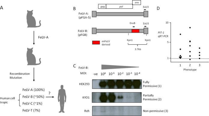
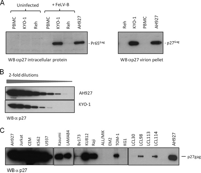
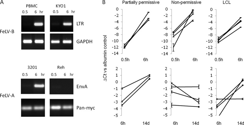
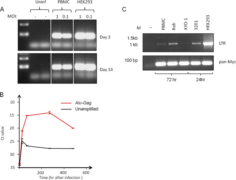
The human genome displays a rich fossil record of past gammaretrovirus infections, yet no current epidemic is evident, despite environmental exposure to viruses that infect human cells in vitro Feline leukemia viruses (FeLVs) rank high on this list, but neither domestic nor workplace exposure has been associated with detectable serological responses. Nonspecific inactivation of gammaretroviruses by serum factors appears insufficient to explain these observations. To investigate further, we explored the susceptibilities of primary and established human cell lines to FeLV-B, the most likely zoonotic variant. Fully permissive infection was common in cancer-derived cell lines but was also a feature of nontransformed keratinocytes and lung fibroblasts. Cells of hematopoietic origin were generally less permissive and formed discrete groups on the basis of high or low intracellular protein expression and virion release. Potent repression was observed in primary human blood mononuclear cells and a subset of leukemia cell lines. However, the early steps of reverse transcription and integration appear to be unimpaired in nonpermissive cells. FeLV-B was subject to G→A hypermutation with a predominant APOBEC3G signature in partially permissive cells but was not mutated in permissive cells or in nonpermissive cells that block secondary viral spread. Distinct cellular barriers that protect primary human blood cells are likely to be important in protection against zoonotic infection with FeLV.IMPORTANCE Domestic exposure to gammaretroviruses such as feline leukemia viruses (FeLVs) occurs worldwide, but the basis of human resistance to infection remains incompletely understood. The potential threat is evident from the human genome sequence, which reveals many past epidemics of gammaretrovirus infection, and from recent cross-species jumps of gammaretroviruses from rodents to primates and marsupials. This study examined resistance to infection at the cellular level with the most prevalent human cell-tropic FeLV variant, FeLV-B. We found that blood cells are uniquely resistant to infection with FeLV-B due to the activity of cellular enzymes that mutate the viral genome. A second block, which appears to suppress viral gene expression after the viral genome has integrated into the host cell genome, was identified. Since cells derived from other normal human cell types are fully supportive of FeLV replication, innate resistance of blood cells could be critical in protecting against cross-species infection.

Keywords: APOBEC; feline leukemia virus; restriction factors; zoonosis.


Figures

FIG 1
FIG 1
(A) Rationale for focusing on FeLV-B as the most likely zoonotic agent. The percentage of natural isolates containing each FeLV subgroup is given in parentheses. (B) Outline of the molecular structure of FeLV-B proviruses that are derived by recombination between FeLV-A and endogenous (en) FeLV-related proviruses in the feline genome. The hybridization probes (ExU3, EnvB) used to detect FeLV sequences in human cells are indicated. (C) The three basic patterns of susceptibility of cultured human cells to infection with FeLV-B. Cells were exposed to FeLV-B at various multiplicities of infection and were passaged for 14 days before the extraction of high-molecular-weight DNA and analysis of FeLV proviral DNA content by Southern blot hybridization of KpnI-digested DNA with the ExU3 probe. -ve, uninfected control. (D) Relative expression of mRNA encoding the FeLV-B receptor PIT-1 in a panel of cell lines sorted by the pattern of FeLV-B spread (phenotype 1, 2, or 3, as exemplified in panel C).
FIG 2
FIG 2
Human hematopoietic cells display two patterns of susceptibility to FeLV-B replication. (A) (Left) Western blot (WB) analysis of the intracellular Gag precursor protein (Pr65Gag) in partially permissive (phenotype 2) FeLV-B-infected KYO-1 cells shows expression levels similar to those in fully permissive feline fibroblasts (AH927). In contrast, expression of Pr65Gag is barely detectable in nonpermissive Reh cells and human PBMCs. (Right) This pattern is reflected in the levels of virus particle release, which are similar in partially permissive KYO-1 cells and permissive AH927 cells, as illustrated by Western blot analysis of pelleted virion preparations from equal volumes of culture medium. (B) Results of a separate experiment where Western blot analysis of serial 2-fold dilutions of virions released from AH927 and KYO-1 cells again showed levels of particle release from KYO-1 cells within a range similar to that for AH927 cells. (C) Western blot analysis of pelleted virions harvested from cells 14 days after infection with FeLV-B. Equal volumes of cell supernatant (2 ml) were sampled 24 h after medium change and were processed as described in Materials and Methods.
FIG 3
FIG 3
Permissive and nonpermissive human cells support FeLV-B entry and early proviral DNA synthesis at similar levels. (A) Initial studies using standard PCR with primers based on FeLV LTR or envelope sequences showed marked increases in proviral DNA levels between 0.5 and 6 h after the exposure of cells to FeLV-B at an MOI of 1. Because no PIT-1-negative cell lines were identified, a receptor-negative control was generated for this assay by comparing the FeLV-A infection of permissive feline cells (3201 cells) and of Reh cells, which lack detectable expression of mRNA for the human homologue of FeLV-A receptor THTR1. Loading controls used human glyceraldehyde-3-phosphate dehydrogenase (GAPDH) or pan-Myc primers, which efficiently detect human and feline DNA. (B) A wider analysis of FeLV-B infection of human hematopoietic cell lines. Proviral DNA levels were analyzed by quantitative PCR. The line graphs show changes in FeLV DNA concentrations (detected by EnvB primers) relative to those of a housekeeping gene control (β2-microglobulin) between 0.5 and 6 h (top) or between 6 h and 14 days (bottom) after infection. The y axis shows the threshold cycle (CT) value relative to that of the β2-microglobulin control (taken as zero). Partially permissive cells were CEM, Jurkat, and Raji cells. Nonpermissive cells were PBMCs (n = 2), Reh cells, and ALL/MIK cells. Lymphoblastoid cell lines were LCL30, LCL98, LCL113, and LCL114.
FIG 4
FIG 4
Persistence of integrated FeLV-B DNA in human PBMCs. (A) Semiquantitative PCR reveals dose-dependent persistence of FeLV DNA in human PBMCs at 14 days postinfection. Fully permissive HEK293 control cells show stable levels by 3 days postinfection. (B) Results of a quantitative PCR time course analysis for FeLV sequences in PBMC DNA, with and without a preamplification step using primers to FeLV Gag and consensus human Alu sequences. The threshold cycle (CT) value is inversely proportional to the DNA content. (C) Results of semiquantitative PCR for LTR circles in FeLV-B-infected cells 3 days (PBMC, Reh, and KYO-1 cells) or 24 h (3201 and HEK293 cells) after infection. While single and double LTR circle forms are readily detected in productively infected HEK293 and 3201 cells, they are barely detectable in Reh cells and PBMCs relative to levels in permissive HEK293 control cells. The loading control was provided by PCR amplification with conserved Myc primers (pan-Myc).
FIG 5
FIG 5
Hypermutation of the FeLV-B genome and APOBEC3G mRNA expression in human cells. FeLV genomic sequences (segments of Gag and Pol) were cloned by PCR from infected human cells, and individual templates were sequenced and compared to the reference input virus (pFGB clone). APOBEC3G mRNA expression was determined in the same cells (prior to infection) by quantitative real-time PCR (with SYBR green). (A) Representative plots of hypermutation visualized by the online HYPERMUT program (www.hiv.lanl.gov/content/sequence/HYPERMUT/hypermut.html), where sequence changes relative to the reference FeLV-B genome are color coded (red, GG→AG; cyan, GA→AA; green, GC→AC; magenta, GT→AT; black, non-G→A). (B) X/Y plots of G→A mutation (per kilobase) against APOBEC3G mRNA levels (where the level in HEK293 cells is taken as 1) with cell lines sorted according to FeLV restriction phenotype. Results for LCLs, which are discordant by virtue of their low levels of infectious virion release (Table 2) but postinfection accumulation of proviral DNA (Fig. 3), are enclosed by a dashed oval. (C) Percentage of G→A mutations that conform to the A3G signature (42) for all cell lines in which significant levels of mutations were detected. Blue-gray bars, hematopoietic cells; yellow bars, nonhematopoietic cells. (D) Relative levels of APOBEC3G mRNA (on a log10 scale, with the level in HEK293 cells taken as 1) for all the cell lines tested, sorted into hematopoietic (blue circles) and nonhematopoietic (green circles) cell lines. Nonpermissive cells (nonspreading, with low virion release) are represented by black circles.

References

    1. Lander ES, Linton LM, Birren B, Nusbaum C, Zody MC, Baldwin J, Devon K, Dewar K, Doyle M, FitzHugh W, Funke R, Gage D, Harris K, Heaford A, Howland J, Kann L, Lehoczky J, LeVine R, McEwan P, McKernan K, Meldrim J, Mesirov JP, Miranda C, Morris W, Naylor J, Raymond C, Rosetti M, Santos R, Sheridan A, Sougnez C, Stange-Thomann Y, Stojanovic N, Subramanian A, Wyman D, Rogers J, Sulston J, Ainscough R, Beck S, Bentley D, Burton J, Clee C, Carter N, Coulson A, Deadman R, Deloukas P, Dunham A, Dunham I, Durbin R, French L, et al. ; International Human Genome Sequencing Consortium. 2001. Initial sequencing and analysis of the human genome. Nature 409:860–921. doi: 10.1038/35057062. - DOI - PubMed
    1. Gifford R, Kabat P, Martin J, Lynch C, Tristem M. 2005. Evolution and distribution of class II-related endogenous retroviruses. J Virol 79:6478–6486. doi: 10.1128/JVI.79.10.6478-6486.2005. - DOI - PMC - PubMed
    1. Olah Z, Lehel C, Anderson WB, Eiden MV, Wilson CA. 1994. The cellular receptor for gibbon ape leukemia virus is a novel high affinity sodium-dependent phosphate transporter. J Biol Chem 269:25426–25431. - PubMed
    1. Xu W, Stadler CK, Gorman K, Jensen N, Kim D, Zheng H, Tang S, Switzer WM, Pye GW, Eiden MV. 2013. An exogenous retrovirus isolated from koalas with malignant neoplasias in a US zoo. Proc Natl Acad Sci U S A 110:11547–11552. doi: 10.1073/pnas.1304704110. - DOI - PMC - PubMed
    1. Alfano N, Kolokotronis SO, Tsangaras K, Roca AL, Xu W, Eiden MV, Greenwood AD. 2016. Episodic diversifying selection shaped the genomes of gibbon ape leukemia virus and related gammaretroviruses. J Virol 90:1757–1772. doi: 10.1128/JVI.02745-15. - DOI - PMC - PubMed

Publication types

Substances

LinkOut - more resources