. 2017 Apr 20;45(7):3724-3737.
doi: 10.1093/nar/gkw1288.

The replication initiator of the cholera pathogen's second chromosome shows structural similarity to plasmid initiators

Affiliations

The replication initiator of the cholera pathogen's second chromosome shows structural similarity to plasmid initiators

Natalia Orlova et al. Nucleic Acids Res. .

Abstract

The conserved DnaA-oriC system is used to initiate replication of primary chromosomes throughout the bacterial kingdom; however, bacteria with multipartite genomes evolved distinct systems to initiate replication of secondary chromosomes. In the cholera pathogen, Vibrio cholerae, and in related species, secondary chromosome replication requires the RctB initiator protein. Here, we show that RctB consists of four domains. The structure of its central two domains resembles that of several plasmid replication initiators. RctB contains at least three DNA binding winged-helix-turn-helix motifs, and mutations within any of these severely compromise biological activity. In the structure, RctB adopts a head-to-head dimeric configuration that likely reflects the arrangement in solution. Therefore, major structural reorganization likely accompanies complex formation on the head-to-tail array of binding sites in oriCII. Our findings support the hypothesis that the second Vibrionaceae chromosome arose from an ancestral plasmid, and that RctB may have evolved additional regulatory features.


Figures

Figure 1.
Figure 1.
Architecture of the origin of DNA replication of the secondary chromosome (oriCII) from Vibrio cholerae. oriCII contains five distinct binding sites for the RctB initiator, named for their length: 12-mer (pink), 11-mer (cyan), 39-mer (black), 29-mer (purple) and the rctA-39-mer (teal). The oriCII-min segment contains six direct repeats of the 12-mer site, and is sufficient to direct replication initiation in the presence of RctB. The other RctB binding sites are proposed to serve regulatory purposes. The origin also contains binding sites for DnaA (light orange) and integration host factor (IHF, light green), and an A-T rich region (white) that is the locus of the initial melting at the origin. The numbered black bars represent the probes used for DNA binding electrophoretic mobility shift assay.
Figure 2.
Figure 2.
Sequence conservation, domain architecture and structures of the RctB initiator protein. (A) A BLAST alignment (63) consisting of 99 RctB orthologues was converted to a numerical conservation score where equivalence of amino acid at each position was established using a normalized BLOSUM62 matrix (64). Conservation score is plotted against the primary sequence (gray lines). Reds dots represent positions with greater 90% sequence conservation. (B) Domain architecture of RctB as deduced from mass spectrometric analysis of proteolytic digestion products. A precise boundary for the fourth domain of RctB could not be obtained owing to this region's sensitivity to limited proteolysis. The cleavage sites revealed by our analysis are depicted with scissors. (C) The crystal structures of RctB domain 1 and domains 2–3 are shown in a cartoon representation. The coloring scheme employed corresponds to that in Figure 2B. The dotted line represents one protomer of the RctB dimer. Full-length RctB forms a dimer, and the dimerization interface localizes to domain 2.
Figure 3.
Figure 3.
The structures of the central domains of RctB resemble the structures of plasmid initiators π, RepE and RepA. The structure of the central domains (domains 2–3) of RctB was aligned to the π (Z-score = 9.0, RMSD = 4.9 Å), RepE (Z-score = 8.4, RMSD = 6.1 Å) and RepA (Z-score = 7.7, RMSD = 2.9 Å) plasmid replication initiators. Depicted here is an alignment of secondary structure elements extracted from this alignment. Elements shared by each protein are colored in cyan. Elements unique to RctB are colored in orange, while elements present in the plasmid initiators, but not in RctB, are colored in black. Grey circles represent regions of the various structures that were not modeled. Depiction of RepA is limited to the one available domain. A schematic in the bottom right corner shows that RctB middle portion (domains 2 and 3) aligns with the entire structure of π, therefore, the secondary structure alignment is shown only for domains 2–3 of RctB and structures of the plasmid initiators.
Figure 4.
Figure 4.
RctB has three DNA binding domains. Structural comparisons predicted the presence of at least three distinct DNA binding domains in RctB. To evaluate these predictions, three mutant forms of RctB were studied: (A) RctB Q83A-R84A-R86A (domain 1), (B) RctB K271A-K272A-S274A (domain 2) and (C) RctB R420A-R423A (domain 3). The top of each panel depicts a ribbon representation (colored blue) of the predicted DNA binding domain of RctB modeled onto a DNA molecule taken from a structural homolog bound to its DNA target. The residues selected for analysis are shown in a ball and stick representation (colored orange). The middle portion of each panel summarizes, in a sequence alignment format, the structural alignment of the DNA binding domains that emerged from database searches. The residues tested in this study are shown in orange, and labeled with orange stars. Shown in green are positions implicated in DNA binding by other studies (,,–70). The lower portion of each panel shows the binding affinity for oriCII-min and performance in the transformation assay by wild-type and the mutant RctB proteins.
Figure 5.
Figure 5.
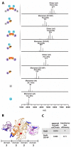
RctB adopts a head-to-head dimeric configuration in solution. (A) Native mass spectrometric analysis of the oligomeric state of full length and truncated constructs of RctB, including variants that harbored the D314P mutation (wild-type: red, mutant: black). Spectra for the wild-type and D314P entities are grouped together. To the left of each spectrum appears a schematic, colored as in Figure 2B, of the configuration revealed by the analysis. (B) The head-to-head dimer of RctB seen in the crystal. The Asp314 residue on each protomer is depicted as a red sphere. Residues shown to be involved in contacts to DNA are depicted in the ball-and-stick representation and colored dark blue. The two monomers of RctB are colored in varying shades (domain 2: orange, domain 3: purple). One of the RctB monomers is outlined with a dashed line. (C) Binding affinity for oriCII-min and performance in the transformation assay for wild-type and RctB-D314P.
Figure 6.
Figure 6.
Incompatibility of a head-to-head dimer structure with origin binding. RctB middle fragment structure is shown as a ribbon representation. Domain 2 is colored in different shades of orange, domain 3 is colored in different shades of purple. The top RctB monomer is shown by dotted line. The bottom monomer is modeled to be bound to its site on the origin, according to our findings about the residues involved in the DNA binding and structural alignments; the two winged-helix-turn-helix domains contact two adjacent major grooves of the DNA, the DNA binding residues are shown as red sticks. When one of the monomers is bound to DNA, the DNA binding residues of the second monomer are located very far away from the DNA, and they can not interact with the following binding site on the DNA. Therefore a head-to-head RctB dimer is incompatible with origin binding not only because of the binding site orientation (direct repeats), but also because of the molecule geometry that does not allow the second monomer to contact the same DNA molecule.
Figure 7.
Figure 7.
Model for the interaction of RctB with DNA. (A) The 12-mer binding site is of insufficient length to accommodate the three DNA binding domains of RctB. On the left is a ribbon representation of the experimental structure of the π initiator (domains of π are colored in light orange and purple) bound to its 22 bp iteron DNA target. Each domain of the π plasmid initiator binds to ∼10 bp of DNA. On the right is shown a schematic of the three DNA binding domains of RctB (domain1: cyan, domain 2: orange, domain 3: purple) and its 12-mer binding site (colored in red), drawn approximately to scale. We propose that domains of RctB (tentatively domains 1 and 2) will make contacts to positions in a 12-mer binding site on either side of the major groove, and that domain 3 (tentatively) will make contacts to positions in the ‘spacer’ sequence between the 12-mers. (B) Linear representation of a putative head-to-tail RctB oligomer formed on the array of 12-mer sites at oriCIImin. One of the members of the RctB oligomer is outlined with a dashed line. (C) Schematic of the putative organization of the RctB oligomer on the 12-mer array in a DNA loop configuration to facilitate melting of the A-T-rich region of oriCIImin. This model is constructed by analogy with that proposed for the plasmid initiator (47).

Similar articles

Cited by

References

    1. Mott M.L., Berger J.M.. DNA replication initiation: mechanisms and regulation in bacteria. Nat. Rev. Microbiol. 2007; 5:343–354. - PubMed
    1. Wolanski M., Donczew R., Zawilak-Pawlik A., Zakrzewska-Czerwinska J.. oriC-encoded instructions for the initiation of bacterial chromosome replication. Front. Microbiol. 2014; 5, Article 735, 1–14. - PMC - PubMed
    1. Leonard A.C., Grimwade J.E.. Regulation of DnaA assembly and activity: taking directions from the genome. Annu. Rev. Microbiol. 2011; 65:19–35. - PMC - PubMed
    1. Ozaki S., Noguchi Y., Hayashi Y., Miyazaki E., Katayama T.. Differentiation of the DnaA-oriC subcomplex for DNA unwinding in a replication initiation complex. J. Biol. Chem. 2012; 287:37458–37471. - PMC - PubMed
    1. Robinson A., van Oijen A.M.. Bacterial replication, transcription and translation: mechanistic insights from single-molecule biochemical studies. Nat. Rev. Microbiol. 2013; 11:303–315. - PubMed

LinkOut - more resources