. 2017 Feb 10;292(6):2110-2119.
doi: 10.1074/jbc.M116.754796. Epub 2016 Dec 27.

Force Spectroscopy of the Plasmodium falciparum Vaccine Candidate Circumsporozoite Protein Suggests a Mechanically Pliable Repeat Region

Affiliations

Force Spectroscopy of the Plasmodium falciparum Vaccine Candidate Circumsporozoite Protein Suggests a Mechanically Pliable Repeat Region

Aditya Prasad Patra et al. J Biol Chem. .

Abstract

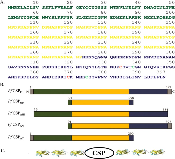
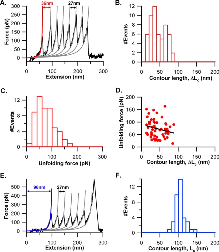
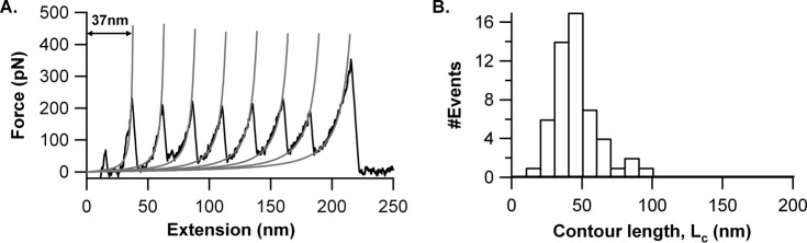
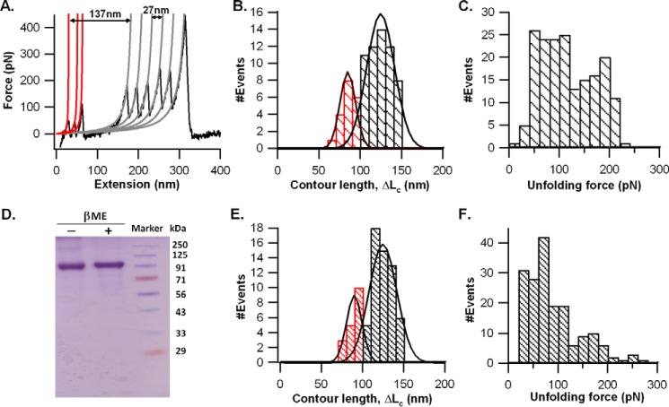
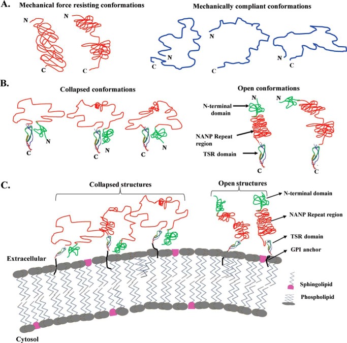
The most effective vaccine candidate of malaria is based on the Plasmodium falciparum circumsporozoite protein (CSP), a major surface protein implicated in the structural strength, motility, and immune evasion properties of the infective sporozoites. It is suspected that reversible conformational changes of CSP are required for infection of the mammalian host, but the detailed structure and dynamic properties of CSP remain incompletely understood, limiting our understanding of its function in the infection. Here, we report the structural and mechanical properties of the CSP studied using single-molecule force spectroscopy on several constructs, one including the central region of CSP, which is rich in NANP amino acid repeats (CSPrep), and a second consisting of a near full-length sequence without the signal and anchor hydrophobic domains (CSPΔHP). Our results show that the CSPrep is heterogeneous, with 40% of molecules requiring virtually no mechanical force to unfold (<10 piconewtons (pN)), suggesting that these molecules are mechanically compliant and perhaps act as entropic springs, whereas the remaining 60% are partially structured with low mechanical resistance (∼70 pN). CSPΔHP having multiple force peaks suggests specifically folded domains, with two major populations possibly indicating the open and collapsed forms. Our findings suggest that the overall low mechanical resistance of the repeat region, exposed on the outer surface of the sporozoites, combined with the flexible full-length conformations of CSP, may provide the sporozoites not only with immune evasion properties, but also with lubricating capacity required during its navigation through the mosquito and vertebrate host tissues. We anticipate that these findings would further assist in the design and development of future malarial vaccines.

Keywords: NANP; NANP repeats; Plasmodium falciparum; atomic force microscopy (AFM); circumsporozoite protein (CSP); force spectroscopy; malaria; malaria parasite; plasmodium; proline-rich peptides; proline-rich protein; single-molecule biophysics; sporozoite; structural biology.


Conflict of interest statement

The authors declare that they have no conflicts of interest with the contents of this article

Figures

FIGURE 1.
FIGURE 1.
Sequence and constructs of P. falciparum CSP used in SMFS experiments. A, protein sequence of P. falciparum CSP, which has an N-terminal domain (shown in green) and a C-terminal domain containing TSR domain and GPI anchor peptide (blue). The repeat region at the center (residues 101–272) has 43 NANP repeats (shown in orange). The cysteines of the two disulfide bonds (Cys334–Cys369, Cys338–Cys374) in the TSR domain are marked with different colors. B, schematic representation of different CSP constructs chosen for mechanical study: P. falciparum (Pf) CSPFL, CSPrep, CSPΔHP, CSPΔN, and CSPΔC. C, schematic representation of CSP chimeric polyproteins along with the I27 domain, (I27)3-CSP-(I27)3, for SMFS experiments. Here CSP represents: CSPFL, CSPrep, CSPΔHP, CSPΔN, or CSPΔC. The I27 structure used in the graphic is taken from Protein Data Bank (PDB) code: 1TIT.
FIGURE 2.
FIGURE 2.
Purification of CSP chimeric polyproteins confirmed by SDS-PAGE and Western blotting. A, Coomassie Brilliant Blue-stained gel of the tagged (I27)3-CSPrep-(I27)3 protein purified by FPLC (lanes F1 and F2). MWM, molecular weight markers. B, Western blot of (I27)3-CSPrep-(I27)3 using anti-(NANP)5 monoclonal antibody (lanes F1 and F2). C, Coomassie Brilliant Blue-stained gel of the tagged (I27)3-CSPΔHP-(I27)3 protein purified by FPLC (lanes F1 and F2). D, Western blot of (I27)3-CSPΔHP-(I27)3 using anti-(NANP)5 monoclonal antibody (lanes F1 and F2).
FIGURE 3.
FIGURE 3.
Mechanical unfolding of (I27)3-CSPrep-(I27)3 in SMFS experiments. A, representative FX trace of the protein at a pulling speed of 1000 nm/s. 60% of traces showed a distinct force peak for the CSPrep. The force peaks in the trace were fitted to the WLC model of polymer elasticity (shown as solid lines in red and gray). The force peaks with a contour length change (ΔLc) ∼27 nm correspond to the unfolding of I27 (colored in gray), whereas the force peak with ΔLc ∼36 nm corresponds to CSPrep (colored in red). B, distribution of ΔLc (44 ± 20 nm, average ± S.D.), n = 55, of CSPrep upon unfolding. C, distribution of the unfolding forces of CSPrep. The unfolding force of CSPrep is 73 ± 35 pN (average ± S.D.), n = 55. D, scatter plot of ΔLc and unfolding force of CSPrep. The line represents a linear fit with correlation coefficient, r = −0.21 (p value = 0.12). E, the FX behavior of 40% of cases where CSPrep does not yield any discernible force peak but the FX traces have a long spacer preceding the unfolding sawtooth pattern of I27. F, Lc of the spacer preceding the first I27 unfolding force peak is 104 ± 13 nm (average ± S.D.), n = 37.
FIGURE 4.
FIGURE 4.
Mechanical unfolding of (I27)7 in SMFS experiments. A, representative FX trace of the (I27)7 protein at a pulling speed of 1000 nm/s. B, Lc of the first I27 force peak is 45 ± 16 nm (average ± S.D.), n = 54.
FIGURE 5.
FIGURE 5.
Mechanical unfolding of (I27)3-CSPΔHP-(I27)3 in SMFS experiments. A, FX trace of the CSPΔHP chimera in reducing conditions (5 mm β-mercaptoethanol (βME)) obtained at a pulling speed of 1000 nm/s. The force peaks in the FX trace were fitted to the WLC model. The sawtooth pattern of force peaks with ΔLc ∼27 nm and unfolding force ∼200 pN correspond to the unfolding of I27 (WLC fits are shown in gray), whereas the multiple peaks preceding the I27 sawtooth pattern correspond to the unfolding of CSPΔHP (WLC fits are shown in red). Corresponding ΔLc and unfolding force data are shown in B and C. B, ΔLc between the first force peak of CSPΔHP and the first I27 force peak is found to be a bimodal distribution with Gaussians at 89 ± 8 nm (average ± S.D.), n = 19, and 128 ± 10 nm (average ± S.D.), n = 56. C, distribution of the unfolding forces of all the force peaks of CSPΔHP. The measured unfolding force is 116 ± 54 pN (average ± S.D.), n = 180 (see “Results” for details). D, Coomassie Brilliant Blue-stained SDS-PAGE gel of the (I27)3-CSPΔHP-(I27)3 proteins with and without the reducing agent βME (lanes 1 and 2) showed no significant difference in their mobility. The ΔLc and unfolding force data of the protein in oxidizing conditions (i.e. without βME) are shown in E and F. E, ΔLc between the first force peak of CSPΔHP and the first I27 force peak is found to be a bimodal distribution with Gaussians at 90 ± 8 nm (average ± S.D.), n = 18, and 125 ± 10 nm (average ± S.D.), n = 57. F, distribution of the unfolding forces of all force peaks of CSPΔHP. The measured unfolding force is 88 ± 52 pN (average ± S.D.), n = 177 (see “Results” for details).
FIGURE 6.
FIGURE 6.
Structural model of CSP on the P. falciparum sporozoite surface. A, diagrams showing the potential conformations of CSPrep. Left, the conformations that give observable force peaks might have short stretches of NANP repeats in the resisting structure. Right, conformations that do not give any discernible force peaks in SMFS experiments as they do not have structures that can resist stretching forces. However, there might be a wide distribution of end-to-end lengths giving rise to a wide variation in spacers preceding the unfolding of force marker in chimeric polyproteins, N, N terminus; C, C terminus. B, diagrams showing the potential conformations of CSPΔHP. Left, collapsed conformations where the repeat region acts as a loop. Right, open conformations where the repeat region acts as an elongated rod-like structure. C-terminal TSR domain structure is taken from PDB code: 1VEX. C, schematic representation of CSP conformations anchored on the membrane of sporozoites.

References

    1. World Health Organization (2015) World Malaria Report 2015, WHO Publications, Geneva, Switzerland
    1. Moorthy V. S., and Ballou W. R. (2009) Immunological mechanisms underlying protection mediated by RTS,S: a review of the available data. Malar. J. 8, 312. - PMC - PubMed
    1. Cohen J., Nussenzweig V., Nussenzweig R., Vekemans J., and Leach A. (2010) From the circumsporozoite protein to the RTS, S/AS candidate vaccine. Hum. Vaccin. 6, 90–96 - PubMed
    1. RTS,S Clinical Trials Partnership (2014) Efficacy and safety of the RTS,S/AS01 malaria vaccine during 18 months after vaccination: a phase 3 randomized, controlled trial in children and young infants at 11 African sites. PLoS Med. 11, e1001685. - PMC - PubMed
    1. Kaslow D. C., and Biernaux S. (2015) RTS,S: toward a first landmark on the Malaria Vaccine Technology Roadmap. Vaccine 33, 7425–7432 - PubMed

Publication types

MeSH terms

Substances